


 March 8, 2023
 Phil Good, DVM
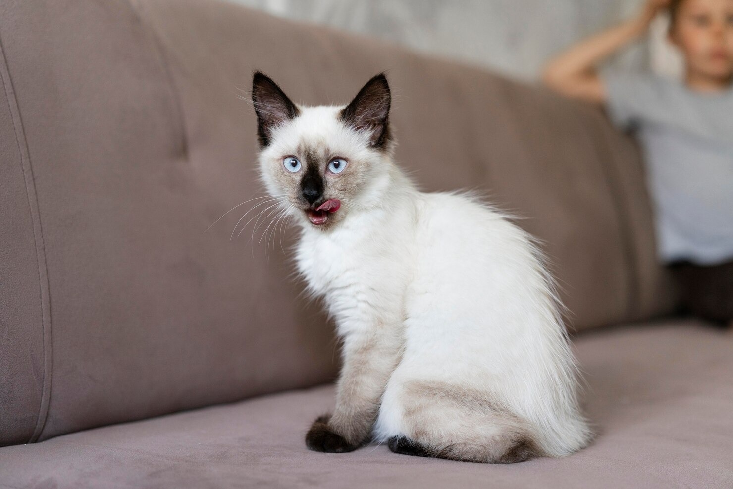
What is Aural Hematoma in Cats?
This content was prepared with AI assistance and reviewed by a licensed professional for accuracy.
Introduction
If you have recently noticed a sudden, fluid-filled swelling on your feline companion’s ear flap, you may be wondering: what is an aural hematoma in cats? Often referred to simply as an ear hematoma or a cat hematoma, this condition occurs when one or more blood vessels within the delicate structure of the ear rupture. The ear flap, or pinna, is constructed of a central layer of rigid auricular cartilage sandwiched tightly between two layers of skin and connective tissue. When a blood vessel shears or breaks beneath the skin, the leaking blood has nowhere to go. It forcefully pools between the skin and the cartilage, creating a distinctive, painful, balloon-like pocket of fluid[1].
An aural hematoma is generally not viewed as a primary disease in itself, but rather as a glaring clinical symptom of an underlying medical issue. Cats are remarkably resilient creatures, but the intense discomfort caused by a rapidly expanding pocket of blood in their ear will almost always trigger significant behavioral changes. The pressure from the pooled fluid stretches the sensitive nerve endings in the skin of the pinna, leading to localized throbbing, heat, and severe pain[2]. Without prompt veterinary intervention, this condition can cause permanent damage to the structural integrity of the ear.
The progression of an untreated aural hematoma is a primary concern for veterinary professionals. As the pooled blood sits stagnant in the pocket, the body attempts to heal the space by producing thick fibrin and scar tissue. This inflammatory cascade leads to the severe crinkling, folding, and thickening of the ear flap, commonly referred to in veterinary medicine as a “cauliflower ear.” This irreversible fibrotic deformation can severely obstruct the ear canal, predisposing the cat to lifelong chronic ear infections[3]. Therefore, obtaining immediate emergency care is crucial not only for alleviating your pet’s acute pain but also for preserving the long-term health and functional anatomy of the ear.
Types of Aural Hematomas in Cats
In feline medicine, aural hematomas are typically classified based on their duration, the nature of the fluid trapped within the pinna, and their anatomical location. The most common classification system divides hematomas into two primary categories: acute and chronic. Understanding the difference between these two stages is essential, as the physical characteristics of the hematoma dictate the most appropriate and effective treatment plan[4].
An acute aural hematoma is characterized by a sudden, rapid onset of swelling. This usually occurs within a matter of hours or days following a traumatic event or a severe bout of scratching. In the acute phase, the swelling is highly fluctuant, meaning it feels like a soft, fluid-filled water balloon when gently palpated. The ear is typically warm to the touch due to active inflammation, and the fluid inside is predominantly fresh, bright red whole blood or serosanguineous (blood-tinged) serum. Cats with an acute hematoma often display high levels of distress, frequently shaking their heads aggressively or pawing frantically at the side of their face[5].
Conversely, a chronic aural hematoma represents a swelling that has been present for a longer duration, usually several weeks or more. Over time, the body attempts to resorb the liquid portion of the blood, leaving behind a dense, organizing clot. Fibrinogen converts into a tough fibrin meshwork, and granulation tissue begins to form within the dead space. As a result, a chronic hematoma feels firm, thickened, and less fluid-like. By this stage, the irreversible process of cartilage deformation has often begun, leading to a shriveled, thickened appearance of the ear flap. Treating a chronic hematoma surgically is generally more complicated because the scar tissue has securely adhered the skin layers in unnatural, distorted positions[6].
Additionally, veterinarians will note the specific location and extent of the swelling. An aural hematoma can be unilateral (affecting only the right or left ear) or bilateral (affecting both ears simultaneously). Bilateral hematomas strongly suggest a severe, generalized underlying condition, such as a massive ear mite infestation or a severe systemic allergic reaction. The swelling may also be localized to the base of the ear, the distal tip, or it may entirely engorge the entire pinna, completely obscuring the normal concave shape of the cat’s ear flap[7].
Causes of Aural Hematoma in Cats

Aural hematomas do not materialize spontaneously without a triggering event. They are the direct mechanical result of physical damage to the vascular network within the ear flap. To properly cure the hematoma and prevent a frustrating recurrence, the veterinarian must play detective to uncover exactly what caused the cat to traumatize its own ear. The underlying triggers can range from simple parasitic infestations to complex immune-mediated diseases. The causes can generally be categorized into the following primary groups.
Physical Trauma
One of the most prevalent causes of aural hematomas in cats is trauma. The physical trauma required to rupture the blood vessels does not necessarily have to come from an external source, such as an impact or a bite wound from another animal, although cat fights and blunt force injuries certainly can cause hematomas. More often than not, the trauma is entirely self-inflicted through vigorous head shaking or relentless scratching[8].
When a cat violently shakes its head to relieve an itch or dislodge debris, the ear flap acts somewhat like a whip. The intense centrifugal force generated at the tip of the ear creates a massive shearing stress between the skin and the underlying cartilage. The delicate capillary beds and small arterioles running through the ear simply cannot withstand this force, causing them to snap and hemorrhage. Similarly, the sharp claws on a cat’s hind legs can easily tear through the internal blood vessels when the cat vigorously scratches at its ear to relieve the deep, maddening itch of an inner ear issue[9].
Parasites
Microscopic parasites are a leading culprit behind the intense pruritus (itchiness) that leads to self-inflicted ear trauma. Infestations of ear mites, scientifically known as Otodectes cynotis, are exceptionally common in cats, particularly in kittens and outdoor cats. These microscopic, crab-like organisms live deep within the external ear canal, where they feed voraciously on epidermal tissue debris and lymphatic fluids[10].
As the ear mites feed and reproduce, their saliva and feces trigger an intense, localized hypersensitivity reaction within the cat’s ear canal. This results in severe inflammation, the overproduction of dark, crumbly, coffee-ground-like ear wax, and an overwhelming urge to scratch. The cat’s frantic efforts to alleviate the microscopic crawling sensation almost inevitably lead to the violent head shaking and scratching that mechanically ruptures the blood vessels of the pinna, thereby creating the hematoma[11].
Allergic Reactions
Feline allergies frequently manifest as dermatological issues, with the ears being a primary target for inflammation. Cats can suffer from atopic dermatitis triggered by environmental allergens such as pollen, mold spores, or dust mites. When an allergic cat inhales or comes into contact with these triggers, their immune system overreacts, releasing large amounts of histamine from mast cells located in the skin. This histamine release causes profound erythema (redness) and severe itching, particularly around the face, neck, and ears[12].
Furthermore, adverse cutaneous food reactions are a significant consideration. In fact, many cats may scratch their ears excessively due to allergies in food. Proteins in commercial cat foods, such as chicken, beef, fish, or dairy, can trigger an immune response that manifests as chronic, bilateral ear inflammation (otitis externa). The constant discomfort and itchiness stemming from these allergic reactions force the cat to traumatize its own ears, creating the perfect environment for a hematoma to form[13].
Underlying Health Conditions
Any primary disease process that causes discomfort or obstruction within the ear canal can lead to the development of an aural hematoma. Otitis externa, which is an infection of the outer ear canal caused by bacteria (such as Staphylococcus or Pseudomonas) or yeast (such as Malassezia pachydermatis), is a very common instigator. These infections cause the lining of the ear canal to swell, ulcerate, and produce foul-smelling purulent discharge, leading to intense pain and head shaking[14].
Additionally, structural abnormalities deep within the ear can be to blame. Feline inflammatory polyps, which are benign inflammatory growths that originate in the middle ear or nasopharynx, can grow outward into the external ear canal. These polyps create a physical blockage, causing a feeling of fullness and pain that drives the cat to paw at its ear. Similarly, older cats may develop ceruminous gland adenomas or other forms of neoplasia (tumors) within the ear canal. Foreign bodies, such as grass awns or foxtails that become lodged in the ear, also create severe acute irritation that predictably leads to self-trauma and hematoma formation[15].
Unidentifiable Causes
Despite thorough diagnostic investigations, there are instances where the exact cause of the aural hematoma remains elusive. In these idiopathic cases, the veterinarian cannot find any evidence of ear mites, infections, allergies, polyps, or trauma. Current veterinary literature suggests that some of these unexplained hematomas may actually be the result of a localized immune-mediated disease. In these rare scenarios, the cat’s own immune system mistakenly produces autoantibodies that attack the auricular cartilage, causing it to weaken and fracture spontaneously, which then shears the adjacent blood vessels[16].
Furthermore, structural genetics may play a role. Certain cats might be predisposed to aural hematomas based on their breed anatomy. Cats with unusually large, heavy, or naturally folded ear cartilage may experience different mechanical stresses during normal grooming or head movement, making their auricular blood vessels more susceptible to rupture. Regardless of the underlying cause, the mechanical reality of the pooled blood remains the same, requiring swift clinical intervention.
Symptoms of Cat Ear Hematoma

Identifying an aural hematoma is usually straightforward for a pet owner, as the clinical signs are highly visible and the cat’s behavioral cues are difficult to ignore. The most prominent symptom is the sudden appearance of a distinct, fluid-filled swelling on the outer flap of the ear. This swelling can vary drastically in size; it might present as a small, localized bulge near the base of the ear, or it may entirely engorge the pinna, making the ear look like a thick, distended, heavy balloon[17].
When you gently touch the affected ear, it will often feel unusually warm due to the localized inflammatory response. In the acute stage, the swelling will feel fluctuant and soft, squishing slightly beneath your fingers. The sheer weight of the pooled blood will often cause the affected ear to droop downward heavily, completely altering the cat’s normal alert facial symmetry.
Beyond the physical appearance, the cat will display a multitude of behavioral symptoms directly related to the pain and pressure of the swelling. You will likely observe the cat engaging in incessant, violent head shaking or holding its head at a severe tilt, favoring the heavy, painful side. Cats will frequently use their hind legs to aggressively scratch or paw at the problematic ear, which can cause secondary excoriations, hair loss (alopecia), and bleeding on the surface of the skin[1].
Because the jaw joint (temporomandibular joint) is located very close to the base of the ear canal, the pressure from a large hematoma or an underlying deep ear infection can make opening the mouth incredibly painful. Consequently, the cat may experience a decreased appetite, dropping food, or refusing to eat hard kibble entirely. You may also notice significant changes in the cat’s behavior, such as hiding in dark spaces, vocalizing in distress when approached, or exhibiting uncharacteristic aggression if you attempt to touch their head. In cases where the underlying cause is a severe bacterial infection or a ruptured tympanic membrane, you may also see foul-smelling pus or blood-tinged discharge leaking from the opening of the ear canal[2].
Diagnosis of Ear Hematoma in Cats

When you bring your cat to the clinic, the veterinarian will execute a comprehensive, step-by-step diagnostic protocol. The goal is twofold: accurately confirm that the swelling is indeed an aural hematoma, and relentlessly hunt down the primary underlying cause that triggered the cat to traumatize its ear in the first place.
Clinical Assessment
The diagnostic process begins with a thorough clinical assessment. The veterinarian will carefully inspect the outer surface of both ear flaps, checking for asymmetry, erythema, and excoriations from scratching. Through gentle digital palpation, the vet will assess the texture, temperature, and fluctuance of the swelling to distinguish an aural hematoma from other potential conditions, such as a localized skin abscess resulting from a bite wound, an insect sting reaction, an edematous allergic flare, or a solid neoplastic tumor. Checking the patient’s overall body temperature, heart rate, and lymph nodes helps rule out systemic infectious processes[3].
Otoscopic Evaluation
To identify the root cause of the self-trauma, an otoscopic evaluation is strictly mandatory. Using a specialized handheld instrument called an otoscope, which provides bright magnification and illumination, the veterinarian will carefully navigate deep into the vertical and horizontal ear canals. They will look for heavy accumulations of dark cerumen (wax), strictures, foreign bodies like grass awns, crawling ear mites, and inflammatory polyps. Importantly, the vet will attempt to visualize the tympanic membrane (eardrum) to ensure it is intact and to check for signs of a deeper middle ear infection, known as otitis media[4].
Aspiration and Cytological Analysis
If the physical diagnosis is ambiguous, the veterinarian may perform a Fine Needle Aspirate (FNA). By inserting a sterile, small-gauge needle into the swelling and pulling back the syringe plunger, the vet can extract a sample of the trapped fluid. If the fluid is bright red blood or a clear, blood-tinged serum, an aural hematoma is confirmed. If the fluid is thick, white, or green pus, an abscess is diagnosed instead.
Simultaneously, the vet will use a cotton swab to collect debris from deep inside the ear canal. This material is rolled onto a glass microscope slide, heat-fixed, and stained with a specialized compound like Diff-Quik. Through cytological analysis under a high-powered microscope, the vet can identify exactly which microorganisms are thriving in the ear, differentiating between rod-shaped bacteria (like Pseudomonas), round bacteria (cocci like Staphylococcus), and budding yeast organisms (like Malassezia). A mineral oil preparation may also be examined to look for live mites and their eggs[5].
Imaging Techniques
While an aural hematoma itself does not require an x-ray, advanced imaging techniques are heavily utilized when the veterinarian suspects severe underlying middle ear or inner ear disease. If the cat exhibits a severe head tilt, facial nerve paralysis, or nystagmus (flicking of the eyes), it implies that an infection or mass has deeply penetrated the skull. In these complex cases, the vet may perform a series of skull radiographs to evaluate the tympanic bullae (the bony shells enclosing the middle ear) for thickening or fluid accumulation. For even greater precision, a CT scan or an MRI can provide high-resolution, three-dimensional views to perfectly map out deep-seated tumors or invasive inflammatory polyps prior to surgical removal[17].
Probing for Underlying Causes
Diagnosing an aural hematoma goes far beyond the ear itself. If the cat has a history of chronic skin issues, excessive grooming, or recurrent bilateral ear infections, the vet will probe for systemic, underlying causes. This might involve blood tests to rule out endocrine disorders, such as feline hyperthyroidism, which can affect skin and coat health. If an allergic basis is strongly suspected, the veterinarian may recommend initiating a strict, 8-to-12-week hydrolyzed protein food trial or performing environmental allergy testing. Uncovering and successfully treating these systemic trigger factors is the only reliable way to guarantee that the cat will stop scratching, thereby preventing future hematomas on either ear[18].
Treating Aural Hematoma in Cats

The successful management of a feline aural hematoma requires a dual-pronged approach: the physical removal of the pooled blood to alleviate the painful pressure, and the simultaneous, aggressive medical treatment of the underlying ear disease that caused the initial scratching. Leaving the hematoma to heal on its own is medically inappropriate, as it practically guarantees massive scar tissue formation and lifelong disfigurement. Always consult your veterinarian before making any changes to your pet’s care, as the optimal treatment method varies based on the size, age, and chronicity of the hematoma.
Needle Aspiration
For very small, acute hematomas, or in cases where the patient is a poor candidate for general anesthesia due to advanced age or severe systemic illness, a veterinarian may attempt a conservative procedure known as fine needle aspiration. During this minimally invasive procedure, the skin over the hematoma is clipped and surgically prepped. A large-gauge needle attached to a syringe is inserted directly into the pocket to suction out the fluid and fresh blood. While this provides immediate, profound relief of pain and pressure, it has a glaring flaw: the dead space between the cartilage and the skin remains intact, and the ruptured blood vessel is not ligated[6].
Because the internal bleeding often continues, the pocket frequently refills within 24 to 48 hours. Needle aspiration alone has a notoriously high recurrence rate. To combat this, the veterinarian may attempt to place a tight, compressive head bandage over the ears to mechanically force the skin and cartilage together, though cats are notoriously intolerant of head wraps and often manage to remove them. Therefore, aspiration is often viewed as a temporary palliative measure rather than a definitive cure.
Surgical Intervention
Surgical intervention remains the absolute gold standard for treating aural hematomas in cats, offering the lowest recurrence rate and the best cosmetic outcome. Under full general anesthesia, the surgical site is sterilized, and the veterinary surgeon makes a precise incision directly over the hematoma on the concave (inner) surface of the ear flap. This incision—which may be a straight line, an S-shape, or a series of small circular holes created with a biopsy punch—allows for the complete evacuation of the liquid blood and the meticulous removal of any solid, organizing fibrin clots[8].
Once the pocket is thoroughly flushed and cleaned, the surgeon must eliminate the dead space so the ear cannot refill with blood. This is achieved by placing multiple, staggered mattress sutures completely through the ear flap. These sutures are placed parallel to the major blood vessels of the ear to avoid cutting off circulation, and they essentially quilt the two layers of skin tightly against the central cartilage plate. A passive latex drain (like a Penrose drain) or a small active vacuum drain may be temporarily left in place to siphon off ongoing inflammatory fluid. The ear is typically bandaged, and the sutures remain in place for two to three weeks to allow dense, healthy scar tissue to permanently bond the layers back together[14].
Corticosteroid Injections
In certain scenarios, a veterinarian may choose to utilize corticosteroid therapy, either as an alternative to surgery or as an adjunctive treatment to needle aspiration. After drawing out the fluid with a syringe, the vet may inject a long-acting depot steroid, such as triamcinolone or methylprednisolone, directly into the empty cavity. Corticosteroids are powerful anti-inflammatory agents that drastically reduce the localized swelling, suppress the immune response that leads to excessive fibrin production, and encourage the tiny, damaged capillaries to seal themselves off[20].
Some practitioners may also prescribe a tapering dose of oral systemic steroids, like prednisolone, to calm the generalized itchiness and inflammation associated with severe allergies or widespread otitis. While this method is less invasive and less expensive than major surgery, it does carry potential systemic side effects—such as increased thirst, increased urination, and mild immunosuppression—which must be monitored carefully.
Treatment of the Underlying Causes
No surgical or medical treatment of the ear flap will be successful in the long term if the underlying cause is ignored. If the cytology reveals a bacterial or yeast infection, the vet will prescribe potent medicated otic drops containing a combination of antibiotics, antifungals, and glucocorticoids to be applied directly into the ear canal daily. If an ear mite infestation is confirmed, highly effective topical or systemic parasiticides belonging to the isoxazoline or macrocyclic lactone classes will be administered to eradicate the mites. If deep polyps are found, they must be pulled out via traction or removed via a deeper surgical procedure called a ventral bulla osteotomy[11].
During the entire recovery period, the cat must wear a rigid Elizabethan collar (the “cone of shame”) to absolutely prevent any further scratching or trauma to the healing ear. Follow-up visits are mandatory to remove sutures, check for fluid re-accumulation, and perform repeat cytology to ensure the underlying ear canal infection is completely resolved.
Prevention of Feline Aural Hematoma
Preventing an aural hematoma from forming is vastly easier, safer, and less expensive than subjecting your cat to corrective surgery. Prevention fundamentally relies on the vigilant management of the various underlying health conditions that cause ear irritation. By implementing a proactive, comprehensive health care strategy, you can protect your feline friend from this painful affliction.
Regular Vet Check-ups
Routine, comprehensive veterinary wellness exams, ideally conducted on an annual or biannual basis, are the absolute bedrock of preventive care. During these visits, your veterinarian doesn’t just administer vaccines; they use an otoscope to look deep into your cat’s ear canals. This routine screening can identify microscopic, subclinical inflammation, early-stage yeast overgrowth, or tiny, developing polyps long before they cause enough pain to trigger violent head shaking. Early pharmaceutical intervention can stop an infection in its tracks before a hematoma ever has the chance to form[1].
Parasite Control
Maintaining a strict, year-round parasite control regimen is arguably the easiest and most effective way to prevent ear mite-induced hematomas. Modern veterinary medicine offers highly safe, broad-spectrum topical liquids and oral chewables that reliably kill Otodectes cynotis, alongside fleas, ticks, and heartworms. It is a dangerous misconception that strictly indoor cats do not need parasite prevention; mites and fleas can easily be tracked inside on human clothing or introduced by other visiting pets. Consistent use of these preventatives ensures your cat’s ears remain pest-free[19].
Regular Ear Checks at Home
Pet owners play a vital role in early detection. You should establish a weekly routine of gently inspecting your cat’s ears for signs of infection. A healthy feline ear should look clean, pale pink, and be entirely free of strong, offensive odors. If you lift the ear flap and notice a buildup of thick, dark, coffee-ground-like debris, bright yellow pus, intense redness, or if the ear emits a distinctly yeasty, sweet, or foul odor, you must contact your vet immediately. Do not attempt to dig into the ear canal with cotton swabs, as this can pack debris deeper against the eardrum and cause the exact irritation you are trying to avoid.
Safe Environment
Trauma prevention requires maintaining a safe, calm, and enriched environment. If you live in a multi-cat household, inter-cat aggression and dominance scuffles can easily result in bites or claw tears to the delicate ear flaps. Providing adequate vertical space, multiple litter boxes, and utilizing synthetic feline facial pheromone diffusers can dramatically reduce household tension. Furthermore, keeping your cat’s claws routinely trimmed will minimize the soft tissue damage they can inflict on themselves if they do happen to experience a minor, fleeting itch[7].
Monitor for Allergies
If your cat is diagnosed with a generalized atopic skin disease or an adverse food reaction, meticulous management is required to prevent secondary ear issues. You must strictly adhere to the veterinary-prescribed hypoallergenic or hydrolyzed protein diets, ensuring no unauthorized treats or table scraps are consumed. For environmental allergies, your vet may recommend daily oral antihistamines, immunomodulatory medications like cyclosporine, or allergen-specific immunotherapy injections. Understanding how to properly care for your cat during seasonal allergy flare-ups will drastically reduce their urge to scratch and traumatize their ears[12].
Frequently Asked Questions
Can a cat’s ear hematoma heal on its own?
While an untreated aural hematoma will eventually cease expanding as the bleeding stops internally, it will never heal properly on its own. The fluid trapped inside will slowly be reabsorbed by the body over several painful weeks, but the empty space will be violently replaced by dense, contracted scar tissue. This process causes the ear cartilage to warp, crinkle, and fold in on itself, resulting in a permanent, unsightly deformity known as a cauliflower ear. This deformity can block the ear canal, predisposing the cat to lifelong, chronic ear infections. Therefore, veterinary treatment is absolutely necessary to alleviate the acute pain and preserve the structural integrity of the ear.
How long does it take for an aural hematoma to heal after surgery?
The standard recovery timeline following surgical intervention for a feline aural hematoma is generally between two to three weeks. During this critical healing phase, the specialized mattress sutures placed by the veterinarian remain in the ear flap to hold the skin tight against the cartilage, completely eliminating the dead space. Throughout this entire period, the cat must continuously wear a rigid Elizabethan collar to prevent any scratching, rubbing, or disruption of the surgical site. Once the veterinarian removes the sutures at the follow-up appointment, the ear may still have a mildly thickened appearance, but it should be fully healed and pain-free.
Are certain cat breeds more prone to developing aural hematomas?
While any cat of any breed, age, or sex can develop an aural hematoma if they experience trauma or an underlying ear infection, certain anatomical features may increase the risk. Cats with naturally pendulous or folded ears, such as the Scottish Fold, or cats with excessively large, heavy pinnae, like the Maine Coon or Savannah cat, may be slightly more predisposed. The extra weight and unique cartilage angles in these ears can create more severe mechanical whip-like forces during head shaking, increasing the likelihood of blood vessel rupture. However, chronic otitis and ear mites remain the primary driving factors across all breeds.
References
- American Veterinary Medical Association. Ear Infections and Hematomas. AVMA, 2023.
- Merck Veterinary Manual. Diseases of the Pinna in Cats. Merck & Co., 2022.
- Journal of Feline Medicine and Surgery. Pathophysiology and Management of Aural Hematomas. JFMS, 2018.
- VCA Animal Hospitals. Hematoma of the Ear in Cats. VCA, 2021.
- Cornell University College of Veterinary Medicine. Ear Mites Fact Sheet. Cornell Feline Health Center, 2023.
- Veterinary Record. Conservative Medical Management of Aural Hematomas. Vet Rec, 2011.
- ASPCA. Common Cat Diseases and Parasites. ASPCA, 2022.
- Journal of Small Animal Practice. Surgical Treatment of Aural Hematoma. JSAP, 2013.
- Veterinary Surgery. Comparison of Incisional Drainage Techniques for Aural Hematomas. Vet Surg, 2012.
- Merck Veterinary Manual. Otodectes cynotis (Ear Mites). Merck & Co., 2023.
- Veterinary Clinics of North America: Small Animal Practice. Otology and Feline Ear Disease. VCNA, 2019.
- Veterinary Dermatology. Feline Allergic Skin Disease. Vet Dermatol, 2016.
- Journal of Feline Medicine and Surgery. Adverse Food Reactions in Cats. JFMS, 2018.
- Journal of the American Veterinary Medical Association. Retrospective Study of Feline Otitis Externa. JAVMA, 2004.
- Veterinary Pathology. Inflammatory Polyps in the Feline Ear. Vet Pathol, 2017.
- Veterinary Information Network. Management of the Cauliflower Ear. VIN, 2008.
- Veterinary Radiology & Ultrasound. Imaging the Feline Tympanic Bulla. VRU, 2015.
- Veterinary Dermatology. Diagnosis and Management of Cutaneous Adverse Food Reactions. Vet Dermatol, 2017.
- Centers for Disease Control and Prevention. Animals and Parasitic Diseases. CDC, 2022.
- Journal of Veterinary Internal Medicine. Corticosteroid Therapy and Fibrinolysis in Hematomas. JVIM, 2021.


