


 March 11, 2023
 Phil Good, DVM
What is Urethral Obstruction in Cats?
This content was prepared with AI assistance and reviewed by a licensed professional for accuracy.
Introduction
A urethral obstruction in cats, commonly referred to as a blocked urethra or urinary blockage, is an acute, agonizing, and immediately life-threatening medical emergency. This condition occurs when the urethra—the narrow, tubular structure responsible for carrying urine from the bladder out of the body—becomes completely or partially occluded. When a cat cannot empty its bladder, the continuous production of urine by the kidneys causes the bladder to distend to dangerous levels. Without prompt and aggressive veterinary intervention, this mechanical backup of urine leads to severe metabolic disturbances, acute kidney injury, and eventually death within 48 to 72 hours.[1]
Feline urethral obstruction is predominantly observed in male cats due to their specific reproductive anatomy. While female cats have a relatively short, wide, and distensible urethra, male cats possess a much longer urethra that tapers significantly as it passes through the penis. This anatomical bottleneck creates an ideal environment for microscopic crystals, mucus, blood clots, or inflammatory debris to become wedged, halting the flow of urine.[2] The inability to void urine not only causes profound physical pain and distress but also prevents the body from eliminating toxic waste products, such as urea, creatinine, and potassium, which rapidly accumulate in the bloodstream.
Recognizing the early warning signs of a urinary blockage can mean the difference between life and death for a beloved feline companion. Pet owners must understand the underlying mechanisms, risk factors, and clinical presentations associated with this severe condition. Immediate stabilization, comprehensive medical diagnostics, and precise therapeutic strategies are required to relieve the obstruction, restore urinary patency, and reverse the catastrophic biochemical imbalances that occur during an obstructive episode.[3] By exploring the complex pathophysiology behind these blockages, caretakers can better partner with their veterinary team to ensure long-term management and prevention.
What Causes Feline Urethral Blockage?

Feline urethral blockage is rarely a simple, isolated event; rather, it is typically the culmination of underlying disease processes within the urinary system, collectively known as Feline Lower Urinary Tract Disease (FLUTD). The actual mechanical blockage is usually caused by physical material becoming lodged in the narrow penile urethra, though severe muscle spasms can also contribute to functional obstruction. Understanding the specific culprits behind these blockages is essential for preventing recurrence.[4]
One of the most frequent causes of a blocked urethra is the formation of urethral plugs. Unlike solid stones, a urethral plug is a soft, toothpaste-like substance composed of a protein-rich matrix mixed with mineral crystals and cellular debris. The protein matrix is often formed by Tamm-Horsfall mucoproteins, which are secreted by the kidneys, alongside inflammatory proteins leaking from an irritated bladder wall. When a cat is suffering from inflammation, these proteins bind with microscopic minerals—most commonly struvite crystals—forming a pliable but dense plug that perfectly molds to the tapering shape of the male urethra, creating an impenetrable seal.[5]
Urolithiasis, or the formation of true bladder stones (uroliths), is another primary cause of urinary blockage. These are rock-hard mineral concretions that form over weeks or months inside the bladder. If a small stone is swept into the urethra during voiding and is too large to pass, it will become lodged, abruptly halting urine flow. The two most common types of urinary stones in cats are struvite (magnesium ammonium phosphate) and calcium oxalate. The formation of these stones is heavily influenced by the cat’s urine pH, hydration status, and dietary mineral intake. Highly concentrated urine, common in cats fed exclusively dry kibble, provides a supersaturated environment where these minerals easily precipitate into solid stones.[6]
A significant underlying trigger for the formation of both plugs and stones is Feline Idiopathic Cystitis (FIC). FIC is a complex, poorly understood syndrome characterized by chronic, sterile inflammation of the bladder wall. Cats with FIC experience neurogenic inflammation, often exacerbated by environmental stress, which damages the protective glycosaminoglycan (GAG) layer of the bladder. This allows harsh urine components to irritate the underlying tissue, triggering severe muscle spasms and the shedding of inflammatory debris that ultimately leads to obstruction.[7]
Other, less common causes of urethral obstruction include physical strictures (scar tissue formation from previous trauma or urinary catheterization), severe bacterial urinary tract infections (which are rare in young male cats but can cause pus and debris buildup), and neoplasia (tumors of the bladder or urethra, such as transitional cell carcinoma). Furthermore, certain lifestyle factors drastically increase a cat’s risk. Sedentary, indoor-only overweight cats are heavily predisposed, as their lack of activity and infrequent trips to the litter box lead to prolonged urine retention and increased crystal formation. Stressful environments, inter-cat conflict, and inadequate water consumption further compound these risks, making environmental and dietary management critical components of long-term care.[8]
What are the Signs of Urethral Obstruction?
The clinical signs of a urethral obstruction often progress rapidly, transitioning from subtle behavioral changes to severe, systemic illness in a matter of hours. Early detection relies heavily on an owner’s vigilance regarding their cat’s litter box habits. Initially, a cat with a partial or impending blockage will exhibit signs typical of lower urinary tract inflammation. They will make frequent, unproductive trips to the litter box, a condition known as pollakiuria. Owners often observe the cat posturing to urinate for extended periods, passing only a few drops of urine, or none at all. This severe straining, or dysuria, is frequently mistaken by pet owners as constipation.[9]
As the obstruction solidifies and pressure within the bladder builds, the pain becomes excruciating. Cats will often vocalize loudly—crying, howling, or growling—while in the litter box or when their abdomen is touched. You may notice the Presence of blood in urine (hematuria), either in the few drops the cat manages to pass or spotted on the floor, bedding, or around the cat’s perineal area. In a desperate attempt to soothe the intense localized pain, the cat will excessively lick their genital region, sometimes to the point of causing trauma to the penis. Periuria, or urinating outside the litter box in inappropriate places like bathtubs, sinks, or laundry baskets, is also common as the cat associates the litter box with pain.[10]
If the blockage is not relieved within 24 hours, the condition progresses from a localized urinary issue to a systemic, life-threatening metabolic crisis. As the kidneys are unable to filter and excrete waste, toxins rapidly build up in the bloodstream—a condition known as uremia. The cat will become profoundly lethargic, hiding in unusual places and refusing all food and water. The buildup of toxins often stimulates the chemoreceptor trigger zone in the brain, leading to severe nausea, drooling, and vomiting.[11]
In the final, most critical stages of an obstruction (typically 48 to 72 hours post-blockage), the cat will experience extreme weakness, collapse, and an altered state of consciousness, appearing comatose or unresponsive. This extreme deterioration is largely driven by hyperkalemia—a lethal spike in blood potassium levels. Potassium, normally excreted in the urine, builds up and profoundly disrupts the electrical conduction system of the heart, leading to a dangerously slow and irregular heart rate (bradycardia). Without immediate emergency stabilization, this will progress to fatal cardiac arrest. Any cat exhibiting the inability to pass urine accompanied by lethargy and vomiting must be transported to an emergency veterinary hospital immediately.[12]
Diagnosis of Urethral Obstruction in Cats

Diagnosing a urethral obstruction requires swift, decisive action. Because the condition rapidly deteriorates into a systemic metabolic crisis, the diagnostic process is often carried out simultaneously with emergency stabilization. Veterinarians utilize a combination of physical assessments, laboratory analyses, and diagnostic imaging to confirm the blockage, determine its underlying cause, and evaluate the extent of the internal damage caused by urine retention.
Physical Examination
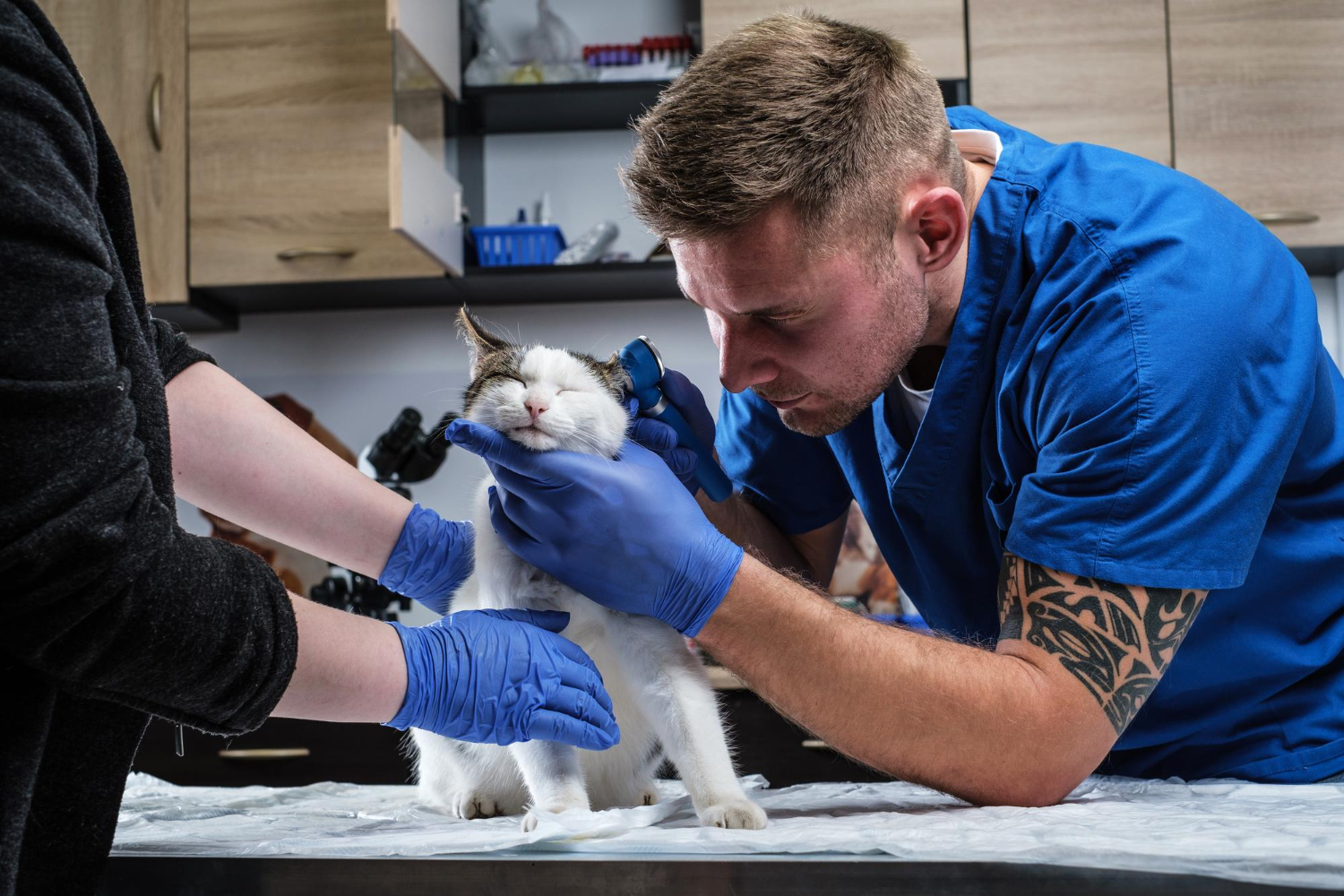
The diagnostic protocol begins with a rapid but thorough physical examination. The veterinary team will immediately assess the cat’s cardiovascular stability, focusing on heart rate, pulse quality, and mucous membrane color. A heart rate below 140 beats per minute in a highly stressed, painful cat is a major red flag for hyperkalemia. The vet will closely observe the cat’s behavior and mentation, noting signs of severe uremic depression or altered consciousness. Body temperature is evaluated, as obstructed cats often present with significant hypothermia due to poor perfusion and shock. The physical exam quickly establishes the urgency of the intervention required.[13]
Palpation
The hallmark diagnostic step for a urethral obstruction is the careful abdominal palpation performed by the veterinarian. In a normal cat, the bladder feels like a small, soft, pliable water balloon. In an obstructed cat, the bladder becomes massively distended, firm, and turgid, often described as feeling like a hard peach or grapefruit in the caudal abdomen. Crucially, the bladder cannot be manually expressed. The veterinarian must perform this palpation with extreme gentleness; aggressive squeezing or excessive pressure on an obstructed, compromised bladder wall can lead to catastrophic bladder rupture (uroabdomen), spilling toxic, bacteria-laden urine directly into the abdominal cavity.[14]
Urinalysis
Once the obstruction is relieved via catheterization, collecting a sterile urine sample for comprehensive urinalysis is paramount. In blocked cats, cystocentesis (drawing urine directly from the bladder with a needle) is generally contraindicated prior to catheterization due to the high risk of bladder rupture. The urinalysis evaluates the urine specific gravity (USG), which indicates how well the kidneys are concentrating urine. A microscopic evaluation of the urine sediment is crucial for identifying the presence and type of crystals (such as struvite or calcium oxalate), red blood cells (hematuria), white blood cells (pyuria indicating inflammation or infection), and bacterial rods or cocci. Additionally, evaluating the urine pH helps dictate future dietary management, as struvite crystals thrive in alkaline urine, while calcium oxalate prefers acidic environments.[15]
Blood Tests
Comprehensive bloodwork, including a complete blood count (CBC), serum chemistry panel, and venous blood gas analysis, is absolutely vital. The chemistry panel will typically reveal profound post-renal azotemia, characterized by massive elevations in Blood Urea Nitrogen (BUN) and creatinine, reflecting the kidneys’ inability to filter waste. The most critical parameter evaluated is the serum potassium level (hyperkalemia). Normal potassium ranges from 3.5 to 5.5 mEq/L; blocked cats frequently present with levels exceeding 8.0 mEq/L, which causes life-threatening cardiac arrhythmias. Blood tests also evaluate phosphorus levels (hyperphosphatemia) and acid-base balance, often revealing severe metabolic acidosis that requires specific therapeutic correction during fluid therapy.[16]
Imaging
Diagnostic imaging is required to visualize the lower urinary tract and determine the physical nature of the blockage. Abdominal radiographs (X-rays) are standard procedure to identify radiopaque uroliths (calcium oxalate and struvite stones) located in the bladder or lodged within the urethra. Radiographs also help assess the overall size of the bladder and the size of the kidneys. Abdominal ultrasonography may be utilized to detect radiolucent stones (like urate), evaluate the thickness and irregularity of the bladder wall (cystitis), and check for hydronephrosis (swelling of the kidneys due to back-pressure). Together, these imaging modalities provide a comprehensive picture of feline lower urinary tract disease, guiding whether surgical or medical management is the most appropriate next step.[17]
Treatment for Urinary Blockage in Cats

The treatment of a feline urethral obstruction is a multi-step, intensive care process that requires hospitalization, advanced medical therapies, and continuous monitoring. Because this is a life-threatening crisis, the treatment protocol is rigorous and highly structured to ensure the cat survives the initial metabolic shock, successfully regains urinary patency, and recovers kidney function without permanent damage. Below are the standard therapeutic steps taken by veterinary emergency teams.
Urethral Catheterization
The definitive mechanical treatment for a blocked cat is the placement of an indwelling urinary catheter to bypass the obstruction. Because the procedure is painful and the cat is highly stressed, sedation or general anesthesia is required. To protect the compromised cardiovascular system, a sacrocaudal epidural (a local anesthetic block placed near the tail base) is often administered to numb the penis and urethra, allowing for lighter systemic sedation. The veterinarian carefully extrudes the penis and attempts to pass a sterile, open-ended Tomcat catheter. If a hard plug is encountered, the “retropulsion” technique is utilized. The vet uses a syringe of sterile saline attached to the catheter to forcefully but carefully flush the plug backward into the bladder, clearing the urethral lumen. Once patency is achieved, a softer, longer-term catheter (such as a Slippery Sam or Mila catheter) is guided into the bladder and sutured securely to the prepuce to prevent removal.[18]
Fluid Therapy
Intravenous fluid therapy is the cornerstone of metabolic recovery. Upon presentation, most blocked cats are severely dehydrated, acidotic, and highly toxic. An IV catheter is placed in a peripheral vein, and aggressive fluid resuscitation begins using balanced intravenous electrolyte solutions. Once the urinary catheter is placed, it is attached to a closed collection system (a specialized urinary bag) to accurately measure urine output. Following unblocking, cats frequently experience “post-obstructive diuresis,” a phenomenon where the kidneys, in their effort to flush out accumulated toxins, produce massive, life-threatening volumes of urine. Intravenous fluids must be meticulously matched to the urine output—sometimes requiring hundreds of milliliters per hour—to prevent the cat from quickly dehydrating again and to support the healing of the renal tubules.[19]
Pain Management
Urethral obstruction is widely recognized as one of the most painful conditions in veterinary medicine, making multimodal pain management a critical component of treatment. Initially, strong prescription pain medication is administered intravenously to provide profound systemic analgesia without further depressing the cardiovascular system. Because the kidneys are severely compromised (low glomerular filtration rate), prescription non-steroidal anti-inflammatory drugs (NSAIDs) are strictly contraindicated during the acute phase, as they can precipitate permanent renal failure. Once the cat is stable, nerve-pain medication prescribed by your veterinarian is introduced to manage neuropathic pain and anxiety. Furthermore, prescription urinary muscle relaxants (alpha-1 adrenergic antagonists) are frequently prescribed to relax the smooth muscle of the urethra, reducing painful urethral spasms and lowering the risk of re-obstruction.[20]
Bladder Expression
Following a period of hospitalization—typically 48 to 72 hours—the indwelling urinary catheter is removed once the urine is clear of heavy blood and debris, and kidney blood values have normalized. However, the treatment does not end there. Because the bladder wall was stretched well beyond its normal capacity, the tight junctions between the smooth muscle cells can become damaged, leading to a condition called detrusor atony. In this state, the bladder becomes flaccid and lacks the muscle tone to contract and empty itself, even though the urethra is clear. Veterinary staff must carefully monitor the cat’s voiding post-catheter removal. If the cat cannot urinate voluntarily, gentle manual bladder expression by a trained professional may be required to prevent urine retention. In some cases, a prescription medication is utilized to stimulate bladder muscle contractions.[21]
Surgery
While the vast majority of feline urethral obstructions are managed medically, surgical intervention becomes necessary when medical management fails, when an obstruction cannot be cleared via catheterization, or when a cat suffers from frequent, recurrent blockages despite aggressive preventative care. The gold-standard surgical procedure is the Perineal Urethrostomy (PU). This complex surgery involves the complete amputation of the penis and the removal of the narrowest portion of the pelvic urethra. The surgeon then sutures the wider, more proximal urethra directly to the skin of the perineum, essentially reconstructing the male’s anatomy to resemble the wider, shorter urethra of a female cat. While a PU drastically reduces the risk of future life-threatening obstructions, it is a major surgery with potential complications, including surgical site strictures, bleeding, and an increased lifelong risk of ascending bacterial urinary tract infections.[22]
Emergency Treatment
While the steps above outline the specific therapies and procedures utilized, it is crucial to understand that urethral blockage is managed as a highly coordinated, comprehensive emergency treatment protocol from the moment the cat arrives at the hospital. Before any sedatives are administered or catheterization is attempted, the critical metabolic emergencies must be stabilized. If the cat presents with severe bradycardia and ECG abnormalities (like absent P waves and spiked T waves) due to hyperkalemia, emergency medications are immediately administered. Intravenous emergency cardiac-protecting medication is given slowly to protect the heart muscle and stabilize the cardiac resting membrane potential. Simultaneously, specific intravenous metabolic therapies are administered to drive the excess potassium out of the bloodstream and back into the cells. Only after the heart is protected and the metabolic crisis is temporarily buffered can the clinical team proceed safely to the steps of anesthesia and mechanical unblocking. This immediate, life-saving emergency phase is what allows the subsequent treatments to be successful.[23]
Prevention of Urinary Blockage in Cats
Because the recurrence rate of feline urethral obstruction is frustratingly high, comprehensive, lifelong preventative care is non-negotiable once a cat has been discharged from the hospital. The primary goals of prevention are to dilute the urine, manage the pH to prevent crystal formation, and reduce the systemic stress that triggers bladder inflammation (FIC). A multifaceted approach, known as Multimodal Environmental Modification (MEMO), is the gold standard for maintaining a healthy urinary tract.
The single most effective preventative measure is promoting sufficient hydration to ensure the production of dilute urine. Cats are descended from desert-dwelling ancestors and naturally have a very low thirst drive, meaning they rely on their food for moisture. Pet owners should reconsider exclusive dry food diets, as kibble contains only 5-10% moisture, leading to highly concentrated urine that is prone to supersaturation and crystal formation. Transitioning the cat to a high-quality, exclusively canned or wet food diet (which contains 70-80% moisture) drastically increases total water intake. Furthermore, placing multiple wide, shallow ceramic water bowls or continuously flowing pet water fountains throughout the home can entice hesitant cats to drink more frequently.[24]
Nutritional management is equally critical. Based on the cat’s urinalysis results, the veterinarian will likely prescribe a therapeutic urinary diet carefully formulated for your pet’s needs. These meticulously formulated diets are designed using Relative Supersaturation (RSS) methodology. They control the precise levels of magnesium, phosphorus, and calcium, while artificially modulating the urine pH to create an environment where struvite crystals actively dissolve and calcium oxalate stones are prevented from forming. Many cats that are genetically predisposed to urolithiasis, such as Persians or Himalayans, require these prescription diets for the rest of their lives.
Environmental stress plays a massive role in triggering idiopathic cystitis. Cats are highly sensitive to changes in their environment, inter-cat aggression, boredom, and dirty resources. To manage stress levels, owners must provide an enriched indoor environment featuring vertical climbing spaces, secure hiding spots, and regular interactive play to mimic hunting behaviors. The use of synthetic feline facial pheromones can help reduce generalized anxiety. Furthermore, strict litter box husbandry is essential. The general rule is to have one litter box per cat, plus one extra, distributed in quiet, easily accessible locations across different floors of the house. Boxes should be scooped daily, washed weekly, and filled with unscented, fine-grained clumping litter, as many cats will actively withhold urine if forced to use a dirty or strongly scented box.[25]
Finally, pet owners must remain continually observant. Keep track of urination and litter box habits daily. Schedule regular vet check-ups to monitor kidney values and perform routine urinalyses to ensure the prescribed diet is maintaining the correct pH and specific gravity. Always consult your veterinarian before making any changes to your pet’s care, diet, or medication regimen to ensure you are not inadvertently increasing the risk of another blockage.
Frequently Asked Questions
How long can a cat survive with a blocked urethra?
A complete urethral obstruction is rapidly fatal. Once a cat is entirely unable to pass urine, acute kidney failure begins within 24 hours. By 48 hours, the buildup of potassium in the bloodstream (hyperkalemia) begins to profoundly affect the heart’s electrical system. Without emergency veterinary intervention, a cat will typically suffer fatal cardiac arrest or bladder rupture within 48 to 72 hours. Immediate emergency care is required at the first sign of straining.
Is a blocked urethra painful for a cat?
Yes, a urethral obstruction is considered one of the most agonizing and painful conditions a cat can experience. The extreme distension of the bladder wall stretches nerves to their limits, while the physical scraping of crystals or plugs against the sensitive lining of the urethra causes severe burning and localized trauma. This intense pain is why blocked cats frequently vocalize, howl, and obsessively lick their genital area in an attempt to find relief.
Can a female cat get a urethral obstruction?
While extremely rare due to their anatomy, female cats can theoretically experience a urethral obstruction. The female urethra is significantly shorter, wider, and more flexible than the male’s, allowing them to pass most small stones, crystals, and mucus plugs without issue. When a female cat does become blocked, it is almost exclusively due to a very large bladder stone lodging in the urethra, or due to physical compression from a severe tumor (neoplasia) in the lower urinary tract.
Protect Your Cat’s Urinary Health Today
If you suspect your cat is experiencing a urethral obstruction or has a history of urinary tract issues, immediate action is critical. Don’t wait until it’s too late.
References
- American Veterinary Medical Association (AVMA). Feline Lower Urinary Tract Disease. AVMA, 2023.
- VCA Animal Hospitals. Urethral Obstruction in Cats. VCA Hospitals, 2022.
- Gerber B, et al. Evaluation of clinical signs and causes of lower urinary tract disease in European cats. Journal of Small Animal Practice, 2015.
- Merck Veterinary Manual. Feline Lower Urinary Tract Disease. Merck & Co., 2023.
- Kruger JM, et al. Urethral plugs in feline lower urinary tract disease. Veterinary Clinics of North America: Small Animal Practice, 2011.
- Veterinary Information Network (VIN). Urolithiasis in Cats. VIN, 2021.
- Buffington CA. Idiopathic cystitis in domestic cats—beyond the lower urinary tract. Journal of Veterinary Internal Medicine, 2011.
- ASPCA. Feline Urinary Tract Health. ASPCA Pet Care, 2022.
- Texas A&M School of Veterinary Medicine. Feline Lower Urinary Tract Disease. TAMU Vet Extensions, 2020.
- Gunn-Moore DA. Feline lower urinary tract disease. Journal of Feline Medicine and Surgery, 2014.
- VCA Animal Hospitals. Uremia in Cats. VCA Hospitals, 2021.
- Lee JA, et al. Retrospective evaluation of the management of feline urethral obstruction. Journal of Veterinary Emergency and Critical Care, 2003.
- Merck Veterinary Manual. Feline Idiopathic Cystitis. Merck & Co., 2023.
- Segev G, et al. Uroabdomen in dogs and cats: a retrospective study. Journal of Veterinary Internal Medicine, 2010.
- VCA Animal Hospitals. Urinalysis in Pets. VCA Hospitals, 2022.
- Pouzot C, et al. Hyperkalemia in feline urethral obstruction. Journal of Veterinary Emergency and Critical Care, 2012.
- Biller DS, et al. Ultrasound of the feline lower urinary tract. Veterinary Clinics of North America, 2017.
- Veterinary Information Network (VIN). Management of Feline Urethral Obstruction. VIN, 2020.
- Rieser TM. Urinary tract emergencies. Veterinary Clinics of North America: Small Animal Practice, 2005.
- Dorsch R, et al. Feline lower urinary tract disease in a German cat population. Tierärztliche Praxis, 2014.
- Bartges JW. Therapeutics of feline lower urinary tract disease. Veterinary Clinics of North America, 2008.
- VCA Animal Hospitals. Perineal Urethrostomy Surgery in Cats. VCA Hospitals, 2021.
- Macintire DK. Pediatric intensive care. Veterinary Clinics of North America (Emergency Protocols in Cats), 2004.
- Merck Veterinary Manual. Nutritional Management of Urinary Tract Disease. Merck & Co., 2022.
- Ohio State University College of Veterinary Medicine. Indoor Pet Initiative: Environmental Enrichment. OSU Vet Extensions, 2023.


