


 March 10, 2023
 Phil Good, DVM
What is Mast Cell Tumor in Cats?
This content was prepared with AI assistance and reviewed by a licensed professional for accuracy.
Introduction
Receiving a cancer diagnosis for a beloved feline companion can be an incredibly overwhelming and frightening experience for any pet owner. When a veterinarian mentions a mast cell tumor in cats, it immediately brings to mind a flood of questions and anxieties about the future. While a localized cutaneous mastocytoma may appear as a simple lump on the surface, understanding the underlying biology of this complex disease—and its potential to act as a rare skin cancer with systemic implications—is essential for making informed treatment decisions. Pet owners like Samantha, who discovered an unusual nodule on her cat Fluffy while brushing her, often feel lost in the sea of medical terminology. However, gathering accurate, up-to-date information is the first step toward taking control of your cat’s health journey. [1]
Fundamentally, a Mast cell tumor in cats is a cancer that originates from a specific type of white blood cell known as a mast cell. These cells are integral components of the feline immune system, strategically positioned throughout the body’s connective tissues to act as first responders against environmental threats and parasites. However, when the regulatory mechanisms that control cellular division and lifecycle fail, these normally protective cells begin to multiply uncontrollably, forming neoplastic masses or tumors. [2]
Feline mast cell tumors are unique in the veterinary world and differ significantly from their canine counterparts. While dogs frequently develop highly aggressive skin tumors, cats present with a much more diverse clinical picture. The severity and biological behavior of a feline mast cell tumor are heavily dictated by its anatomic location. Tumors can manifest externally on the skin, within the deeper subcutaneous fat layers, inside the gastrointestinal tract, or within critical blood-filtering organs like the spleen. [3] Each of these locations carries its own distinct prognosis, ranging from entirely benign and curable to highly malignant and life-threatening.
Navigating this complex oncological landscape requires a deep dive into the clinical characteristics of the disease. Recognizing the early warning signs, understanding the necessity of comprehensive diagnostic staging, and exploring the multifaceted treatment options—including surgery, radiation, and targeted chemotherapy—are paramount. By equipping yourself with clinical knowledge, you can work collaboratively with your veterinary oncology team to manage abnormal cell growth, mitigate uncomfortable symptoms, and ultimately preserve your cat’s overall quality of life and well-being for as long as possible. [4]
What is a Feline Mast Cell?
To truly comprehend how a mast cell tumor affects a cat’s body, one must first understand the remarkable biology of the normal feline mast cell. Mast cells are highly specialized immune system components originating from CD34+ hematopoietic stem cells located deep within the bone marrow. [5] Unlike other white blood cells that circulate freely and continuously in the bloodstream, mast cell precursors migrate outward into the peripheral connective tissues. There, they mature and take up permanent residence at the interfaces between the cat’s internal environment and the external world, such as the skin, the lining of the respiratory tract, and the mucosal layers of the gastrointestinal system.
The primary function of a healthy mast cell is to act as a sentinel. They play a crucial role in the body’s defense mechanisms, particularly in mediating inflammatory responses, fighting off parasitic infections (such as intestinal worms or external pests like fleas), and facilitating tissue repair and wound healing. [6] The hallmark characteristic of a mast cell is its cytoplasm, which is densely packed with microscopic, dark-staining secretory vesicles known as granules. These granules are essentially chemical armories, loaded with highly potent bioactive compounds that the cell can deploy at a moment’s notice.
When a mast cell’s surface receptors—specifically the high-affinity IgE receptors—are triggered by a perceived threat or allergen, the cell undergoes a rapid process called degranulation. [7] During degranulation, the mast cell explosively releases its stored chemical payload into the surrounding tissue space. The most well-known of these substances is histamine. Histamine causes localized blood vessels to dilate and become leaky, which creates the classic signs of inflammation: redness, swelling, heat, and itching. This allows other immune cells to quickly exit the bloodstream and enter the tissue to fight the localized threat.
In addition to histamine, mast cell granules contain heparin, a powerful natural anticoagulant that prevents blood from clotting and ensures continuous blood flow to the inflamed area. They also release a variety of proteolytic enzymes, such as tryptase and chymase, which help break down connective tissue matrices to clear the way for immune cells and initiate the wound-healing cascade. Furthermore, mast cells synthesize and secrete an array of cytokines and chemokines—chemical messengers that recruit eosinophils, neutrophils, and macrophages to the site of the reaction. [8]
In a healthy feline body, this complex chemical orchestration is highly controlled and localized. However, when a mast cell undergoes a malignant transformation and becomes a tumor cell, this regulatory control is lost. The neoplastic mast cells continue to produce and store these potent chemicals but may release them spontaneously or inappropriately in massive quantities. This uncontrolled degranulation is responsible for many of the severe, systemic, and sometimes life-threatening symptoms associated with mast cell tumors, transforming a localized skin lump into a complex, whole-body medical crisis. [9]
What are the Types of Mast Cell Tumors in Cats?
The clinical presentation and biological behavior of mast cell tumors (MCTs) in felines are incredibly diverse. Unlike in humans or dogs where the disease may follow a more predictable pattern, the prognosis for a cat is heavily dependent on the tumor’s specific anatomic location and histologic subtype. Veterinary oncologists generally classify feline mast cell tumors into several distinct categories based on where they originate within the body’s tissues. [10]
Cutaneous Mast Cell Tumors
The cutaneous form is the most frequently diagnosed presentation, accounting for a significant portion of all dermatological malignancies in feline medicine. In fact, it is considered the second leading cause of skin tumor in cats, trailing only behind basal cell tumors. Cutaneous mast cell tumors predominantly affect the superficial dermal layers of the skin. They are most commonly found on the head, particularly around the ears and base of the nose, as well as on the neck and trunk. [11]
Clinically, cutaneous mastocytomas often appear as solitary, firm, raised nodules. They are frequently hairless (alopecic), pale pink or red in color, and may occasionally ulcerate or bleed due to the cat scratching at them, as they can be intensely pruritic (itchy). While they generally present as a single mass, roughly 20% of cats will develop multiple cutaneous tumors simultaneously. Veterinary pathologists further divide cutaneous feline MCTs into two distinct histologic subtypes: the mastocytic form and the histiocytic form. [12]
The mastocytic form is by far the most common and typically affects older cats (averaging 10 years of age). This form is further sub-categorized into well-differentiated tumors, which behave relatively benignly, and pleomorphic (poorly differentiated) tumors, which exhibit aggressive characteristics, a high rate of cellular division, and a greater potential to spread to local lymph nodes. Conversely, the histiocytic form is a unique, atypical variant that almost exclusively affects young Siamese cats under four years of age. These tumors often present as multiple small, firm nodules that, remarkably, may undergo spontaneous regression and disappear entirely over a period of several months without any medical intervention. [13]
Subcutaneous Mast Cell Tumors
Subcutaneous mast cell tumors originate in the fatty connective tissue layer situated directly beneath the dermis. Because they are located deeper within the tissue planes, their outward appearance is distinctly different from the classic hairless, pink nodules seen in the cutaneous form. Subcutaneous MCTs are often characterized by soft, mobile masses that can easily be mistaken during a routine physical examination for benign fatty tumors, known as lipomas. [14]
These tumors may not cause the overlying skin to lose hair or ulcerate, making them visually deceptive. The overlying skin usually remains mobile and normal in appearance. However, the malignant potential of subcutaneous mast cell tumors can vary widely. While many act in a benign fashion, remaining localized and slow-growing, a subset can be highly aggressive, invading the underlying muscle fascia or spreading via the lymphatic system. Because they mimic benign lesions so closely, visual inspection alone is completely inadequate for diagnosis. A definitive tissue sample through cytology or biopsy is absolutely critical to ascertain the true nature of the mass and to formulate an appropriate, evidence-based surgical plan. [15]
Gastrointestinal Mast Cell Tumors
Gastrointestinal (GI) mast cell tumors represent a highly aggressive and dangerous visceral form of the disease. They arise within the mucosal layers of the gastrointestinal tract, most commonly taking root in the small intestine—specifically the jejunum and ileum—though they can occasionally be found in the stomach or the large intestine. GI mast cell tumors are the third most common intestinal cancer in cats, following lymphoma and adenocarcinoma. [16]
The biological behavior of feline GI MCTs is notoriously malignant. These tumors frequently induce a dense, fibrous reaction in the surrounding intestinal tissue, a process known as sclerosis. This sclerosing effect causes the intestinal wall to become rigid, thickened, and severely narrowed, frequently leading to partial or complete, life-threatening bowel obstructions. The clinical signs associated with intestinal tumors are severe and systemic. Cats typically suffer from chronic, intractable vomiting, profuse diarrhea, dramatic weight loss, profound anorexia, and significant abdominal pain. [17] Due to the location and aggressive nature of these cells, gastrointestinal mast cell tumors carry a guarded to poor prognosis, as metastasis to the mesenteric lymph nodes and the liver has often already occurred by the time the clinical symptoms become severe enough to prompt a veterinary visit. Surgical resection is the cornerstone of treatment, but the long-term outlook remains challenging.
Splenic Mast Cell Tumors
Splenic mast cell tumors are another primary form of visceral mast cell disease in felines, and remarkably, the spleen is the most common internal organ targeted by this cancer in cats. The spleen functions as a massive, highly vascularized filter for the blood and an integral part of the immune system. When mast cells undergo malignant transformation within the spleen, they proliferate rapidly, causing the organ to become massively enlarged—a condition clinically termed splenomegaly. [18]
Cats suffering from a splenic MCT often present with vague, non-specific symptoms that reflect systemic illness rather than localized pain. Owners may notice their cat has become deeply lethargic, has lost interest in food (anorexia), and is experiencing recurrent episodes of vomiting. Upon physical examination, a veterinarian may palpate a large, firm, asymmetric mass within the cranial abdomen. A unique hallmark of splenic mast cell disease in cats is “mastocythemia,” a condition where large numbers of neoplastic mast cells break free from the spleen and circulate openly within the peripheral bloodstream. [19]
Despite the inherently systemic nature of a tumor that is freely shedding cancer cells into the blood, the clinical outcome for feline splenic MCTs can be surprisingly positive with prompt intervention. The surgical removal of the affected spleen (splenectomy) serves to rapidly debulk the primary source of the tumor burden and drastically reduces the systemic levels of circulating histamine and heparin. Cats that undergo a splenectomy for this condition frequently experience rapid clinical improvement and can enjoy an extended median survival time of 12 to 19 months, a stark contrast to the poor prognosis associated with the gastrointestinal variant. [20]
Causes of Mast Cell Tumor in Cats

The definitive, singular cause behind the oncogenesis (the development of tumors) of mast cell tumors in cats remains a subject of ongoing veterinary research and debate. Unlike certain infectious cancers in felines, such as those caused by the Feline Leukemia Virus (FeLV), there is no evidence to suggest that mast cell tumors are viral or contagious in nature. Instead, the emergence of MCTs is believed to be multifactorial, stemming from a complex interplay of genetic mutations, cellular abnormalities, and potentially environmental triggers. [21]
- Genetic Factors: Veterinary oncologists place significant emphasis on the genetic underpinnings of feline mast cell tumors. Breed predispositions strongly suggest a hereditary component. Siamese cats, Burmese, and Sphynx cats show a markedly higher statistical incidence of developing both the cutaneous and visceral forms of MCTs compared to the general feline population. This pronounced breed clustering heavily implies a probable genetic predisposition to this type of neoplasia. [22] Furthermore, advanced molecular biology has identified specific mutations in the c-KIT proto-oncogene in a subset of feline mast cell tumors. The c-KIT gene encodes for the stem cell factor receptor (KIT), a critical protein that regulates the survival, proliferation, and differentiation of mast cells. When this gene mutates (specifically documented in exons 8 and 9 in cats, unlike the exon 11 mutations seen in dogs), the receptor becomes permanently “turned on” without needing the external stem cell factor ligand. This continuous, unregulated signaling drives the mast cells to divide and multiply endlessly, forming a tumor. [23]
- Environmental Triggers: While the scientific link is less concrete than the genetic evidence, environmental exposure is frequently theorized as a contributing factor. Continuous exposure to certain external elements, industrial chemicals, airborne pollutants, ultraviolet radiation, or long-term contact with specific topical carcinogens might act as a catalyst for cellular mutation. Although definitive identification of a single environmental causative agent for feline MCTs remains pending, minimizing a pet’s exposure to known toxins is a prudent standard of care. [24]
- Immune System Irregularities: The fundamental role of the mast cell is to participate in immune responses. Therefore, chronic irregularities or dysfunctions within the immune system itself may precipitate the development of tumors. Changes in normal immune surveillance—the process by which the body identifies and destroys abnormal cells before they can form a tumor—could fail, allowing early neoplastic mast cells to evade detection and proliferate unchecked. This failure of immune regulation creates a permissive environment for oncogenesis. [25]
- Inflammatory Conditions: There is a well-documented link in oncology between chronic inflammation and the promotion of cancer. Chronic inflammatory conditions, whether localized in the skin due to chronic allergies or systemic within the gastrointestinal tract due to inflammatory bowel disease, constantly stimulate the recruitment, activation, and division of mast cells. This continuous cycle of cellular turnover increases the statistical probability of a DNA replication error occurring, which can lead to the initial genetic mutations required for a mast cell tumor to develop. [26]
While these risk factors provide a theoretical framework for understanding MCTs, the precise causative mechanisms that transform a healthy cell into the second most common skin tumor in cats still require comprehensive scientific study. Continued molecular and epidemiologic research is imperative to map out the exact pathways of disease. Understanding these underlying causes is vital, not only for answering why these tumors appear—whether as a solitary cutaneous nodule, multiple dermal masses, or life-threatening infiltrations of the spleen and intestines in older cats—but also for developing next-generation, targeted medical therapies that can intercept the cancer at a genetic level. [27]
Symptoms of Mast Cell Tumor in Felines

The clinical manifestations of a mast cell tumor in cats are incredibly variable and are directly dictated by two main factors: the physical location of the tumor mass and the downstream effects of the biologically active chemicals (histamine, heparin, and proteases) released by the neoplastic cells. These systemic symptoms, caused by the circulating biochemicals rather than the physical tumor itself, are known as paraneoplastic syndromes. [28] Identifying these signs promptly is crucial for early intervention.
- Skin Manifestations: The most direct and visible symptoms occur with cutaneous and subcutaneous tumors. Owners may discover various abnormal formations such as firm lumps, raised plaques, discrete nodules, or ill-defined subcutaneous masses appearing anywhere on the cat’s body. These lesions can fluctuate dramatically in physical appearance, size, and texture over short periods of time. A tumor may appear small and flat one day, and swollen and angry the next. [29]
- Inflammation and Amplification (Darier’s Sign): A hallmark clinical feature of a mast cell tumor is localized swelling and redness that exacerbates when the mass is touched, rubbed, or manipulated. In human medicine and veterinary dermatology, this phenomenon is referred to as Darier’s sign. Mechanical agitation of the tumor causes the fragile neoplastic mast cells to degranulate simultaneously, dumping large amounts of histamine into the local tissue bed. This results in immediate vasodilation, profound erythema (redness), and tissue edema (fluid swelling) directly over and around the tumor site. [30]
- Itching, Scratching, and Self-Trauma: The massive release of histamine and other pro-inflammatory cytokines is notoriously pruritic. Cats suffering from cutaneous MCTs will frequently lick, vigorously scratch, or persistently bite at the tumor site in response to the intense discomfort and irritation. This localized allergy-like response not only causes distress to the animal but can severely damage the surrounding healthy skin. [31]
- Ulceration and Bleeding: Because the tumors produce substantial quantities of heparin (an anticoagulant) and proteolytic tissue-destroying enzymes, the overlying skin often breaks down. This leads to open, weeping sores, chronic ulceration, and localized bleeding that stubbornly refuses to clot or heal normally. These open wounds are particularly common in high-grade, fast-growing, or highly aggressive cutaneous malignancies and pose a significant risk for secondary bacterial skin infections. [32]
- Gastrointestinal Indicators (Paraneoplastic GI Ulcers): When mast cell tumors are located within the visceral organs (spleen, intestines) or when a high-grade skin tumor releases massive amounts of systemic histamine, the stomach and intestines suffer. Systemic histamine binds to H2 receptors situated on the parietal cells lining the stomach wall. This inappropriate binding commands the stomach to overproduce massive, unchecked quantities of hydrochloric gastric acid. [33] The resulting extreme acidity erodes the stomach lining, leading to severe gastric ulceration. Symptoms include chronic vomiting, vomiting of partially digested blood (which resembles dark coffee grounds), profound loss of appetite, acute abdominal discomfort, and the passage of dark, black, tarry stools (melena), which is indicative of digested blood passing through the GI tract.
- Systemic Symptoms: In cases of widespread, metastatic, or advanced visceral disease (such as a massive splenic tumor), cats will exhibit generalized, whole-body signs of severe illness. Profound lethargy, sudden muscle weakness, drastically diminished activity levels, pale mucous membranes due to internal bleeding or anemia, and a complete refusal to eat or drink may present rapidly. These systemic signs require immediate, emergency veterinary stabilization. [34]
Because the symptoms of degranulation so closely mimic severe allergic responses, parasitic infections, and primary gastrointestinal diseases, clinical signs alone are never sufficient for a definitive diagnosis. A thorough, systematic diagnostic workup by an experienced veterinarian, combining physical examination, cytology, and advanced imaging, is absolutely pivotal to ascertain the true origin of these symptoms, confirm the presence of a mast cell tumor, and accurately map out the extent of the disease process. [35]
Diagnosis of Feline Mast Cell Cancer

Confirming a diagnosis of feline mast cell disease requires a highly structured, step-by-step clinical approach. Because these tumors originate from a specific type of white blood cell and have the potential to spread systematically through the lymphatic and vascular systems, veterinary professionals at an animal hospital leverage an array of sophisticated diagnostic and staging techniques. The goals of diagnostics are threefold: confirm the presence of mast cells, grade the malignant potential of the tumor, and stage the extent of the disease throughout the body. [36]
Physical Assessment
The diagnostic journey always begins with a comprehensive, nose-to-tail physical examination. The veterinarian will meticulously inspect the cat’s entire body surface, separating the fur to look for hidden skin masses, tiny dermal lumps, or ulcerated lesions that the owner may have missed. Each identified mass is carefully measured using clinical calipers to establish a baseline size, and its shape, texture, mobility, and exact anatomical location are thoroughly documented in the medical record. Crucially, the veterinarian will carefully palpate the regional lymph nodes (such as the submandibular, prescapular, and popliteal nodes). Firm, enlarged, or asymmetrical lymph nodes serve as a major clinical red flag, potentially indicating that the tumor has breached its local boundaries and begun to metastasize through the lymphatic channels. [37]
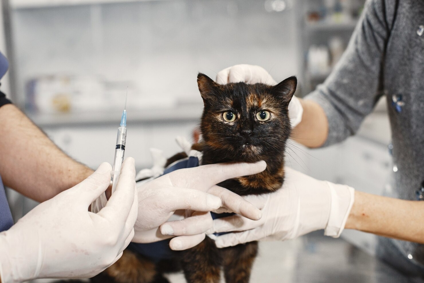
Fine Needle Aspiration (FNA)
The cornerstone of initial tumor diagnostics in veterinary oncology is Fine Needle Aspiration (FNA) cytology. This rapid, minimally invasive, and relatively painless procedure involves inserting a small-gauge sterile needle attached to a syringe directly into the suspect tumor. The veterinarian applies negative pressure to suction a small sample of cells and interstitial fluid into the needle hub. The acquired cellular material is then carefully expelled and spread onto glass microscope slides. [38] These slides are air-dried and treated with specific metachromatic cytological stains, such as Wright-Giemsa or Toluidine blue. A trained veterinary pathologist or skilled clinician then analyzes the slides under high magnification. Normal and neoplastic mast cells are typically easy to identify on cytology; they appear as distinct round cells whose cytoplasm is densely packed with striking, dark purple or magenta granules. Cytology provides a rapid confirmation of “mast cell tumor,” but it cannot evaluate the tissue architecture needed to determine the tumor’s grade or invasiveness. [39]
Biopsy
When FNA cytology is inconclusive—which can happen if the mast cells are poorly differentiated and lack their characteristic purple granules—or when the oncologist requires a definitive tumor grade to formulate a surgical plan, a tissue biopsy becomes necessary. A biopsy is a surgical procedure that involves extracting a solid piece of the suspected tumor tissue. Depending on the size and location of the mass, the veterinarian may perform an incisional biopsy (using a small punch tool or scalpel to remove a wedge of tissue while leaving the bulk of the tumor behind) or an excisional biopsy (attempting to surgically remove the entire visible mass at once). [40] The intact tissue sample is immediately preserved in formalin fixative and dispatched to a specialized veterinary pathologist for detailed histopathological analysis.
Histopathology
Histopathology is the undisputed gold standard for diagnosing and grading mast cell tumors. This sophisticated method involves a board-certified veterinary pathologist embedding the tissue sample in paraffin wax, slicing it into microscopically thin sections, and examining the architectural relationship between the tumor cells and the surrounding healthy stroma. The pathologist rigorously probes the tissue to assess the degree of cellular atypia, nuclear variation, and pleomorphism. Crucially, they calculate the mitotic index, which is the number of actively dividing cells seen in a specific area (usually 10 high-power microscopic fields). A high mitotic index correlates strongly with a highly aggressive, malignant biological behavior. [41] Furthermore, if an excisional biopsy was performed, the pathologist will evaluate the surgical margins—the very edges of the removed tissue—to determine if they are “clean” (free of cancer cells) or “dirty” (cancer cells extend to the edge, indicating tumor was left behind in the cat). Histopathology provides the foundational data necessary for predicting prognosis and selecting follow-up therapies.
Imaging
Once a mast cell tumor is confirmed via cytology or biopsy, “staging” the patient is the next critical phase. Staging refers to a battery of tests designed to determine if and where the cancer has spread beyond the primary tumor site. In instances where metastasis or visceral involvement is suspected, advanced imaging techniques are heavily utilized. Thoracic X-rays (radiographs) are performed to evaluate the lungs, cardiovascular structures, and thoracic lymph nodes. While feline mast cell tumors rarely metastasize directly to the lung tissue, X-rays are vital for assessing overall cardiopulmonary health prior to general anesthesia. [42]
Abdominal ultrasonography is an indispensable imaging modality in feline MCT staging. The sonographer conducts a detailed sweep of the abdominal cavity, looking for characteristic “Swiss cheese” mottling or massive enlargement of the spleen and liver. The ultrasound provides detailed, real-time images of internal organs, allowing the clinician to gauge tumor size, precisely map locations, and evaluate the mesenteric lymph nodes for metastatic spread. If abnormalities are seen on the ultrasound, the clinician may perform an ultrasound-guided FNA of the spleen or liver to confirm metastatic disease. [43] In complex cases, advanced 3D imaging modalities like computed tomography (CT) scans or magnetic resonance imaging (MRI) may be employed to comprehensively evaluate tumors located in difficult anatomical regions, such as the skull, deep neck, or spinal cord, to plan precise surgical or radiation treatments.
Complementing the imaging, comprehensive systemic laboratory workup is mandatory. Complete blood counts (CBC), buffy coat smears (to look for mast cells circulating in the blood), and comprehensive serum biochemistry panels are performed to check red and white blood cell levels, assess kidney filtration rates, and monitor liver enzyme function. These metrics are vital to detect paraneoplastic complications like anemia from GI bleeding and to ensure the cat’s internal organs are robust enough to withstand anesthesia and the metabolic demands of aggressive chemotherapy protocols. [44]
Collectively, these meticulously executed diagnostic tests are critical in accurately diagnosing, grading, staging, and determining the most appropriate, medically sound treatment options for mast cell tumors in cats. By combining these advanced diagnostic tools with the deep clinical expertise of veterinary oncology professionals, a customized, patient-specific approach for managing MCTs and significantly enhancing the cat’s long-term prognosis can be successfully developed and implemented. [45]
Treatment for Mast Cell Tumors in Cats

The therapeutic approach and medical strategy for combating mast cell tumors (MCTs) in cats are highly individualized. The ideal treatment protocol relies on a complex matrix of factors, including the histologic grade of the tumor, the clinical stage of the disease, the exact anatomical location of the mass, the presence or absence of metastasis, and the cat’s concurrent systemic health and age. Working closely with a veterinary oncologist, the treatment plan may propose a multimodal approach utilizing one or more of the following advanced treatment options. [46]
Surgery
Aggressive, wide surgical excision remains the absolute cornerstone and primary front line treatment for localized mast cell tumors in felines. The surgical goal is curative: to completely remove the visible tumor mass along with a substantial peripheral margin of completely healthy, normal-appearing tissue to eradicate microscopic tendrils of cancer cells and minimize the risk of local recurrence. In canine MCT surgery, extremely wide 3-centimeter margins are standard; however, feline cutaneous MCTs tend to be slightly less locally invasive, and veterinary surgeons often achieve clean excision with 1 to 2-centimeter lateral margins and one deep fascial plane. [47]
When dealing with visceral MCTs, surgery takes on a different scope. For splenic mast cell tumors, a complete splenectomy (removal of the entire spleen) is the gold standard of care. Because handling the spleen during surgery can trigger massive degranulation and cause life-threatening hypotensive shock, specialized anesthetic protocols and premedication with antihistamines are strictly required. For intestinal mast cell tumors, an intestinal resection and anastomosis (R&A) is performed to remove the diseased segment of the bowel and reconnect the healthy ends. Furthermore, in cases of both cutaneous and visceral tumors, prophylactic or therapeutic regional lymph node extirpation (removal) may be highly recommended if the preoperative staging indicates potential metastatic spread. [48]
Radiation Therapy
Radiation therapy (radiotherapy) is a highly specialized oncological modality that utilizes targeted, high-energy ionizing radiation (such as X-rays or electron beams) to damage the DNA of cancer cells, thereby destroying their ability to divide and multiply. In feline MCT management, radiation therapy is most frequently suggested for specific, challenging clinical situations. It is the primary adjuvant treatment of choice when a cutaneous tumor has been surgically removed but the biopsy report indicates “dirty” or incomplete margins, and performing a second, wider surgery is anatomically impossible (such as tumors located on the distal lower limbs, the face, or the eyelids). [49]
Radiation endeavors to systematically annihilate microscopic residual cancer cells left behind in the surgical bed. Depending on the intent, a definitive (curative) radiation protocol may involve 15 to 19 fractionated daily treatments, while a palliative protocol—designed to diminish tumor size, control pain, and relieve clinical symptoms in massive, inoperable tumors—may involve only a few larger doses of radiation. A specialized form of topical radiation therapy may also be employed for very small, superficial cutaneous lesions. [50]
Chemotherapy
Systemic chemotherapy is implemented in challenging clinical scenarios where the mast cell tumor exhibits highly aggressive biological behavior, when the histopathology report indicates a high mitotic index, or when advanced diagnostics confirm that the cancer has metastasized to the lymph nodes, liver, or bone marrow. Chemotherapy utilizes powerful pharmacological agents that circulate throughout the entire body to hunt down and eliminate rapidly dividing cancer cells. [51]
In modern veterinary oncology, the use of targeted biological therapies, specifically Tyrosine Kinase Inhibitors (TKIs), has revolutionized feline MCT treatment. These targeted prescription medications work by specifically binding to and blocking the mutated c-KIT receptors on the surface of the mast cells. By cutting off this aberrant molecular signaling pathway, the therapy starves the tumor of the commands it needs to grow and build new blood vessels (angiogenesis). Traditional cytotoxic chemotherapy protocols may also be employed, utilizing specific chemotherapy agents tailored to your pet, often administered intravenously or orally in conjunction with high doses of prescription corticosteroids to induce mast cell apoptosis and boost overall treatment effectiveness. [52]
Supportive Care
While surgery and chemotherapy target the cancer cells directly, intensive supportive care measures hold paramount significance in managing the severe, life-threatening paraneoplastic syndromes caused by mast cell degranulation. Without aggressive supportive care, a cat can succumb to the chemical mediators long before the tumor itself becomes fatal. [53]
Supportive pharmacotherapy relies heavily on blocking histamine receptors. Antihistamines are administered routinely to control systemic allergic reactions, severe pruritus, and localized tissue swelling. Crucially, targeted antacids and acid-reducing medications are prescribed to block histamine from stimulating the stomach acid pumps, thereby preventing or healing severe gastrointestinal ulcerations. Additional supportive medications may include prescription gastroprotectants to coat existing ulcers, potent anti-nausea medications prescribed by your veterinarian to control severe nausea and vomiting, prescription appetite stimulants, and robust analgesic protocols utilizing prescription pain medication administered by your veterinarian to alleviate the profound abdominal or incisional pain associated with the tumor or its aggressive surgical treatment. [54]
Ultimately, the comprehensive treatment strategy for MCTs in cats must be highly personalized and dynamically adjusted over time. Medical decisions are made collaboratively, carefully weighing the cat’s overall health status, the specific microscopic characteristics of the tumor, the financial and logistical capabilities of the owner, and the ultimate goals for the patient’s quality of life. Routine, rigorous follow-up examinations, scheduled imaging, and continuous clinical monitoring are absolutely essential to accurately assess the tumor’s response to therapy, to rapidly tackle any emerging pharmacological complications, and to vigilantly screen for local tumor recurrence or distant metastasis. [55]
Because the landscape of veterinary oncology is highly complex and constantly evolving with new drug approvals and surgical techniques, ongoing consultation with a board-certified veterinary oncologist or a highly experienced primary care veterinarian is crucial. They will expertly guide the entire treatment process, interpret complex diagnostic data, and provide apt, compassionate recommendations based entirely on the unique, individual circumstances of the cat’s mast cell disease.
Prevention for Mast Cell Tumors in Cats
Cancer prevention in veterinary medicine is an incredibly challenging frontier. Because the exact, definitive instigators of most feline mast cell tumors (MCTs) remain obscured by complex genetics and cellular biology, there is no single vaccine, medication, or specific dietary intervention that can guarantee a cat will never develop this disease. However, pet owners are not entirely powerless. There exist several vital, proactive health management measures and husbandry practices that pet parents can vigorously employ to potentially curtail environmental risks, promote robust immune health, and, most importantly, detect developing MCTs in their earliest, most highly treatable stages: [56]
- Routine, Comprehensive Veterinary Visits: The foundation of preventative oncology is the annual or bi-annual wellness exam. Organize regular check-ups with your veterinarian, particularly as your cat enters its senior years (typically over the age of 7). These visits enable the veterinary team to conduct comprehensive, hands-on physical examinations, palpate internal organs, and track any subtle weight or health changes in your cat over time. Routine geriatric blood panels can assist in the early detection of underlying systemic irregularities long before visible masses appear. [57]
- Vigilant Skin and Tumor Monitoring: Pet owners are the first line of defense. Regularly inspect and massage your cat’s skin at home, focusing deeply on areas where cutaneous MCTs typically manifest, such as the head, the base of the ears, the neck, the limbs, and the ventral abdomen. Be intensely vigilant for any new lumps, small bumps, thickened plaques, or unexplained skin lesions that do not heal. Swiftly report any dermatological changes to your veterinarian. Remember that because mast cells can cause dramatic local swelling, a lump that rapidly changes size or becomes red and angry indicates the potential presence of a degranulating tumor that requires immediate cytological evaluation. [58]
- Environmental Mindfulness and Toxin Reduction: While the direct link is still under scientific investigation, it is wise to limit your cat’s daily exposure to potential environmental mutagens that might elevate the risk of cellular DNA damage and subsequent MCTs. This includes strictly curtailing their interaction with potential carcinogens, secondhand tobacco smoke, harsh industrial cleaning chemicals, lawn herbicides, and toxic agricultural pesticides. Keeping cats strictly indoors significantly reduces their exposure to these uncontrolled environmental variables and harmful ultraviolet radiation. [59]
- Prompt Veterinary Intervention and Behavioral Tracking: As a pet owner, you know your cat’s daily routines better than anyone. Seek veterinary attention immediately if you observe any worrisome systemic signs or symptoms. This includes not only the emergence of new cutaneous masses but also subtle shifts in your cat’s behavior, such as increased hiding, uncharacteristic lethargy, sudden aggression when touched, unexplained weight loss, or changes in litter box habits. Because visceral MCTs hide internally, behavioral changes and chronic vomiting are often the only early clues. A swift, aggressive diagnostic workup and immediate surgical treatment can significantly enhance the long-term clinical outcome of MCTs and ensure that localized cancer has been fully removed before it can spread. [60]
- Responsible Genetic Considerations: The breed predispositions seen in Siamese, Burmese, and Sphynx cats strongly point toward heritability. If you are contemplating purchasing a pedigree cat or breeding your own feline, liaise extensively with a reputable, health-focused breeder or a veterinary geneticist to endorse responsible breeding practices. Removing cats with a known history of early-onset mast cell tumors from the active breeding pool is essential to slowly diminish the risk of genetic predisposition to MCTs in future feline generations. [61]
While these preventative measures and husbandry guidelines may aid in reducing environmental risk factors or facilitate the early, life-saving identification of MCTs, it is a difficult medical reality that not all cancer cases can be prevented. Regular, high-quality veterinary care, nutritional support, and unwavering owner attentiveness are the absolute keys to successfully monitoring your cat’s lifelong health and addressing any oncological concerns promptly. In the complex case of mast cell tumors located internally within the spleen or intestines, prompt attention becomes exponentially more critical as the required surgical treatment can be inherently risky for your cat. Therefore, being intensely mindful of the tumor’s location, recognizing that severe systemic itching occurs because the tumors produce massive quantities of specific vasoactive substances, and understanding that the prognosis for advanced visceral disease is often less than one year after treatment, underscores why early detection is the ultimate goal. Always remember to consult your veterinarian before making any changes to your pet’s care, diet, or medication regimens.
Furthermore, it is essential to remember that when a neoplastic mast cell is accidentally agitated or released during a biopsy, it possesses volatile granules that can exacerbate the localized condition and trigger systemic shock. This clinical danger underlines the immense importance of utilizing proper surgical techniques and conducting meticulous histopathological margin evaluations to confirm that the cancer has been fully and cleanly removed, particularly since a significant percentage of poorly differentiated mast cell tumors can reoccur locally in the skin form of the feline condition if even microscopic tumor cells are left behind in the surgical bed. [62]
Frequently Asked Questions
Are mast cell tumors in cats always fatal?
No, a diagnosis of a mast cell tumor in a cat is not a universal death sentence. The prognosis is highly variable and depends entirely on the tumor’s location and histologic grade. The majority of cutaneous (skin) mast cell tumors in cats are biologically benign, well-differentiated, and can be completely cured with a single, straightforward surgical excision. However, mast cell tumors located in the gastrointestinal tract, or those categorized by a pathologist as high-grade or pleomorphic, are highly aggressive, metastasize rapidly, and carry a much poorer prognosis, often requiring complex chemotherapy protocols to extend survival.
Can I touch or squeeze my cat’s mast cell tumor?
You should absolutely avoid touching, squeezing, massaging, or unnecessarily manipulating any suspected mast cell tumor on your cat. Mast cell tumors are incredibly fragile. Mechanical agitation causes the cancer cells to undergo massive, spontaneous degranulation, releasing huge quantities of histamine and heparin into the surrounding tissues and bloodstream. This phenomenon, known as Darier’s sign, can cause the tumor to suddenly swell, turn bright red, become intensely itchy, and bleed. In severe cases, squeezing a large tumor can release enough histamine to trigger a life-threatening systemic anaphylactic shock, causing sudden drops in blood pressure and severe vomiting.
How do mast cell tumors in cats differ from those in dogs?
Feline mast cell tumors differ from canine mast cell tumors in several significant ways. Firstly, while MCTs are the most common skin cancer in dogs, they are the second most common in cats. Secondly, cats are uniquely predisposed to developing visceral mast cell tumors, specifically within the spleen and the gastrointestinal tract, which is less common in dogs. Thirdly, canine skin MCTs are notoriously locally invasive and require massive surgical margins (up to 3 cm) for clean removal, whereas feline cutaneous MCTs tend to be more well-circumscribed, often requiring much smaller surgical margins (1 to 2 cm) for successful curative excision. Finally, the specific genetic mutations driving the cancer (such as the c-KIT mutations) occur on different exons in cats compared to dogs, meaning targeted chemotherapy drugs may have different efficacy rates between the two species.
Take Action for Your Cat’s Health
Early detection and intervention are crucial when dealing with potential mast cell tumors. If you have noticed any unusual skin lumps, changes in your cat’s behavior, or unexplained gastrointestinal symptoms, do not hesitate to seek professional care. Schedule an appointment with your veterinarian today for a comprehensive examination and personalized treatment plan.
References
- American Veterinary Medical Association (AVMA). Cancer in Pets. AVMA, 2023.
- Merck Veterinary Manual. Tumors of the Skin in Cats. Merck & Co., Inc., 2022.
- Sabattini S, et al. Histologic grading of feline mast cell tumor. Veterinary Pathology, 2015.
- VCA Animal Hospitals. Mast Cell Tumors in Cats. VCA Hospitals, 2023.
- Welle MM, et al. Mast cell tumors in dogs and cats: a review of the pathogenesis and diagnosis. Veterinary Pathology, 2006.
- Galli SJ, et al. The development of mast cells and their roles in immunology. Nature Reviews Immunology, 2020.
- Blackwood L, et al. Feline mast cell tumours: a review. Journal of Feline Medicine and Surgery, 2009.
- Cornell Feline Health Center. Feline Cancer. Cornell University College of Veterinary Medicine, 2021.
- Merck Veterinary Manual. Paraneoplastic Syndromes. Merck & Co., Inc., 2023.
- Kiupel M, Camus M. Diagnosis and Prognosis of Canine and Feline Mast Cell Tumors. Veterinary Clinics: Small Animal Practice, 2019.
- Melville K, et al. Feline Cutaneous Mast Cell Tumours. Veterinary Record, 2015.
- Johnson TO, et al. Histologic subclassification of feline cutaneous mast cell tumors. Veterinary Pathology, 2002.
- Lepri E, et al. Histiocytic mast cell tumor in a Siamese cat. Journal of Veterinary Diagnostic Investigation, 2004.
- VCA Animal Hospitals. Feline Skin Masses. VCA Hospitals, 2022.
- Dobromylskyj P, et al. Subcutaneous mast cell tumours in cats: a retrospective study of 200 cases. Journal of Feline Medicine and Surgery, 2011.
- Barrett LE, et al. Gastrointestinal mast cell tumors in 36 cats: a retrospective study. Veterinary Surgery, 2015.
- Veterinary Information Network (VIN). Feline Gastrointestinal Tumors. VIN, 2020.
- Kraus KA, et al. Splenic mast cell tumors in cats: 38 cases. Journal of the American Veterinary Medical Association, 2006.
- Piviani M, et al. Feline systemic mastocytosis: prevalence and prognostic indicators. Veterinary Clinical Pathology, 2012.
- Evans SE, et al. Splenectomy for treatment of splenic mast cell tumors in cats. Journal of Feline Medicine and Surgery, 2016.
- ASPCA. Common Cat Diseases and Cancers. ASPCA, 2022.
- Litster AL, et al. Breed predispositions to feline mast cell tumors. Journal of Veterinary Internal Medicine, 2013.
- Isotani M, et al. Mutations of the c-kit gene in feline mast cell tumors. Journal of Veterinary Medical Science, 2011.
- Baker Institute for Animal Health. Cancer Genetics in Animals. Cornell University, 2022.
- London CA, Seguin B. Mast cell tumors in the dog and cat. Veterinary Clinics of North America: Small Animal Practice, 2003.
- Coussens LM, Werb Z. Inflammation and cancer. Nature, 2002.
- Munday JS, et al. Feline cutaneous neoplasia: a retrospective study. Veterinary Dermatology, 2010.
- Bergman PJ. Paraneoplastic syndromes in oncology. Veterinary Clinics: Small Animal Practice, 2012.
- Sabattini S, Bettini G. Grading feline mast cell tumors. Veterinary Pathology, 2013.
- Merck Veterinary Manual. Mast Cell Tumors in Animals. Merck & Co., Inc., 2022.
- Steffan J, et al. Pruritus in feline oncology. Veterinary Dermatology, 2008.
- VCA Animal Hospitals. Mast Cell Tumors and Skin Ulcerations. VCA Hospitals, 2021.
- Fox LE. Gastrointestinal ulceration associated with mast cell tumors. Veterinary Clinics of North America, 1999.
- Book AP, et al. Systemic signs associated with feline mast cell disease. Journal of Feline Medicine and Surgery, 2017.
- Gieger TL, et al. Diagnostics in veterinary oncology. Veterinary Clinics: Small Animal Practice, 2006.
- American Veterinary Medical Association (AVMA). Diagnosing Cancer in Pets. AVMA, 2021.
- Krick EL, et al. Lymph node evaluation in feline mast cell tumors. Veterinary and Comparative Oncology, 2014.
- Sharkey LC, et al. Cytology of feline mast cell tumors. Veterinary Clinics: Small Animal Practice, 2012.
- Crivellenti LZ, et al. Diagnostic accuracy of cytology in feline oncology. Journal of Veterinary Diagnostic Investigation, 2002.
- American College of Veterinary Surgeons. Biopsy Techniques for Mast Cell Tumors. ACVS, 2021.
- Sabattini S, et al. Prognostic significance of mitotic index in feline mast cell tumors. Veterinary Pathology, 2015.
- Penninck D, et al. Radiographic staging of feline oncology patients. Veterinary Radiology & Ultrasound, 2002.
- Hanson JA, et al. Ultrasonographic appearance of splenic mast cell tumors in cats. Veterinary Radiology & Ultrasound, 2007.
- Cornell Animal Health Diagnostic Center. Clinical Pathology and Hematology. Cornell University, 2022.
- Kiupel M. Comprehensive staging in feline mast cell tumors. Veterinary Clinics: Small Animal Practice, 2019.
- VCA Animal Hospitals. Cancer Treatment Protocols in Cats. VCA Hospitals, 2023.
- Simpson AM, et al. Surgical margins for feline cutaneous mast cell tumors. Veterinary Surgery, 2016.
- Kraus KA, et al. Splenectomy techniques and outcomes. Journal of the American Veterinary Medical Association, 2006.
- Turrel JM, et al. Radiation therapy for incompletely excised feline mast cell tumors. Veterinary Radiology & Ultrasound, 2009.
- Jarvis P, et al. Strontium-90 plesiotherapy for superficial skin tumors. Veterinary and Comparative Oncology, 2013.
- Thamm DH. Tyrosine Kinase Inhibitors in Veterinary Oncology. Veterinary Clinics: Small Animal Practice, 2014.
- Harper A, et al. Use of toceranib phosphate (Palladia) in feline mast cell tumors. Journal of Feline Medicine and Surgery, 2011.
- Merck Veterinary Manual. Supportive Pharmacology in Oncology. Merck & Co., Inc., 2022.
- Marks SL. Gastroprotectants and anti-emetics in small animal practice. Veterinary Clinics: Small Animal Practice, 2011.
- American Veterinary Medical Association (AVMA). Quality of Life for Cancer Patients. AVMA, 2022.
- American Veterinary Medical Association (AVMA). Senior Pet Care Preventative Guidelines. AVMA, 2023.
- Epstein M, et al. AAHA Senior Care Guidelines for Dogs and Cats. Journal of the American Animal Hospital Association, 2005.
- VCA Animal Hospitals. Checking Your Pet for Lumps and Bumps. VCA Hospitals, 2021.
- Reif JS. Animal sentinels for environmental and public health. Public Health Reports, 2011.
- ASPCA. Caring for Older Cats and Recognizing Illness. ASPCA, 2022.
- O’Brien SJ, et al. The promise of feline genetics. Journal of Feline Medicine and Surgery, 2008.
- Shaw T, et al. Recurrence rates of feline cutaneous mast cell tumors. Veterinary and Comparative Oncology, 2015.


