


 March 3, 2023
 Phil Good, DVM
What Are Oral Masses in Dogs?
This content was prepared with AI assistance and reviewed by a licensed professional for accuracy.
Introduction
Discovering lumps found in dogs’ mouths can be an incredibly distressing experience for any devoted pet parent. As a veterinarian, I frequently evaluate patients for canine oral masses, and I understand the immediate anxiety that accompanies the sudden appearance of lumps in mouth tissues. Oral masses in dogs represent a diverse and complex category of veterinary pathology, encompassing everything from entirely benign, self-limiting tissue overgrowths to highly aggressive, life-threatening malignancies that can rapidly spread to regional mouth lymph nodes and beyond. The canine oral cavity is a robust, highly vascularized environment designed for grasping, tearing, and chewing, but this intense cellular turnover and frequent exposure to environmental elements also make it a common site for abnormal cellular proliferation. Recognizing the clinical significance of these growths requires a thorough understanding of oral anatomy, tissue pathology, and the specific behaviors of different tumor types.[1]
The oral cavity is anatomically intricate, lined with mucous membranes and containing the teeth, periodontal ligaments, alveolar bone, hard and soft palates, tongue, and major and minor salivary glands. When an oral mass develops, it can originate from any of these distinct tissue types. For instance, a mass may arise from the gingival (gum) tissue, the buccal mucosa (inside of the cheeks), the structural bone of the maxilla or mandible, or the specialized cells that form the teeth. Because of this anatomical diversity, canine oral masses can exhibit an extraordinarily wide range of visual characteristics and physical textures. Some may present as small, smooth, firm nodules that perfectly blend with the surrounding gum tissue, while others might erupt as large, friable, ulcerated, cauliflower-like lesions that bleed with the slightest provocation. The biological behavior of these masses—whether they remain localized and harmless or aggressively destroy adjacent bone and spread through the lymphatic system—is just as varied as their physical appearance.[2]
Age, breed, and overall health status play significant roles in the development and prevalence of oral growths. While we predominantly diagnose clinically significant oral tumors in middle-aged to senior dogs, juvenile and young adult dogs are certainly not immune and frequently present with virus-induced growths or congenital cysts. Furthermore, certain breeds demonstrate strong genetic predispositions toward specific types of oral masses, a fact that helps guide our clinical suspicion during the initial examination. Regardless of the dog’s age or breed, the discovery of any abnormal tissue in the mouth warrants immediate, comprehensive veterinary investigation. A “wait and see” approach is highly discouraged in veterinary dental oncology, as the mouth’s rich blood supply and intricate lymphatic drainage network can facilitate rapid tumor growth and early metastasis. Prompt identification, precise tissue biopsy, and early clinical staging are the cornerstones of achieving a favorable prognosis and preserving the patient’s quality of life.[3]
It is also crucial to recognize the profound impact that an oral mass can have on a dog’s daily comfort and nutritional intake. The mouth is highly innervated, and expanding masses can cause severe pain, difficulty swallowing (dysphagia), misalignment of the jaw, and secondary infections due to trapped food debris and bacterial proliferation. In many cases, the secondary complications of an oral mass—such as extreme halitosis, drooling, and reluctance to eat—are the very symptoms that prompt a veterinary visit. By exploring the specific classifications, underlying causes, diagnostic pathways, and advanced treatment modalities associated with these growths, pet owners can become empowered, proactive advocates for their canine companions. Navigating a diagnosis of an oral mass requires teamwork between the owner, the primary care veterinarian, and often a board-certified veterinary oncologist or dentist, all working together to formulate a compassionate and effective care plan.[4]
Types of Oral Masses in Dogs
Benign Epulides
The term “epulis” (plural: epulides) is a general clinical descriptor historically used in veterinary medicine to denote any localized, fleshy mass situated on the gingiva (gums). However, modern veterinary pathology has reclassified these growths based on their specific cellular origins, giving us a much more accurate understanding of their behavior. Benign epulides are among the most frequently diagnosed non-cancerous oral masses in dogs. They predominantly arise from the periodontal ligament—the tough, fibrous connective tissue that securely anchors the dog’s tooth within its bony socket. While these growths are strictly benign, meaning they completely lack the biological machinery required to metastasize to the lungs or distant internal organs, their local behavior can still cause significant oral health complications if left unchecked. They are most commonly identified in middle-aged to older dogs, particularly in brachycephalic (flat-faced) breeds like Boxers, Pugs, and Bulldogs, although any breed can be affected.[5]
Veterinary pathologists currently recognize three primary histological categories of benign epulides, each presenting unique clinical challenges. The first category is the Fibromatous Epulis (now more accurately termed peripheral odontogenic fibroma). These masses typically present as smooth, firm, pink nodules that protrude directly from the gum line adjacent to a tooth. They consist primarily of dense, tough fibrous connective tissue. Because they do not possess invasive properties, they do not destroy the underlying mandibular or maxillary bone. However, as they expand, they can physically displace adjacent teeth, alter the dog’s bite alignment, and create deep pseudo-pockets where dangerous periodontal bacteria can thrive, leading to severe localized dental disease.[6]
The second classification is the Ossifying Epulis. This growth is clinically and visually indistinguishable from the fibromatous variety to the naked eye. The defining characteristic of an ossifying epulis is the microscopic presence of osteoid (bone-like) matrix or actual bone tissue developing within the fibrous stroma of the mass. Like their fibromatous counterparts, ossifying epulides are expansive rather than invasive. They push neighboring tissues aside as they grow slowly over time. Because of the calcified internal structure, these masses are exceptionally firm to the touch and are vividly visible on intraoral dental radiographs, appearing as a dense, radiopaque area within the soft tissue profile of the gums. Complete surgical excision, including the removal of the periodontal ligament of origin and often the associated tooth, is typically curative.[7]
The third, and most clinically significant, type is the Acanthomatous Epulis (formally known as canine acanthomatous ameloblastoma). While technically classified as a benign tumor because it does not metastasize to distant organs, its local behavior is notoriously aggressive and destructive. Acanthomatous epulides vigorously invade and destroy the surrounding alveolar bone of the jaw. If a veterinarian merely shaves this mass down to the gum line, it will rapidly recur, often causing even more extensive bony destruction. Radiographically, these tumors present with severe, localized bone lysis (dissolution of the bone), which can sometimes be misdiagnosed as a malignant cancer or a severe tooth root abscess. Because of their highly invasive nature, the standard of care for an acanthomatous epulis requires wide-margin surgical resection, which often necessitates a partial mandibulectomy (removal of a portion of the lower jaw) or maxillectomy (upper jaw) to ensure all microscopic tumor cells are completely eradicated. Alternatively, these specific tumors are uniquely sensitive to radiation therapy, which can be an excellent option for masses located in anatomically difficult areas.[8]
Malignant Tumors
Malignant oral tumors in dogs represent a grave health threat, characterized by unregulated cellular division, profound local tissue destruction, and the ominous potential for systemic metastasis. When cells within the oral cavity undergo catastrophic genetic mutations, they can bypass normal growth constraints and evade the immune system. The oral cavity is the fourth most common site for malignant cancer in dogs, making early detection imperative. Unlike benign growths, malignant tumors frequently present with a rapid onset, accompanied by ulceration, spontaneous bleeding, foul odor, and visible necrosis (tissue death). These cancers not only cause severe, unrelenting pain, but they also utilize the mouth’s extensive lymphatic and vascular networks as highways to spread to the regional lymph nodes and vital organs, most notably the lungs.[9]
Malignant Melanoma holds the unfortunate distinction of being the most frequently diagnosed malignant oral tumor in dogs, accounting for roughly 30% to 40% of all canine oral malignancies. It arises from melanocytes, the cells responsible for producing pigment. While typically presenting as a rapidly expanding, dark brown or black, fleshy mass on the gums, lips, or palate, it is critical to note that up to one-third of oral melanomas are “amelanotic”—meaning they lack pigment entirely and appear deceptively pink and fleshy. Oral melanoma is exquisitely aggressive; by the time the primary mass is discovered by the owner, microscopic metastasis to the regional lymph nodes or the lungs has frequently already occurred. Small breeds with heavily pigmented oral mucosa, such as Scottish Terriers, Poodles, and Dachshunds, exhibit a markedly increased genetic predisposition to this devastating disease. Due to its highly metastatic nature, systemic therapies are almost always required alongside surgical control of the primary tumor.[10]
The second most common oral malignancy is Squamous Cell Carcinoma (SCC). This cancer originates from the squamous epithelial cells that line the entire surface of the mouth. The biological behavior and clinical prognosis of canine SCC are heavily dependent on the tumor’s specific anatomical location. Non-tonsillar SCC, which typically occurs on the rostral (front) gums or the hard palate, is generally locally invasive, aggressively eating away at the underlying jawbone, but it possesses a relatively low rate of distant metastasis. In stark contrast, SCC that originates in the tonsils is a remarkably different biological entity; tonsillar SCC is highly aggressive, frequently metastatic to the regional lymph nodes early in the disease process, and carries a much poorer prognosis. Regardless of location, SCC often presents as a red, inflamed, severely ulcerated, and deeply painful lesion that owners may initially mistake for a severe dental infection or a wound.[11]
Fibrosarcoma ranks as the third most prevalent oral malignancy in dogs. Arising from the fibrous connective tissue beneath the oral mucosa, these tumors generally present as firm, flat, intimately attached masses that are often difficult to visually demarcate from normal surrounding tissue. They most frequently occur on the maxilla (upper jaw) and the hard palate. Canine oral fibrosarcoma presents a unique diagnostic challenge known to oncologists as being “histologically low-grade but biologically high-grade.” This means that under a microscope, the cells may appear relatively normal and slow-growing, misleading the untrained eye, but in the patient’s mouth, the tumor behaves with devastating local invasiveness, rapidly infiltrating deep into the surrounding bone and muscle. Large breed dogs, specifically Golden Retrievers and Labrador Retrievers, are significantly overrepresented for oral fibrosarcoma. While metastasis is less common (occurring in about 10% to 20% of cases), achieving complete surgical margins is notoriously difficult due to their diffuse, poorly defined borders.[12]
Finally, Osteosarcoma, the most common primary bone cancer in dogs, can also originate within the axial skeleton, including the mandible and maxilla. While appendicular osteosarcoma (bone cancer in the limbs) is highly metastatic and rapidly fatal, oral osteosarcoma exhibits a somewhat different clinical course. Tumors arising in the mandible actually carry a surprisingly favorable prognosis if wide surgical excision (mandibulectomy) is performed, with lower rates of metastasis compared to limb tumors. However, osteosarcoma originating in the maxilla carries a much more guarded prognosis due to the extreme difficulty of achieving clean surgical margins without compromising vital structures like the eyes and nasal passages. These tumors manifest as hard, immovable, deeply seated bony swellings that cause significant facial deformity and severe localized pain upon palpation.[13]
Odontogenic Tumors
Odontogenic tumors represent a fascinating, though uncommon, subset of canine oral masses that originate specifically from the embryonic tissues destined to form the teeth. During fetal development, a complex interplay of epithelial and mesenchymal cells coordinates to form enamel, dentin, cementum, and the periodontal ligament. When the genetic signaling in these residual primitive cells goes awry later in the dog’s life, an odontogenic tumor can develop. Because these masses arise strictly from tooth-forming elements, they are exclusively located within the tooth-bearing regions of the mandible and maxilla, never appearing on the tongue, tonsils, or buccal mucosa. While the vast majority of odontogenic tumors are benign in terms of metastatic potential, their expansion within the rigid confines of the jawbone can lead to extensive local tissue morbidity.[14]
Ameloblastoma (specifically distinguishing from the acanthomatous epulis mentioned earlier) is a classic benign odontogenic tumor arising from ameloblasts, the specialized cells responsible for depositing tooth enamel. These masses are generally slow-growing and present as firm, expansile swellings within the jawbone, often causing the overlying gum tissue to stretch tightly. As an ameloblastoma slowly enlarges, it exerts immense pressure on the surrounding alveolar bone, leading to pressure necrosis, cystic cavity formation within the bone, and the subsequent loosening or total displacement of adjacent teeth. Dental radiographs are crucial for diagnosis, typically revealing a well-defined, multilocular (soap-bubble appearance) area of bone loss. Treatment requires complete surgical enucleation or marginal resection to prevent recurrence.[15]
Odontomas are technically classified as hamartomas—which are benign, disorganized malformations of normal tissue—rather than true neoplasms. They contain all the specialized hard and soft tissues of a tooth (enamel, dentin, cementum, and pulp) but arranged in a chaotic, non-functional pattern. Odontomas are further divided into two types: the compound odontoma, which appears radiographically as a cluster of multiple, tiny, malformed tooth-like structures (denticles), and the complex odontoma, which appears as an amorphous, solid mass of calcified dental tissue. These are most commonly discovered incidentally during routine dental radiographs in young dogs, often when a deciduous (baby) tooth fails to fall out or an adult tooth fails to erupt properly. While entirely benign, they should be surgically extracted to prevent the formation of secondary dentigerous cysts.[16]
Though exceedingly rare, malignant variants do exist, such as the Odontogenic Carcinoma and the Odontogenic Sarcoma. These highly aggressive tumors retain some histological characteristics of dental tissues but exhibit rapid, invasive growth, extreme bone destruction, and the capacity for widespread systemic metastasis. They present with severe oral pain, rapid facial swelling, and spontaneous tooth exfoliation. Due to their rarity, treatment protocols are extrapolated from other aggressive oral cancers, typically involving radical surgical resection combined with radiation or chemotherapy, though the long-term prognosis remains poor.[17]
Oral Papillomas (Warts)
Canine oral papillomatosis is a highly contagious, virus-mediated condition characterized by the sudden eruption of benign epithelial tumors, commonly referred to as warts, within the oral cavity. This condition is exclusively caused by the Canine Papillomavirus type 1 (CPV-1), a remarkably resilient DNA virus that exhibits a strict tropism for the squamous epithelial cells of the dog’s mouth, lips, and occasionally the conjunctiva of the eyes. The classic presentation of oral papillomas is almost unmistakable: they emerge as multiple, pale pink to grayish-white, exophytic masses featuring a distinct frond-like, fimbriated, or “cauliflower-like” surface. While they often start as tiny, smooth bumps, they rapidly mature into these larger, textured lesions. They are most frequently attached to the oral mucosa by a narrow stalk (pedunculated) and can cluster in large numbers, sometimes numbering in the dozens, covering the tongue, palate, and inner cheeks.[18]
The disease almost exclusively impacts young dogs, typically those under two years of age, because their cell-mediated immune systems are not yet fully mature. It is also occasionally seen in older dogs suffering from profound immunosuppression, such as those undergoing intense chemotherapy or those with severe systemic illnesses. The virus is transmitted through direct, dog-to-dog contact with an infected individual, or via indirect contact with viral particles lingering in the environment on shared items like water bowls, chew toys, or sticks. It is highly transmissible at dog parks and doggy daycares. Following exposure, the virus exploits minor abrasions in the dog’s oral mucosa to invade the basal cells. The incubation period is remarkably long, taking anywhere from one to two months before the visible warts erupt. Pet owners should be aware that when evaluating masses such as papillomas, the appearance can be alarming, but the condition is almost always self-limiting.[19]
In the vast majority of clinical cases, canine oral papillomatosis requires no medical intervention. As the young dog’s immune system recognizes the viral antigens, it mounts a robust cellular response. Over a period of one to three months, the papillomas will typically undergo spontaneous regression—they will gradually shrink, turn dark gray or black, and slough off, leaving the dog with lifelong immunity to that specific viral strain. However, veterinary intervention becomes necessary in severe, complicated cases. If the warts grow so large and numerous that they interfere with prehension (grasping food), mastication (chewing), or swallowing, they can lead to severe weight loss and malnutrition. Furthermore, if the dog accidentally bites the papillomas while chewing, they can bleed profusely and become secondarily infected with oral bacteria. In these severe instances, treatment options include surgical crushing of the warts (to stimulate an immune response), precise surgical excision, cryotherapy (freezing), or the administration of systemic immunomodulatory drugs prescribed by your veterinarian, which have shown anecdotal efficacy in hastening regression.[20]
Gingival Hyperplasia
Gingival hyperplasia, more accurately described as generalized gingival enlargement or overgrowth, is a non-neoplastic (non-cancerous) condition characterized by the profound, excessive proliferation of the gum tissue. Unlike discrete tumors that form isolated masses, gingival hyperplasia typically presents as a diffuse, widespread thickening of the gingiva that can affect the entire dental arcade. The overgrown tissue is usually firm, pink, and stippled, closely resembling normal healthy gums, but the sheer volume of tissue becomes deeply pathological. In severe cases, the hyperplastic gums can grow so extensively that they almost completely engulf the crowns of the teeth, leaving only the very tips visible, drastically altering the architecture of the oral cavity.[21]
The precise etiology of gingival hyperplasia is multi-factorial, primarily driven by genetics, chronic inflammation, and certain pharmaceutical agents. There is a deeply entrenched genetic predisposition for this condition in specific breeds, most notably Boxers, Bulldogs, Mastiffs, Collies, and Dalmatians. In these breeds, the fibroblasts within the gingival tissue possess an inherent, genetically programmed hyper-reactivity to everyday stimuli. Chronic periodontal disease is the most common trigger; the persistent presence of plaque and calculus causes mild, continuous gingival inflammation, which stimulates the already hyper-reactive tissue to proliferate wildly. Additionally, gingival hyperplasia is a well-documented adverse side effect of several common veterinary medications, specifically certain prescription allergy medications, blood pressure medications, and anti-seizure medications.[22]
The primary clinical consequence of gingival hyperplasia is the formation of deep “pseudopockets.” Normally, the gingival sulcus (the natural pocket between the tooth and gum) is only 1 to 3 millimeters deep. When the gums overgrow upward, they create massive artificial pockets that can measure 10 millimeters or more in depth. These deep, oxygen-deprived crevasses become the perfect incubation chambers for aggressive, anaerobic periodontal bacteria. Food debris, hair, and plaque become deeply impacted within these pockets, leading to severe, chronic periodontal infection, extremely foul halitosis, bleeding gums, and ultimately, the destruction of the alveolar bone and loss of the underlying teeth. Furthermore, the overgrown tissue can be continually traumatized by the opposing teeth during chewing, causing extreme pain and reluctance to eat. Treatment typically involves a surgical procedure known as a gingivectomy, performed using a scalpel, electrosurgery, or a CO2 laser, to meticulously resect the excess tissue and re-establish a normal, cleanable gum line. Strict daily home dental care is mandatory post-operatively to manage the underlying inflammation and delay recurrence.[23]
Oral Cysts
Oral cysts are pathological, fluid-filled, or semi-solid epithelial-lined sacs that develop within the soft tissues or the bone of the canine oral cavity. While strictly benign and entirely non-cancerous, cysts behave as slowly expanding space-occupying lesions. Because fluid constantly secretes into the enclosed cavity of the cyst, the internal hydraulic pressure steadily increases over time. This unrelenting pressure forces the cyst to expand outward, causing significant displacement of normal oral tissues and often leading to severe pressure necrosis and lysis of the surrounding jawbone. If an expanding cyst is ignored, it can silently destroy a massive section of the mandible, leading to an extremely painful pathological fracture of the jaw during normal activities like chewing or playing with a toy.[24]
By far the most common type of oral cyst encountered in veterinary dentistry is the Dentigerous Cyst. This specific cyst originates from the remnants of the enamel organ (the follicular sac) that naturally surrounds the crown of an unerupted developing tooth. If a tooth fails to erupt properly through the gum line—a condition remarkably common in brachycephalic breeds like Pugs, Boston Terriers, and Boxers, particularly concerning the first premolars—the surrounding sac can fill with fluid, initiating the formation of a dentigerous cyst. These cysts often remain entirely asymptomatic and invisible to the naked eye for years, stealthily destroying the surrounding bone until they become large enough to cause visible facial swelling or compromise adjacent healthy teeth. Routine dental radiographs are absolutely critical for identifying unerupted teeth and their associated early-stage cysts.[25]
Other, less common oral cysts include the Radicular Cyst (or periapical cyst), which forms at the apex (root tip) of a dead, infected tooth as a direct result of chronic pulpal necrosis and severe endodontic disease. The chronic inflammatory mediators produced by the dead tooth stimulate latent epithelial cells in the periodontal ligament to form a cystic cavity. Epidermoid and Dermoid Cysts are rare congenital anomalies that occur when skin cells, hair follicles, or sebaceous glands become abnormally trapped beneath the mucosal surface during embryonic development; they often present as soft, fluctuant swellings on the floor of the mouth or under the tongue. Treatment for almost all oral cysts requires a specialized surgical procedure called complete enucleation, which involves meticulously peeling away and removing the entire epithelial lining of the cyst wall, coupled with the extraction of the offending unerupted or dead tooth. If even a microscopic fragment of the cyst lining is left behind, the cyst will inevitably reform.[26]
What Causes Canine Oral Masses?

The exact etiology (root cause) of most canine oral masses, particularly the malignant variants, is considered multifactorial, arising from a complex, poorly understood interplay between genetic mutations, environmental exposures, viral infections, and chronic inflammatory states. While human oncology has made immense strides in identifying specific carcinogens linked to oral cancers (such as alcohol and tobacco chewing), the precise triggers in veterinary medicine remain less definitive but are the subject of intense ongoing research. Unlike many infectious diseases, there is rarely a single “smoking gun” that causes an oral tumor; rather, it is usually a cascade of detrimental cellular events.[27]
- Genetic Predisposition and Breed Susceptibility: Genetics play an undeniable, overwhelmingly powerful role in the development of both benign and malignant oral masses. Decades of epidemiological data highlight that certain breeds inherit mutated tumor suppressor genes (like p53) or hyperactive oncogenes that drastically lower their threshold for developing cancer. For example, the staggering prevalence of malignant melanoma in small, dark-pigmented breeds (Scottish Terriers, Miniature Poodles), the high incidence of fibrosarcoma in large retrievers, and the specific predisposition for acanthomatous epulides in Old English Sheepdogs point heavily to inherited genetic flaws. These inherited traits dictate how a dog’s cells manage DNA damage throughout their life.
- Advancing Age and Cellular Senescence: The most significant overarching risk factor for the development of most oral masses (excluding viral papillomas and congenital cysts) is advanced age. As a dog progresses into its senior years, its cells have undergone millions of cycles of division. With each cycle, the risk of a DNA replication error increases. Concurrently, the senior dog’s immune system undergoes immunosenescence—a natural decline in its ability to detect and destroy rogue, mutated cells before they form a palpable tumor. Thus, an older dog is biologically primed for neoplastic growth.
- Chronic Periodontal Disease and Inflammation: The oral cavity is constantly exposed to a massive microbiome. When plaque and tartar accumulate, it leads to chronic, relentless periodontal disease. The constant influx of inflammatory mediators, cytokines, and immune cells attempting to fight the bacterial infection creates a highly reactive, volatile microenvironment. Chronic inflammation is a well-established driver of cellular mutation and cancer development across many species, as the constant state of cellular injury and rapid repair increases the likelihood of DNA errors, potentially contributing to the development of squamous cell carcinomas and fibromatous epulides.
- Environmental Carcinogens and Tobacco Smoke: Dogs living in environments with heavy exposure to airborne pollutants, particularly secondhand tobacco smoke, face a statistically significant increased risk of developing oral cancers. As dogs groom themselves, they lick their fur, ingesting the carcinogenic particulate matter that has settled on their coat from the environment. These carcinogens concentrate in the saliva and bathe the oral mucosa, leading to direct DNA damage within the squamous epithelial cells. Furthermore, chemicals from urban smog, lawn pesticides, and even certain chemical flea collars have been studied as potential environmental triggers.
- Viral Infections: As discussed extensively, infectious agents like the Canine Papillomavirus type 1 (CPV-1) are the direct causative agents for the development of oral cavity masses such as papillomas. The viral DNA physically integrates into the host cell’s DNA, hijacking the cellular machinery to force rapid, uncontrolled multiplication of the epithelial cells, resulting in the characteristic wart-like growths.
- Chronic Physical Trauma: The persistent, mechanical irritation of the oral mucosa can, over time, provoke neoplastic changes. A dog that constantly chews on highly abrasive objects, or a dog suffering from severe malocclusion where a tooth continuously drives into the hard palate or gums, creates an area of perpetual tissue damage and rapid regeneration. Similarly, repeated trauma from a retained foreign body wedged between the teeth can induce profound inflammatory tissue masses that may undergo malignant transformation if left unresolved for extended periods.
It is vital for pet owners to understand that the presence of one or more of these risk factors does not act as a definitive prophecy that their dog will inevitably develop an oral mass. Many dogs with terrible dental disease never develop cancer, and conversely, dogs with pristine mouths can still fall victim to malignant melanoma. The key takeaway is that these factors increase the statistical probability. Mitigating the controllable factors—such as maintaining excellent oral hygiene, providing a smoke-free home environment, and ensuring regular veterinary dental cleanings—can significantly reduce the overall risk. However, vigilance remains the owner’s best defense, as prompt identification of skin cancer or oral malignancies is the sole factor that allows for curative intervention.[28]
How Does Oral Mass Occur?
The biological genesis of an oral mass, termed tumorigenesis or oncogenesis, is a microscopic process that occurs long before any visible lump appears in the dog’s mouth. At its core, the formation of any mass—whether it is a benign fibroma or a highly aggressive squamous cell carcinoma—represents a catastrophic failure of the body’s normal, tightly regulated cellular control mechanisms. Every healthy cell in the canine body contains a vast library of DNA that issues precise commands regarding when the cell should grow, when it should divide to replace old or damaged tissue, and crucially, when it should undergo apoptosis (programmed, deliberate cell death) because it is no longer functioning correctly.[29]
When an oral mass occurs, this elegant system of checks and balances collapses. The process typically begins when a single cell within the oral mucosa, the periodontal ligament, or the jawbone sustains severe damage to its DNA. This genetic damage can be inflicted by the external factors mentioned previously—such as carcinogenic chemicals from tobacco smoke, oxidative stress from chronic periodontal inflammation, viral integration by a papillomavirus, or simply a spontaneous, uncorrected error during routine DNA replication as the dog ages. If the cell’s internal repair mechanisms fail to fix the mutated DNA, and the immune system’s sentinels (like Natural Killer cells) fail to detect and destroy the rogue cell, the stage is set for a tumor to develop.[30]
The mutated cell begins to ignore the inhibitory signals from its neighbors that would normally tell it to stop growing. Instead, it enters a state of continuous, unchecked replication, producing identical clones of itself that also possess the same defective genetic code. As this microscopic cluster of abnormal cells rapidly multiplies, it begins to form a physical mass. In the case of benign tumors, these cells remain relatively well-differentiated (they still vaguely resemble normal tissue) and they stick tightly together, expanding outward like a balloon but refusing to breach the anatomical borders of surrounding tissues. They do not possess the genetic ability to induce angiogenesis (the creation of new blood vessels) to a degree that allows for systemic spread, nor can they digest the basement membrane to invade the bloodstream.[31]
Conversely, the occurrence of a malignant oral mass involves much more sinister genetic mutations. Malignant cells become highly undifferentiated and chaotic. They secrete powerful proteolytic enzymes that literally digest the surrounding healthy collagen, muscle, and jawbone, allowing the tumor to aggressively invade local tissues like tree roots tearing through concrete. Furthermore, malignant cells release specific growth factors (like VEGF) that force the dog’s body to build new blood vessels directly into the tumor, supplying it with stolen nutrients and oxygen to fuel its explosive growth. Eventually, these poorly adhered malignant cells break off from the primary mass, slipping into the newly formed blood vessels or the rich lymphatic network of the mouth. From there, they travel freely to the submandibular lymph nodes or the capillary beds of the lungs, where they take root and establish deadly metastatic colonies.[32]
Symptoms of Oral Mass in Dogs

Because dogs are instinctively driven to hide signs of pain and vulnerability—a survival mechanism inherited from their wild ancestors—oral masses often grow to a surprisingly large, clinically advanced stage before the owner notices anything is wrong. Furthermore, the symptoms of an oral mass frequently mimic those of routine, albeit severe, dental disease, making regular, thorough oral examinations at home and by a professional absolutely critical. Recognizing the subtle, early clinical signs is paramount for early intervention. If your dog exhibits any combination of the following symptoms, an immediate veterinary consultation is highly warranted:[33]
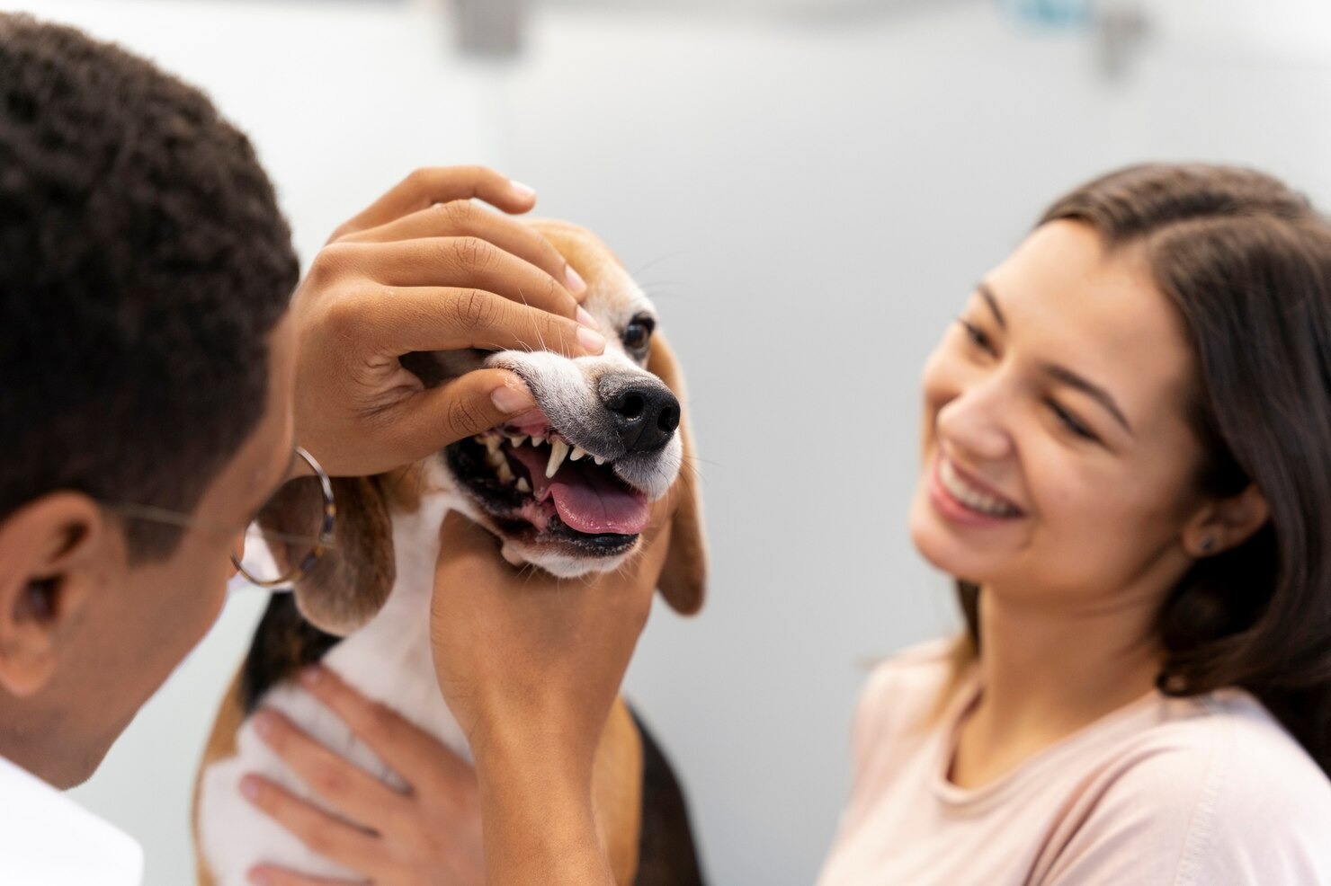
- Presence of a Visible Mass or Lump: The most definitive sign is the direct observation of abnormal tissue. This could be a small, pink swelling on the gum line, a dark, fleshy growth on the palate, or an irregular, cauliflower-like lesion on the tongue. Owners should make a habit of gently lifting their dog’s lips weekly to inspect the teeth and gums.
- Persistent, Severe Bad Breath (Halitosis): While mild “doggy breath” is common, an overpowering, profoundly foul, rotting odor is a major red flag. This smell is typically caused by the necrosis (death) of the rapidly growing tumor tissue, which outgrows its blood supply and rots within the mouth, combined with massive secondary bacterial infections trapped in the crevices of the mass.
- Excessive Drooling (Ptyalism): A mass can physically prevent the dog from closing its mouth fully or swallowing its saliva normally. Additionally, oral pain stimulates the salivary glands to overproduce saliva. The drool may frequently be thick, ropy, and blood-tinged.
- Difficulty Eating, Chewing, or Swallowing (Dysphagia): As the tumor enlarges, it acts as a physical obstruction. The dog may approach the food bowl enthusiastically but abruptly drop the food, cry out in pain when biting down, or exhibit exaggerated, awkward swallowing motions.
- Bleeding from the Mouth: Finding unexplained blood on chew toys, in the water bowl, or smeared on the dog’s front legs after grooming is highly concerning. Malignant tumors and ulcerated masses are highly vascular and fragile, bleeding profusely with minimal friction from food or the tongue.
- Loose, Shifting, or Missing Teeth: Tumors that originate in or invade the alveolar bone—such as acanthomatous epulides, squamous cell carcinomas, or ameloblastomas—rapidly destroy the bony socket and periodontal ligaments, causing otherwise healthy teeth to become incredibly loose, shift out of alignment, or fall out entirely.
- Unexplained Weight Loss and Muscle Wasting: The combination of chronic, severe oral pain, inability to properly chew food, and the massive metabolic drain caused by cancer cachexia (the tumor stealing the body’s nutrients) can lead to rapid, profound weight loss and a decline in body condition.
- Facial Swelling, Asymmetry, or Exophthalmos: A mass growing deep within the jawbone or the sinus cavities may not be visible inside the mouth. Instead, it will cause a hard, firm swelling on the outside of the muzzle or lower jaw. If a maxillary tumor grows upward behind the eye socket, it can cause exophthalmos—a highly alarming condition where the eyeball is physically pushed outward, bulging from the socket.
- Swollen Regional Lymph Nodes: Enlargement of the submandibular or prescapular mouth lymph nodes (felt as hard, firm lumps under the dog’s jaw or in front of the shoulder blades) strongly indicates robust localized inflammation or, more concerningly, that a malignant oral tumor has actively metastasized to the lymphatic system.
- Altered Behavior and Lethargy: Chronic, unrelenting oral pain takes a massive psychological and physiological toll. Dogs may become deeply lethargic, withdrawn, irritable, head-shy (pulling away when you try to pet their head), or completely lose interest in playing or interacting with the family.
- Preference for Chewing on One Side: A dog experiencing localized pain from a mass will quickly learn to shift all of their chewing activity to the opposite, unaffected side of the mouth. This often leads to a massive, asymmetrical build-up of heavy tartar on the painful side, as that side is no longer benefiting from the mechanical cleaning action of chewing.
Diagnosis of Oral Masses in Canine

Arriving at a definitive diagnosis for an oral mass in a dog is an intricate, multi-step process that requires a systematic, highly detailed approach. The goal is not merely to confirm the presence of a lump, but to definitively identify its exact cellular origin, grade its aggressiveness, assess its local invasiveness, and crucially, determine if it has metastasized to other areas of the body. This comprehensive process, known as clinical staging, is the absolute foundation upon which all subsequent treatment decisions and prognostic expectations are built. A rushed or incomplete diagnosis can lead to disastrous surgical failures or missed opportunities for life-saving interventions.[34]
Step 1: The Initial Physical and Conscious Oral Examination. The diagnostic journey begins in the exam room. The veterinarian will take a thorough medical history, noting the onset of symptoms, changes in eating habits, and any relevant breed predispositions. A comprehensive physical exam follows, assessing the dog’s overall body condition, heart and lung health, and palpating the entire body. The veterinarian will carefully palpate the regional lymph nodes (submandibular and medial retropharyngeal) to check for size, firmness, and asymmetry, which could indicate metastasis. The conscious oral exam is then attempted. While limited by the dog’s compliance and pain level, the vet will gently lift the lips to visually assess the size, color, location, and ulceration status of the mass. However, a conscious exam is never sufficient for a full evaluation, as the caudal (back) regions of the mouth, the tonsillar crypts, and the sublingual (under the tongue) areas are almost impossible to visualize in an awake, painful dog.
Step 2: Oral Examination Under General Anesthesia. To truly evaluate the mass and formulate a surgical plan, the dog must be placed under general anesthesia. Once the patient is unconscious and intubated, the veterinarian can perform a meticulous, unhurried inspection of the entire oral cavity. They will probe the margins of the mass, assess tooth mobility in the affected area, map the exact anatomical structures involved, and thoroughly check the tonsils and the roof of the mouth for secondary lesions. Anesthesia also provides the necessary immobility and pain control to proceed with the critical imaging and sampling steps.
Step 3: Diagnostic Imaging (Radiographs and Advanced CT Scans). Visual inspection only reveals the “tip of the iceberg.” Determining the true extent of the mass requires looking beneath the gum line. Intraoral dental radiographs (X-rays) are mandatory. They provide highly detailed, high-contrast images of the teeth and the surrounding alveolar bone, revealing crucial information: is the mass merely sitting on the gums, or is there active lysis (destruction) of the underlying jawbone? While dental X-rays are excellent, a Cone Beam Computed Tomography (CBCT) or a conventional CT scan of the head is the gold standard for staging oral tumors. A CT scan provides three-dimensional, cross-sectional imaging, allowing the oncologist to precisely map the tumor’s invasion into the nasal cavity, the orbital space, or deep into the neurovascular canals of the mandible, which is absolutely vital for planning complex surgical resections.
Step 4: Thoracic Radiographs and Systemic Staging. Because malignant oral tumors—particularly melanomas, osteosarcomas, and tonsillar squamous cell carcinomas—are notorious for rapidly metastasizing to the lungs, a three-view thoracic (chest) radiograph is a mandatory staging step. If pulmonary metastatic nodules are visible on the chest X-rays, the prognosis drastically changes, and aggressive local surgery of the mouth may no longer be appropriate. Additionally, comprehensive blood tests (Complete Blood Count and Serum Chemistry) and a urinalysis are performed to evaluate organ function (liver and kidneys), check for paraneoplastic syndromes (like hypercalcemia), and ensure the patient is a safe candidate for prolonged anesthesia and aggressive treatments.
Step 5: Tissue Sampling (FNA and Biopsy). Ultimately, the definitive diagnosis of what the tumor is requires microscopic evaluation by a board-certified veterinary pathologist. A Fine Needle Aspirate (FNA) involves using a small needle to extract a cluster of cells from the mass or an enlarged lymph node for cytological review. While quick and minimally invasive, FNAs of oral masses are notoriously unreliable because the surface of the tumor is often heavily inflamed, necrotic, or infected, leading to an inconclusive sample. Therefore, a tissue biopsy is almost always required. A small, representative wedge or core of the tumor tissue is surgically removed (incisional biopsy) and sent to the laboratory for histopathology. It is critical that the veterinarian performs an incisional biopsy rather than attempting an excisional biopsy (trying to remove the whole mass at once) before knowing what the tumor is. Attempting to remove an aggressive malignancy without proper wide margins will fail, leaving microscopic cancer cells behind and severely complicating future, definitive surgical attempts.
Treatment for Canine Oral Masses

The treatment of canine oral masses is a highly specialized, multidisciplinary endeavor, heavily dependent on the precise histological diagnosis, the clinical stage of the disease, the tumor’s specific anatomical location, and the overall health and age of the canine patient. Because the biological behavior of these tumors ranges from localized nuisance to rapidly fatal systemic disease, there is no “one size fits all” approach. A comprehensive treatment plan often incorporates a combination of surgery, radiation, and medical therapies, meticulously tailored by a veterinary oncologist and a veterinary dental specialist. The primary objectives of treatment are to completely eradicate the local tumor, prevent or delay systemic metastasis, aggressively manage pain, and preserve a high quality of life, including the dog’s ability to eat comfortably.[35]
Surgical Removal
Surgical excision remains the absolute cornerstone and the most reliably curative treatment modality for the majority of localized oral masses, both benign and malignant. The fundamental principle of oncologic surgery is to achieve “clean margins”—meaning the surgeon must remove the visible tumor along with a significant perimeter of seemingly healthy surrounding tissue to ensure that no microscopic tendrils of cancer cells are left behind. For benign growths like fibromatous epulides, marginal excision (removing the mass and the immediate adjacent periodontal tissue) is often sufficient. However, for aggressive lesions like acanthomatous epulides, squamous cell carcinomas, and melanomas, radical surgery is mandatory. This frequently necessitates advanced maxillofacial surgery, such as a partial or total mandibulectomy (removal of the lower jaw) or maxillectomy (removal of the upper jaw). While the prospect of removing a portion of a dog’s jaw sounds horrific to owners, it is crucial to understand that dogs adapt astoundingly well to these procedures. Because they do not possess the same psychological attachment to their facial aesthetics as humans, and because removing the cancer eliminates the source of their agonizing pain, most dogs return to eating independently and enjoying life within a remarkably short period post-operatively. Feeding tubes (such as esophageal tubes) may be placed temporarily to ensure proper nutrition while the surgical site heals.
Chemotherapy
Unlike in human medicine, where chemotherapy is often intended to be curative, veterinary chemotherapy is primarily utilized as an adjunctive systemic therapy to manage, delay, or prevent the spread of metastatic disease after the primary oral tumor has been surgically addressed. Standard maximum-tolerated dose (MTD) chemotherapy—using intravenous chemotherapy medications—is frequently deployed for highly metastatic cancers such as tonsillar squamous cell carcinoma, osteosarcoma, and high-grade sarcomas. Because dogs process these cytotoxic drugs differently than humans, they generally experience far fewer severe side effects; they rarely lose their hair, and nausea is typically well-managed with modern anti-emetics. Furthermore, the advent of targeted therapies, specifically targeted oral chemotherapy medications, has revolutionized veterinary oncology. These oral medications disrupt the specific molecular pathways that tumors use to grow new blood vessels and proliferate, offering a viable, at-home treatment option for certain types of advanced oral carcinomas and sarcomas.
Radiation Therapy
Radiation therapy (radiotherapy) is a highly specialized, intensely powerful treatment modality that utilizes targeted beams of high-energy ionizing radiation (such as X-rays or electrons) to damage the DNA of cancer cells, thereby destroying their ability to divide and causing them to die. In the management of canine oral tumors, radiation therapy serves two distinct roles: definitive (curative intent) and palliative. Definitive radiation is often utilized for tumors that are highly sensitive to radiation—such as the acanthomatous epulis or certain squamous cell carcinomas—especially when the mass is located in an anatomical region where radical surgery is impossible without unacceptable morbidity (e.g., deep in the caudal pharynx). Advanced techniques, such as Stereotactic Radiation Therapy (SRT) or Intensity-Modulated Radiation Therapy (IMRT), allow veterinary radiation oncologists to deliver massive, tumor-killing doses of radiation with pinpoint, sub-millimeter precision, sparing the surrounding healthy brain, eye, and mucosal tissues. This often reduces the treatment schedule from 20 daily fractions to just 3 to 5 intensive sessions.
Palliative Care
When an oral tumor is diagnosed at an advanced, widely metastatic stage, or if the patient’s advanced age or concurrent medical conditions preclude them from undergoing aggressive surgery or radiation, palliative care becomes the primary, compassionate focus of treatment. The goal of palliative care is strictly to maximize the dog’s comfort, minimize pain, and improve the quality of their remaining time, without necessarily attempting to cure the disease. A robust, multimodal pain management protocol is instituted, often combining prescription anti-inflammatory medications to reduce local tumor swelling, nerve-pain medication prescribed by your veterinarian, prescription pain medication administered by your veterinarian for severe discomfort, and advanced prescription pain-relieving therapies. Palliative radiation therapy—delivered in a few large doses—can be exceptionally effective at rapidly shrinking a bulky, painful tumor, reducing bleeding, and dramatically relieving pain for several months. Nutritional support is also paramount; transitioning the dog to a highly palatable, calorie-dense liquid or soft diet ensures they maintain their weight and strength without the agony of chewing.
Immunotherapy
Immunotherapy represents the cutting-edge frontier of veterinary oncology, shifting the paradigm away from directly attacking the tumor to instead educating and weaponizing the dog’s own immune system to recognize and destroy the cancer cells. The most prominent success in this field is a specialized melanoma therapeutic vaccine. Canine oral malignant melanoma is notoriously resistant to traditional chemotherapy and standard radiation protocols. This therapeutic vaccine utilizes a xenogeneic (from another species) DNA sequence—specifically, human tyrosinase DNA. When injected into the dog’s thigh muscle, the dog’s immune system detects this foreign human protein and mounts a massive immune response. Because human and canine tyrosinase are structurally similar, this aggressively activated immune response cross-reacts and begins hunting down and destroying the dog’s own melanoma cells that are expressing canine tyrosinase. This therapeutic vaccine is primarily used as an adjunctive treatment following surgical removal of the primary tumor, and it has been shown to significantly extend the survival times of dogs with stage II and III oral melanoma by delaying or preventing pulmonary metastasis.
Cryotherapy or Electrocautery
For very specific, early-stage, and strictly benign oral lesions, less invasive alternative techniques such as cryotherapy or electrocautery may be deemed appropriate by the veterinarian. Cryotherapy involves the precise application of extremely cold liquid nitrogen or nitrous oxide directly onto the mass. The intense cold rapidly freezes the intracellular fluid, causing the cells to rupture and die. Over the following days, the treated tissue becomes necrotic and simply sloughs off. Electrocautery, conversely, uses a heated electrical probe to burn and vaporize the abnormal tissue while simultaneously sealing the local blood vessels, resulting in an almost bloodless procedure. These modalities are most frequently employed for the management of small, viral oral papillomas that are failing to regress, or tiny, localized areas of hyperplastic gingival tissue. However, they are contraindicated for any mass suspected of being malignant, as these techniques destroy the tissue architecture, making a definitive histopathological diagnosis impossible and failing to provide the wide, deep margins required to cure cancer.
Follow-up Care
The journey does not end when the surgery is complete or the final radiation fraction is delivered; rigorous follow-up care is an absolutely vital component of the overarching treatment strategy. Follow-up protocols are meticulously designed to monitor the surgical site for proper healing, manage any delayed side effects of radiation or chemotherapy, and, most importantly, provide vigilant surveillance for the early detection of local tumor recurrence or distant metastasis. For dogs that have undergone aggressive maxillofacial surgery, initial follow-ups focus on pain management, monitoring for infection, and assessing the dog’s adaptation to eating. Soft food diets and strict restrictions on hard chew toys are typically mandated for several weeks. From an oncological perspective, patients are placed on a strict re-check schedule—often requiring comprehensive physical exams, inspection of the oral cavity under light sedation, and repeat thoracic radiographs every 3 to 6 months for the first two years post-diagnosis. This aggressive surveillance ensures that if the tumor returns, it is caught at a microscopic stage when secondary interventions are still possible and potentially effective.
Prevention of Oral Masses in Dogs
While the genetic mutations that drive the formation of malignant oral cancers cannot be entirely prevented, pet owners possess immense power to mitigate environmental risk factors and drastically improve the odds of early detection. The prevention and early management of canine oral masses require a dedicated, proactive, and lifelong commitment to exceptional oral hygiene and routine veterinary partnerships. By minimizing chronic inflammation and maintaining a pristine oral environment, we eliminate one of the primary catalysts for pathological cellular changes.
- Persistent, Daily Oral Home Care: The absolute gold standard for preventing chronic periodontal disease—a known driver of gingival hyperplasia and potentially neoplastic changes—is daily tooth brushing. Using a soft-bristled toothbrush and a pet-safe, enzymatic toothpaste (never human toothpaste, which contains toxic xylitol and detergents), owners physically disrupt the sticky plaque biofilm before it can mineralize into rock-hard tartar. Supplementing brushing with Veterinary Oral Health Council (VOHC) approved dental chews, specific dental diets, and water additives provides a comprehensive, multi-modal approach to home hygiene. Owners must strictly avoid incredibly hard items like sterilized marrow bones, antlers, and synthetic nylabones, which can cause catastrophic tooth fractures and severe mucosal trauma.
- Professional Comprehensive Oral Health Assessment and Treatment (COHAT): Daily home care, while essential, cannot replace the necessity of routine professional intervention. Dogs require an annual (and sometimes bi-annual for high-risk breeds) COHAT performed under general anesthesia. A COHAT is far more than a simple “cleaning.” It allows the veterinarian to perform subgingival ultrasonic scaling (removing bacteria trapped deep below the gumline), polish the enamel, take full-mouth dental radiographs, and conduct a meticulous, tooth-by-tooth probe and inspection of the entire oral cavity. This is frequently the exact moment when early, asymptomatic oral tumors are discovered and successfully treated.
- Routine Veterinary Inspections and Vigilance: Between annual cleanings, regular wellness exams are crucial. Every visit to the vet should include a thorough evaluation of the head, neck, and mouth. Furthermore, the owner’s daily vigilance is paramount. Making a habit of lifting your dog’s lips, inspecting the roof of the mouth, and checking for bad breath enables the detection of any early signs of trouble. Finding a mass when it is the size of a pea, rather than the size of a golf ball, exponentially increases the chances of a surgical cure.
- Nutritional Excellence and Immune Support: A robust, fully functioning immune system is the dog’s first line of defense against rogue, mutated cells. Providing a highly digestible, biologically appropriate, balanced commercial diet rich in essential fatty acids, antioxidants, and high-quality proteins supports optimal cellular health and immune surveillance. Working closely with your veterinarian to tailor the diet to your dog’s specific age and health status is crucial.
- Strict Avoidance of Environmental Carcinogens: The correlation between environmental toxins and cancer is well-documented. Dogs living in homes with heavy tobacco smokers exhibit a markedly higher incidence of oral malignancies. The carcinogenic particulate matter settles on their fur and is subsequently ingested during grooming. If you smoke, it is imperative to do so outdoors, away from your pet, and to wash your hands before interacting with them. Additionally, minimize your dog’s exposure to lawn chemicals, industrial solvents, and heavily polluted environments.
Ultimately, the key to navigating the complex world of canine oral health is proactive collaboration. Please, always remember to consult your veterinarian before making any changes to your pet’s care, diet, or treatment protocols. Early detection, definitive biopsy, and aggressive, appropriate intervention are the pillars of success in veterinary dental oncology. Never ignore a new lump, a sudden onset of bad breath, or a change in your dog’s eating habits.
Frequently Asked Questions
How much does it cost to remove an oral tumor from a dog?
The financial investment required to remove an oral tumor in a dog varies extraordinarily based on the tumor’s size, anatomical location, and malignant potential. A simple, marginal excision of a small, benign fibromatous epulis performed by a general practitioner may cost between $500 and $1,500, inclusive of anesthesia, surgical time, and necessary histopathology. However, if the mass is a highly invasive malignancy requiring radical maxillofacial surgery (like a mandibulectomy) performed by a board-certified veterinary surgeon or dentist, the costs can rapidly escalate to between $3,000 and $7,000 or more. This higher tier accounts for advanced pre-surgical imaging (CT scans), specialized surgical equipment, intensive intraoperative monitoring, extended hospitalization, and complex post-operative pain management. You must consult with your veterinary team for a precise, itemized estimate based on your dog’s specific staging and surgical needs.
How do you get rid of oral papillomas in dogs?
In the vast majority of cases, canine oral papillomas (warts) are benign, self-limiting viral infections that do not require aggressive medical intervention. Because they typically afflict young dogs with immature immune systems, the most common “treatment” is simply patient observation. Within 1 to 3 months, as the dog’s immune system recognizes and mounts a defense against the papillomavirus, the warts will spontaneously shrink, turn dark, and fall off, conferring lifelong immunity. However, if the papillomas become so massive or numerous that they cause severe bleeding, chronic secondary infections, or physically prevent the dog from eating or swallowing, veterinary intervention is required. In these severe cases, treatment options include surgical excision, cryotherapy (freezing the lesions), or the off-label use of prescription immunomodulatory medications to artificially stimulate the dog’s immune response and hasten regression.
How long can a dog live with oral melanoma?
The prognosis for a dog diagnosed with oral malignant melanoma is heavily dependent on the clinical stage of the disease at the exact time of diagnosis. Oral melanoma is notoriously aggressive, and unfortunately, up to 80% to 90% of cases have already experienced microscopic metastasis to the lymph nodes or lungs by the time the primary tumor is noticed in the mouth. If left entirely untreated, the median survival time is often a grim 1 to 3 months, as the metastatic disease rapidly compromises lung function. However, with aggressive, multimodal treatment—typically involving wide surgical resection of the jaw, followed by the administration of a specialized melanoma therapeutic vaccine and/or radiation therapy—the prognosis can be significantly improved. Dogs receiving comprehensive treatment for early-stage (Stage I or II) melanoma can often achieve survival times ranging from 12 to 18 months, and in some exceptional cases, well over two years, maintaining an excellent quality of life during that period.
Schedule a Veterinary Appointment
If you notice any unusual lumps, swelling, or changes in your dog’s mouth or eating habits, prompt evaluation is essential. Early detection and precise diagnosis are the cornerstones of successful treatment. Contact your primary care veterinarian to schedule a comprehensive oral examination for your pet today.
References
- American Veterinary Medical Association (AVMA). Cancer in Pets. AVMA, 2023.
- Liptak JM, Withrow SJ. Cancer of the Gastrointestinal Tract: Oral Cavity. Withrow and MacEwen’s Small Animal Clinical Oncology, 5th Ed. Elsevier, 2013.
- VCA Animal Hospitals. Oral Tumors in Dogs. VCA Hospitals, 2022.
- Merck Veterinary Manual. Tumors of the Mouth and Pharynx in Dogs. Merck & Co., 2023.
- Verstraete FJM, et al. Clinical signs and histologic findings in dogs with odontogenic tumors: 152 cases (1995-2005). JAVMA, 2011.
- Fiani N, et al. Clinicopathologic characterization of odontogenic tumors and focal fibrous hyperplasia in dogs. JAVMA, 2011.
- Gengler WR, et al. Canine acanthomatous epulis. J Vet Dent, 1995.
- Theon AP, et al. Radiation therapy of canine acanthomatous epulis: 87 cases (1989-1999). JAVMA, 1997.
- Ramos-Vara JA, et al. Retrospective study of 338 canine oral melanomas with clinical, histologic, and immunohistochemical review. Vet Pathol, 2000.
- Bergman PJ, et al. Development of a xenogeneic DNA vaccine program for canine malignant melanoma at the Animal Medical Center. Vaccine, 2006.
- Fulton LM, et al. Squamous cell carcinoma of the oral cavity and oropharynx in dogs. Vet Comp Oncol, 2013.
- Ciekot PA, et al. Histologically low-grade, yet biologically high-grade, fibrosarcomas of the mandible and maxilla in dogs: 25 cases (1982-1991). JAVMA, 1994.
- Straw RC, et al. Maxillectomy and mandibulectomy for the treatment of oral tumors in dogs. J Vet Surg, 1996.
- Bell CM, et al. Clinicopathologic and immunohistochemical characterization of amyloid-producing odontogenic tumors in cats and dogs. Vet Pathol, 2016.
- Murphy BG, et al. Veterinary Oral and Maxillofacial Pathology. Wiley-Blackwell, 2019.
- Poulet FM, et al. A retrospective study of odontomas in dogs. Vet Pathol, 1992.
- Miles CR, et al. Odontogenic carcinoma in a dog. J Vet Diagn Invest, 2004.
- Lange CE, et al. Canine papillomaviruses. Vet Clin North Am Small Anim Pract, 2011.
- Brooks D. Oral Papillomatosis in Dogs. Veterinary Information Network (VIN), 2019.
- Yağci BB, et al. Systemic immunomodulatory therapy of papillomatosis in dogs: a prospective, randomized, double-blinded, placebo-controlled clinical trial. Vet Dermatol, 2008.
- Niemiec BA. Veterinary Periodontology. Wiley-Blackwell, 2013.
- Thomason JM, et al. The prevalence and severity of medication-induced gingival overgrowth. J Clin Periodontol, 1993.
- Lewis JR, Reiter AM. Management of dental and oral diseases. Small Animal Dental, Oral and Maxillofacial Disease. CRC Press, 2013.
- Babbitt SG, et al. Incidence of radiographic cystic lesions associated with unerupted teeth in dogs. J Vet Dent, 2014.
- DuPont G. Radiographic evaluation and treatment of cysts in the canine oral cavity. J Vet Dent, 1998.
- D’Astous J. Odontogenic cysts in the dog and cat. Can Vet J, 2013.
- Reif JS. Animal models of human cancer: environmental epidemiology. Mutat Res, 2011.
- Snyder LA, et al. Environmental tobacco smoke exposure and risk of canine oral cavity cancer. Am J Epidemiol, 2004.
- Hanahan D, Weinberg RA. Hallmarks of cancer: the next generation. Cell, 2011.
- Vail DM, Thamm DH, Liptak JM. Withrow and MacEwen’s Small Animal Clinical Oncology, 6th Ed. Elsevier, 2019.
- Modiano JF, et al. Distinct B-cell and T-cell lymphoproliferative disease prevalence among dog breeds indicates heritable risk. Cancer Res, 2005.
- Fidler IJ. The pathogenesis of cancer metastasis: the ‘seed and soil’ hypothesis revisited. Nat Rev Cancer, 2003.
- Bellows J. Small Animal Dental Equipment, Materials, and Techniques. Wiley-Blackwell, 2004.
- Ehrhart N, et al. Surgical treatment of oral cavity and oropharyngeal tumors. Small Animal Surgical Oncology. Wiley, 2015.
- Lascelles BD, et al. Guidelines for safe and effective use of prescription anti-inflammatory medications in dogs. Vet Ther, 2005.


