


 March 3, 2023
 Phil Good, DVM
15 Canine Skin Masses in Dogs: Melanomas, Papillomas, Hematomas, Histiocytomas, and More
This content was prepared with AI assistance and reviewed by a licensed professional for accuracy.
Introduction
When a dog owner feels an unusual bump while petting their beloved companion, it often triggers immediate anxiety. The discovery of skin masses in dogs, Melanomas, Papillomas, Hematomas, Histiocytomas, and other cutaneous anomalies is one of the most common reasons pet parents seek veterinary care. The integumentary system—the body’s largest organ, comprising the skin, hair, and nails—serves as a vital protective barrier against environmental threats, regulates body temperature, and provides crucial sensory information. Because the skin is highly visible and easily palpated, abnormalities such as a skin mass are frequently detected early in their development, offering a critical window of opportunity for accurate diagnosis and effective medical intervention.[1]
Canine skin lumps can present with an astonishing variety of clinical characteristics. They may be located strictly within the superficial epidermal layer, deeply embedded in the subcutaneous connective tissue, or aggressively invading underlying musculature. They range from microscopic nodules barely discernible to the touch to massive, ulcerated lesions that severely compromise the animal’s quality of life. Furthermore, these growths exhibit diverse morphological features: some are distinctly pigmented, some are hairless and inflamed, while others feel like soft, freely movable pockets of fat beneath the skin. Understanding the sheer diversity of these growths is essential for any dog owner. Many of these masses are ultimately diagnosed as benign or non-cancerous entities. In fact, comprehensive oncological studies indicate that approximately 60% to 80% of all reported canine cutaneous tumors fall into the benign category, posing no systemic threat to the patient’s long-term health.[2]
However, the remaining percentage represents a significant clinical challenge. Malignant tumors, such as high-grade mast cell tumors or aggressive melanomas, pose a severe risk of localized tissue destruction and distant systemic metastasis if they are not rapidly identified and treated. Dogs with specific genetic predispositions, advancing age, or chronic inflammatory conditions are statistically more likely to develop these life-threatening malignancies. Consequently, adopting a “wait and see” approach without veterinary guidance is highly discouraged. Every newly discovered mass, or any existing lump that exhibits sudden changes in size, shape, color, or texture, warrants a comprehensive evaluation by a licensed veterinarian. Advanced diagnostic techniques, ranging from minimally invasive cytology to complex histopathological tissue analysis, are strictly necessary to definitively categorize the mass and formulate an appropriate, potentially life-saving therapeutic strategy.[3]
Common Skin Masses in Dogs
The canine dermatological landscape encompasses a vast array of potential abnormalities. While the macroscopic appearance of a mass can occasionally provide a seasoned clinician with a tentative suspicion, definitive identification invariably requires microscopic cellular analysis. Below is an exhaustive clinical review of fifteen of the most commonly diagnosed skin masses in canine patients, detailing their pathophysiology, clinical presentation, and typical biological behavior.
1. Lipomas
Lipomas represent the quintessential benign cutaneous neoplasm in veterinary medicine. These masses are composed of fully differentiated adipocytes (fat cells) that have undergone localized, non-malignant proliferation. Typically situated within the subcutaneous fat layers, lipomas are clinically characterized by their soft, compliant, and highly mobile nature when palpated. They are enveloped within a thin, fibrous connective tissue capsule, which prevents them from invading surrounding structures and allows them to be easily manipulated beneath the overlying skin.[4]
Epidemiologically, lipomas are overwhelmingly diagnosed in middle-aged to senior dogs, with a highly documented correlation to canine obesity and sedentary lifestyles. Furthermore, specific breeds, notably Labrador Retrievers, Doberman Pinschers, Miniature Schnauzers, and Cocker Spaniels, exhibit a pronounced genetic predisposition to developing these fatty tumors, often presenting with multiple lipomas simultaneously across different anatomical regions. While the vast majority of lipomas develop on the ventrum, thorax, and proximal limbs, they can theoretically emerge anywhere adipocytes are naturally present.[5]
Although standard lipomas are distinctly benign and lack metastatic potential, they can occasionally become problematic purely due to their sheer physical volume or highly specific anatomical location. A massive lipoma situated in the axillary (armpit) or inguinal (groin) region can severely impede a dog’s natural gait, causing secondary orthopedic strain or severe skin chafing. A specific, more problematic subtype known as an “infiltrative lipoma” lacks a definitive fibrous capsule and aggressively intertwines with adjacent skeletal muscle and fascial planes. While infiltrative lipomas do not metastasize, their aggressive local expansion makes complete surgical excision exceptionally challenging, often requiring advanced imaging such as Computed Tomography (CT) to accurately map the extent of muscular invasion prior to surgical intervention.[6]
2. Sebaceous Cysts
What are commonly referred to by pet owners as “sebaceous cysts” are, in precise veterinary dermatological terms, more accurately classified as epidermal inclusion cysts or follicular cysts. True sebaceous cysts—arising directly from the sebaceous glands themselves—are surprisingly rare in dogs. Most of these cyst-like structures actually originate from the hair follicle infundibulum. The pathophysiology involves the occlusion of the follicular pore, which traps the natural holocrine secretions (sebum) and continuously sloughed keratinized epithelial cells within the dermis.[7]
Over time, this trapped material accumulates, gradually distending the follicle into a prominent, well-circumscribed, and usually painless subcutaneous or dermal nodule. Clinical examination often reveals a central pore or comedo on the surface of the cyst. If pressure is applied, or if the cyst spontaneously ruptures, it will exude a highly characteristic thick, caseous (cheese-like), grayish-white material composed of inspissated keratin and lipid debris.[8]
While fundamentally benign and largely representing a cosmetic blemish, these cysts possess a significant propensity for secondary complications. When the thin epithelial lining of the cyst wall ruptures internally—often secondary to the dog scratching the area or from blunt trauma—the extruded keratin acts as a potent foreign body within the dermal tissue. This immediately triggers a massive localized pyogranulomatous inflammatory response, leading to acute swelling, severe erythema (redness), intense pain, and highly probable secondary bacterial pyoderma. Therefore, while unruptured cysts may only require benign neglect, inflamed or ruptured cysts typically necessitate aggressive medical management, including oral antibiotics and anti-inflammatory medications, followed by complete surgical en bloc excision of the entire cyst wall to permanently prevent recurrence.[9]
3. Histiocytomas
The canine cutaneous histiocytoma is a remarkably unique and highly specific neoplastic entity within veterinary oncology. It is technically classified as a benign tumor of Langerhans cells, which are specialized intraepidermal dendritic cells that serve as crucial antigen-presenting immune cells within the skin. Clinically, histiocytomas are infamous for their exceptionally rapid onset, often erupting seemingly overnight. They typically present as solitary, distinctly raised, firmly indurated, hairless (alopecic), and often vividly erythematous dome-shaped nodules, frequently earning the colloquial moniker of “button tumors.”[10]
These tumors display a profound age predilection, being overwhelmingly diagnosed in young dogs under the age of three years, although they can occasionally manifest in older canines. The most frequent anatomical sites of occurrence are the cranial half of the body, particularly the pinnae (ear flaps), muzzle, head, and distal extremities. While their sudden appearance and rapid expansion can be highly alarming to owners, mimicking the behavior of aggressive malignancies, the biological behavior of a typical histiocytoma is inherently benign and heavily regulated by the host’s immune system.[11]
The defining characteristic of the histiocytoma is its spontaneous regression. Over a period of weeks to a few months, the dog’s own cell-mediated immune system—specifically cytotoxic CD8+ T-lymphocytes—infiltrates the tumor microenvironment, recognizes the histiocytic cells as abnormal, and systematically destroys the mass. During this regression phase, the surface of the tumor frequently becomes ulcerated, crusted, and potentially mildly pruritic (itchy). While observation is often a valid clinical strategy for confirmed histiocytomas, veterinary consultation is critical to definitively differentiate this benign growth from far more sinister round cell tumors, such as mast cell tumors or cutaneous lymphomas, utilizing fine-needle aspiration cytology.[12]
4. Mast Cell Tumors
Mast cell tumors (MCTs) stand as the most common malignant cutaneous neoplasm in dogs, accounting for up to 21% of all diagnosed canine skin tumors. Mast cells are highly specialized hematopoietic tissue-dwelling leukocytes that play a central role in allergic responses, anaphylaxis, and defense against parasitic nematode infections. Their cytoplasm is densely packed with secretory granules containing a potent cocktail of bioactive vasoactive amines, most notably histamine, heparin, various proteases, and chemotactic cytokines.[13]
The clinical presentation of an MCT is notoriously variable, earning it the title of “the great pretender” in veterinary oncology. An MCT can mimic virtually any other skin lesion; it may appear as a soft, subcutaneous lipoma-like mass, a raised, hairless pink nodule resembling a histiocytoma, or an aggressive, ulcerated, rapidly expanding, weeping lesion. A classic clinical sign unique to MCTs is “Darier’s sign,” where physical manipulation of the tumor causes immediate localized degranulation, resulting in acute surrounding tissue erythema, edema, and a localized wheal-and-flare reaction. Systemic release of massive quantities of histamine can also lead to severe paraneoplastic syndromes, primarily profound gastrointestinal ulceration, vomiting (often with digested blood resembling coffee grounds), melena (black, tarry stools), and life-threatening hypotensive shock.[14]
The biological behavior and metastatic potential of MCTs are incredibly broad and are heavily predicted by their histological grade, evaluated by a veterinary pathologist using either the Patnaik (Grades I, II, III) or the more contemporary Kiupel (Low-grade, High-grade) grading systems. Furthermore, genetic mutations in the c-KIT proto-oncogene are frequently analyzed to determine the tumor’s likely aggressiveness and its potential responsiveness to targeted tyrosine kinase inhibitor therapies. Treatment typically relies on aggressive, wide-margin surgical resection, often followed by adjunctive radiation therapy or systemic chemotherapy depending on the tumor grade, mitotic index, and the integrity of the surgical margins.[15]
5. Hematomas
A hematoma in dogs is fundamentally a localized, extravascular accumulation of blood confined within an organ, space, or tissue plane. It is almost exclusively the direct result of acute mechanical trauma that leads to the physical rupture of blood vessels. As the blood heavily pools and eventually coagulates, it creates a highly distinct, fluctuating, fluid-filled swelling that can rapidly expand under the skin or within an organ capsule.[16]
In veterinary dermatology, the most frequently encountered subtype is the aural hematoma, which develops specifically within the pinna (ear flap). The pathophysiology involves extreme centrifugal forces generated by violent, repetitive head shaking or intense ear scratching, typically secondary to an underlying, highly irritating condition such as severe otitis externa (bacterial or yeast ear infection), ear mite infestation (Otodectes cynotis), or localized allergic skin disease. This mechanical trauma forcefully shears the delicate capillary beds lying between the auricular cartilage and the overlying skin, causing blood to aggressively fill the resultant dead space. Clinically, the ear flap presents as significantly swollen, warm, pendulous, and incredibly painful.[17]
If left untreated, an aural hematoma will eventually undergo a slow, painful process of organization and subsequent fibrosis. As the blood clot heavily contracts and is replaced by rigid scar tissue, the ear flap will permanently deform, resulting in a significantly crinkled and contracted appearance commonly referred to as a “cauliflower ear.” Effective treatment requires an immediate, dual-pronged approach: the primary underlying cause of the head shaking must be accurately diagnosed and aggressively treated, while the hematoma itself must be surgically drained. Surgical management often involves making a strategic incision to evacuate the organized clot, followed by the placement of numerous full-thickness mattress sutures to securely obliterate the dead space and meticulously appose the skin back against the cartilage during the critical healing phase.[18]
6. Papillomas
Canine papillomas, colloquially recognized as viral warts, are common, exophytic, distinctly benign epithelial neoplasms induced by the highly species-specific Canine Papillomavirus (CPV). There are several distinct strains of this virus, with CPV-1 being the most notorious for causing severe canine viral papillomatosis. The virus selectively targets and infects the stratified squamous epithelial cells of the skin and various mucous membranes. Once successfully inside the host cell, the viral genome forcibly alters normal cellular regulation, inducing rapid, uncontrolled epidermal hyperplasia and forming the characteristic wart-like structures.[19]
Clinically, papillomas possess a highly recognizable, classic “cauliflower-like” or fimbriated (frond-like) morphological appearance. They often present as multiple, distinctly pale, irregular, pedunculated (stalk-like) growths, most frequently colonizing the highly vascularized oral cavity, lips, tongue, gingiva, and occasionally the conjunctiva or haired skin. The virus is highly contagious among canines and is primarily transmitted via direct physical contact with an infected dog or through heavily contaminated fomites in high-traffic environments, such as shared water bowls, toys, or bedding at dog parks and boarding kennels.[20]
Viral papillomatosis exhibits a profound predilection for immunologically naive puppies under two years of age, or conversely, older dogs heavily suffering from acquired immunodeficiency disorders or currently undergoing significant immunosuppressive medical therapy. In the vast majority of healthy young dogs, the immune system eventually generates a robust, specific cell-mediated response against the viral antigens, leading to spontaneous, complete regression of all warts over a span of several months. Consequently, aggressive surgical removal is generally unnecessary unless a specific papilloma becomes severely ulcerated, heavily infected, or is situated in a highly problematic location that mechanically obstructs normal eating, drinking, or respiration. In extremely rare instances, chronic, non-regressing papillomas in older dogs can undergo malignant transformation into aggressive squamous cell carcinomas.[21]
7. Melanomas
Canine melanocytic tumors represent a complex and highly significant category of neoplasms arising from melanocytes—the specialized, neural crest-derived dendritic cells responsible for synthesizing the pigment melanin. When these unique cells undergo rapid neoplastic transformation, the resulting clinical behavior of the tumor is profoundly dictated by its precise anatomical location on the dog’s body. In dogs, differentiating between a benign cutaneous mass and a life-threatening skin cancer variant of melanoma is critical for establishing an accurate prognosis.[22]
Melanomas that arise strictly on the densely haired areas of the skin—often termed melanocytomas—are generally considered biologically benign. They typically present as solitary, distinctly raised, firmly circumscribed, heavily pigmented (dark brown to black) macules or prominent nodules. Surgical excision with relatively conservative tissue margins is usually highly curative for these haired-skin variants. However, a significant clinical diagnostic challenge arises with amelanotic melanomas, which completely lack the characteristic dark pigment, mimicking less sinister pinkish tumors and heavily delaying an accurate diagnosis.[23]
Conversely, melanomas arising from any mucocutaneous junction (such as the lips, vulva, or anal margins), entirely within the oral cavity (gingiva, hard palate, tongue), or deep within the subungual (nail bed) regions are notoriously malignant and highly aggressive. Malignant oral melanoma is characterized by massive local tissue invasion, frequently destroying underlying maxillary or mandibular bone, and exhibits an exceptionally high rate of early metastasis to regional submandibular lymph nodes and eventually the pulmonary parenchyma. The primary treatment strategy involves highly aggressive surgical resection, often requiring complex partial mandibulectomy or maxillectomy. Due to the high metastatic rate, adjunctive systemic treatments are vital. A significant breakthrough in veterinary oncology is the Oncept melanoma vaccine, a highly specialized xenogeneic DNA immunotherapy that specifically stimulates the dog’s immune system to aggressively target and destroy circulating malignant melanocytes, significantly extending survival times in appropriately staged patients.[24]
8. Abscesses
An abscess is not a true neoplasm but rather a localized, highly intensive collection of purulent exudate (pus) heavily walled off within a newly formed tissue cavity. It represents the intense endpoint of a severe localized inflammatory response to a significant bacterial, fungal, or foreign body invasion. In canine patients, the overwhelmingly most common etiology is a direct, penetrating injury, such as a severe bite wound sustained during an altercation with another animal, or the traumatic embedding of a migrating foreign body like a sharp grass awn (foxtail) or a splinter. These traumatic events forcefully breach the protective epidermal barrier, directly inoculating highly pathogenic environmental or oral bacteria deeply into the sterile subcutaneous tissues.[25]
Once pathogenic bacteria, particularly highly destructive species such as Pasteurella multocida or Staphylococcus pseudointermedius, proliferate within the tissue, the dog’s immune system launches a massive defensive counterattack. Massive quantities of neutrophils (white blood cells) rapidly migrate to the site to fiercely engulf and destroy the invading microbes. The resulting collateral tissue damage, combined with dead neutrophils, bacteria, and necrotic tissue debris, heavily constitutes the clinical formation of pus. To prevent the massive systemic spread of the infection, the body rapidly constructs a thick, highly vascularized fibrous capsule heavily surrounding the infectious focus, creating the characteristic, palpable fluctuant mass.[26]
Clinically, a developing subcutaneous abscess presents as a rapidly expanding, intensely painful, significantly swollen, and distinctly warm lump. The overlying skin frequently exhibits profound erythema and may eventually undergo severe necrosis, spontaneously rupturing to release highly foul-smelling, thick purulent material. Systemic clinical signs such as profound lethargy, severe anorexia, and high fever are common as inflammatory cytokines circulate throughout the body. Effective treatment invariably requires aggressive veterinary intervention, specifically surgical lancing to fully drain the heavy accumulation of pus, exhaustive flushing of the cavity with sterile antimicrobial solutions, and often the surgical placement of a passive Penrose drain to continuously prevent premature closure of the skin before the deep tissues heal. This surgical management is heavily supported by the administration of appropriate, often culture-directed, systemic oral antibiotics.[27]
9. Squamous Cell Carcinomas
Squamous cell carcinoma (SCC) is a locally aggressive, heavily destructive malignant neoplasm arising directly from the keratinizing epidermal cells of the skin. This specific type of cancer represents a classic example of environmental oncology, as its primary documented etiologic factor in dogs is severe, chronic exposure to deeply penetrating ultraviolet (UV) radiation from sunlight. Consequently, SCC is disproportionately diagnosed in specific breeds that possess naturally sparse hair coats, lack significant protective cutaneous pigmentation, and are enthusiastic sunbathers, such as Pit Bull Terriers, Dalmatians, Greyhounds, and Beagles.[28]
The pathophysiology of cutaneous SCC heavily involves cumulative actinic (sun) damage to the cellular DNA, leading to crucial, unrepairable mutations in the critical p53 tumor suppressor gene. This fundamentally allows the squamous cells to proliferate uncontrollably. The tumor most frequently originates on the distinctly glabrous (hairless), lightly pigmented regions of the dog’s body that face the sun when resting, specifically the ventral abdomen, medial thighs, groin, and nasal planum. The lesions typically begin insidiously as areas of severe solar dermatitis—characterized by skin thickening, heavy scaling, and persistent redness—which slowly progress into hardened actinic keratoses before finally transforming into overt, highly invasive carcinomas.[29]
Clinically, established SCC lesions frequently present as firm, deeply raised, severely ulcerated, and chronically bleeding plaques or crusted, cauliflower-like masses that completely fail to heal with conventional topical therapies. While cutaneous SCC is highly locally invasive, sending deep, destructive cellular root-like extensions into the surrounding skin and underlying tissues, it fortunately possesses a relatively low rate of distant metastasis compared to other malignant skin tumors. However, SCCs arising in the highly vascularized subungual (nail bed) regions or completely within the oral cavity exhibit significantly higher metastatic potential and worse clinical prognoses. The cornerstone of effective treatment is aggressive, wide-margin surgical resection, potentially combined with highly localized radiation therapy, such as strontium-90 plesiotherapy, or topical chemotherapy agents like imiquimod for early, superficial lesions.[30]
10. Basal Cell Tumors
Basal cell tumors in dogs represent a broad category of largely benign cutaneous neoplasms that originate from the pluripotent basal epithelial cells, which are located at the very deepest layer of the epidermis and heavily line the outer root sheath of the hair follicles. It is highly important to distinguish that while the term “basal cell carcinoma” is heavily utilized in human dermatology to describe a locally invasive malignancy, the vast majority of tumors arising from these specific cells in canine patients are distinctly benign and are more accurately classified by veterinary pathologists as trichoblastomas or purely benign basal cell epitheliomas.[31]
These tumors are highly prevalent in older dogs, with certain breeds, including Poodles, Cocker Spaniels, Kerry Blue Terriers, and Siberian Huskies, showing a marked genetic predisposition. Clinically, basal cell tumors commonly present as solitary, distinctly firm, well-circumscribed, and highly elevated dome-shaped nodules. They can range significantly in size from a few millimeters to several centimeters in diameter. They are most frequently located on the head, neck, and upper thorax. A very common clinical feature is that the overlying skin frequently becomes completely alopecic (hairless), significantly hyperpigmented, and occasionally deeply ulcerated, especially if the dog frequently traumatizes the mass through scratching.[32]
Because they are virtually always benign, possessing essentially no potential for distant metastasis, basal cell tumors carry an excellent clinical prognosis. However, their physical presence can cause significant local irritation, and their tendency to slowly enlarge and ulcerate often necessitates intervention. Complete, conservative surgical excision is almost always fully curative, and recurrence at the specific surgical site is exceptionally rare if the mass is completely removed. Histopathologically, these tumors exhibit highly characteristic “ribbon-like” or heavily lobular cellular arrangements of basaloid cells, often displaying a classic palisading “row of tombstones” appearance at the tumor periphery, which allows the veterinary pathologist to readily confirm the benign diagnosis.[33]
11. Fibrosarcomas
Fibrosarcomas are a highly aggressive subset of mesenchymal tumors that fall under the broader clinical umbrella category of Soft Tissue Sarcomas (STS). These malignant tumors originate directly from heavily modified fibroblasts, which are the primary resident cells responsible for synthesizing the complex extracellular matrix, collagen, and vital structural connective tissues found extensively throughout the entire body. Due to their foundational cellular origin, fibrosarcomas can theoretically arise absolutely anywhere in the subcutaneous or deep dermal tissues.[34]
The clinical hallmark of a canine fibrosarcoma is its insidious, highly deceptive biological behavior. Upon initial palpation, these tumors often feel deceptively benign—they typically present as firm, deeply rooted, non-painful subcutaneous lumps that may seemingly possess a distinct outer capsule. However, this apparent “pseudocapsule” is clinically treacherous; it is actually composed of heavily compressed, non-neoplastic tissue reacting to the growing mass, and the highly malignant, spindle-shaped tumor cells are actively invading deeply through this layer and spreading insidiously like invisible microscopic “octopus tentacles” deeply into the surrounding healthy muscle, fat, and fascial planes.[35]
Because of this profoundly infiltrative growth pattern, simple surgical “shelling out” of the mass is fundamentally contraindicated and will inevitably result in rapid, far more aggressive local tumor recurrence. A successful surgical cure requires a heavily radical excision, demanding the removal of the mass with a minimum of 3-centimeter margins of completely normal-appearing tissue in all lateral directions, and importantly, the complete removal of one full, uninvaded fascial tissue plane deep to the tumor. While their localized destructiveness is severe, the distant metastatic rate of low- to intermediate-grade fibrosarcomas to the lungs or internal organs is relatively low (roughly 10-20%). If clean surgical margins cannot be physically achieved due to the tumor’s problematic anatomical location (such as on a distal limb), aggressive post-operative radiation therapy is strictly required to eradicate the remaining microscopic tumor burden.[36]
12. Epitheliotropic Lymphoma (Mycosis Fungoides)
Epitheliotropic lymphoma, often historically referred to in human medicine as mycosis fungoides, is a rare, complex, and deeply devastating form of cutaneous cancer. It is a highly specific manifestation of cutaneous T-cell lymphoma, characterized by the malignant transformation and aggressive, abnormal proliferation of specific CD8+ cytotoxic T-lymphocytes that exhibit a profound “epidermotropism”—a distinct, highly abnormal biological affinity for infiltrating and destroying the epidermis and its associated adnexal structures, such as hair follicles and sweat glands.[37]
The clinical progression of epitheliotropic lymphoma is notoriously insidious and profoundly mimics severe, chronic inflammatory skin diseases, making early, accurate clinical diagnosis exceptionally challenging. The disease typically evolves through three distinct, progressive clinical stages. The initial “patch stage” presents as generalized scaling, intense erythema (redness), profound alopecia, and severe pruritus (itching) that completely fails to respond to aggressive anti-allergy medications. As the disease advances to the “plaque stage,” the skin becomes deeply thickened, heavily infiltrated, and raised, intensely painful plaques develop, often prominently affecting the highly sensitive mucocutaneous junctions around the lips, eyelids, and prepuce/vulva. The final, terminal “tumor stage” is characterized by the widespread eruption of massive, heavily ulcerated, severely painful dermal nodules across the entire body, accompanied by systemic spread to internal lymph nodes and vital organs.[38]
Definitive diagnosis heavily relies on obtaining multiple deep skin biopsies, where the pathologist will identify classic “Pautrier’s microabscesses”—distinct intraepidermal clusters of malignant T-lymphocytes. Tragically, the long-term clinical prognosis for canine epitheliotropic lymphoma is generally poor. Because it is a widespread systemic disease of the immune system, localized surgical excision is entirely futile. Treatment centers around palliative systemic chemotherapy, primarily utilizing specialized alkylating agents like Lomustine (CCNU), highly targeted radiation therapy for the most painful lesions, and heavily supportive dermatological care to manage massive secondary bacterial skin infections and provide necessary comfort for the afflicted patient.[39]
13. Skin Adenomas
Cutaneous adenomas in dogs are overwhelmingly benign glandular tumors that arise specifically from the various highly specialized secretory glands embedded deeply within the skin. The two most frequently diagnosed clinical variants are sebaceous gland adenomas, which originate from the glands responsible for secreting the lipid-rich sebum that lubricates the hair coat, and perianal gland adenomas (also known as hepatoid gland tumors), which arise from heavily modified sebaceous glands tightly clustered immediately around the dog’s anal sphincter, tail base, and prepuce.[40]
Sebaceous gland adenomas are extraordinarily common in aging dogs, particularly in breeds like the Miniature Schnauzer, Poodle, and Beagle. They typically present as small, distinctly lobulated, pale-pink or heavily melanized, somewhat greasy, “wart-like” or heavily “cauliflower-like” exophytic growths erupting from the skin surface. They frequently exude a cheesy material if squeezed and can become highly inflamed or locally infected if the dog aggressively scratches or chews at them. While purely benign, they are prone to localized bleeding and crusting, prompting many owners to request their surgical removal for hygiene and comfort purposes.[41]
Perianal gland adenomas display a fascinating and highly specific endocrine dependency. The excessive growth of these specific perianal tumors is powerfully stimulated by circulating systemic testosterone. Consequently, they are diagnosed almost exclusively in older, intact (non-neutered) male dogs. They present as single or heavily clustered, firm, raised nodules immediately circling the anus. Due to their location, they frequently ulcerate during defecation, causing severe pain, bleeding, and serious secondary infections. The profoundly effective, primary treatment of choice for perianal gland adenomas is prompt surgical castration. By abruptly eliminating the testicular source of testosterone, the vast majority of these adenomas will shrink dramatically and permanently resolve without ever requiring direct, risky surgical excision near the delicate anal sphincter.[42]
14. Skin Tags (Acrochordons)
Skin tags, medically documented as acrochordons or fibroepithelial polyps, are extremely common, completely harmless, structurally benign cutaneous outgrowths. They are fundamentally composed of a central core of dense fibrovascular connective tissue (collagen and blood vessels) heavily covered by a completely normal, intact layer of outer epidermal skin. Clinically, they are highly recognizable as distinctly soft, fleshy, often highly pedunculated (hanging by a thin stalk) growths that visibly protrude from the normal skin surface.[43]
The exact pathophysiological mechanism behind the formation of skin tags remains heavily debated in veterinary dermatology, but they are overwhelmingly associated with localized, chronic mechanical friction and persistent skin rubbing. Consequently, they are most frequently discovered in heavily creased, high-friction anatomical regions, particularly the axillae (armpits), the ventral groin, the ventral neck where the collar rests, and the sternal area in larger, deep-chested breeds. They are also diagnosed with significantly higher frequency in larger breed dogs and in animals suffering from significant clinical obesity, where heavy skin folds create an ideal environment for chronic frictional trauma.[44]
Acrochordons are entirely benign, possessing absolutely zero malignant potential, and they fundamentally do not metastasize or aggressively invade local tissue. However, because they frequently hang by a narrow, delicate stalk, their vital blood supply can easily become severely compromised if the dog bites the tag, scratches it vigorously, or if it becomes accidentally caught in a grooming brush or harness. This can lead to acute localized necrosis, severe swelling, and heavy bleeding. If a skin tag becomes a chronic source of irritation or frequent trauma, it can be easily and permanently removed by a veterinarian utilizing a very simple, quick surgical snip excision, often performed utilizing only a mild local anesthetic or localized cryotherapy.[45]
15. Sarcoptic Mange
While fundamentally not a true neoplastic mass or tumor, Sarcoptic mange (Canine Scabies) heavily warrants profound inclusion in this comprehensive discussion because the intense, localized, nodular inflammatory reactions it produces on the skin are frequently and profoundly mistaken by concerned owners—and occasionally by inexperienced clinicians—for rapidly erupting skin tumors or severe allergic hives. Sarcoptic mange is a severely debilitating, highly contagious parasitic dermatosis directly caused by the microscopic, specialized burrowing mite known as Sarcoptes scabiei var. canis.[1]
The pregnant female Sarcoptes mite aggressively burrows deeply into the superficial stratum corneum layer of the dog’s epidermis, excavating long, winding microscopic tunnels where she deposits her eggs and copious amounts of highly allergenic feces. The presence of the mite, combined heavily with its saliva and fecal material, triggers a massively profound Type IV delayed hypersensitivity immunological reaction in the dog. This systemic allergic response results in arguably the most intense, unremitting pruritus (itching) observed in clinical veterinary medicine. The dog will furiously scratch, bite, and mutilate its own skin, rapidly leading to the formation of severe erythematous (red) papules, intensely thickened and crusted skin (hyperkeratosis), profound alopecia, and the eruption of numerous hard, crusty nodules that mimic small tumors, particularly prominently located on the delicate ear margins (pinnae), the lateral elbows, the hocks, and the ventral abdomen. A classic diagnostic clinical sign is the “pinnal-pedal reflex”—vigorous rubbing of the ear margin by the clinician often causes the dog’s hind leg to involuntarily thump in a scratching motion.[2]
Diagnosing Sarcoptic mange can be incredibly frustrating. The mites are notoriously difficult to capture on traditional superficial skin scrapings; multiple, broad, and heavily aggressive scrapings must be performed, and even then, false-negative results are exceedingly common. Often, a presumptive clinical diagnosis is made heavily based on the classic distribution of the intense crusting, the profound level of itching, and the immediate, highly positive response to targeted therapeutic trials. Historically, treatment required multiple toxic dips and prolonged regimens, but modern veterinary medicine has revolutionized scabies treatment with the introduction of the heavily effective oral isoxazoline class of systemic parasiticides (such as fluralaner, sarolaner, and afoxolaner), which rapidly and safely eradicate the mite population, allowing the severe nodular skin lesions to eventually heal completely. Because the mite carries significant zoonotic potential, humans in close contact with an infected dog may also develop a highly transient, intensely itchy red rash.[4]
Causes of Skin Masses in Dogs

The foundational root causes driving the spontaneous development of a localized skin mass, whether categorized as a benign hyperplastic growth or a highly aggressive malignant neoplasm, represent a complex, profoundly intertwined multifactorial web of biological triggers. While the precise molecular catalyst for an individual tumor often remains clinically elusive, extensive veterinary oncological research has identified several prominent, heavily documented etiological categories that significantly elevate a dog’s overall risk profile:
- Genetic and Breed-Specific Predispositions: A dog’s inherent genetic code is arguably the single most profound predictive factor in the development of oncological diseases. Certain canine breeds possess a heightened risk for specific types of skin masses, heavily implying deep-seated inherited genetic mutations, particularly involving crucial tumor suppressor genes or active oncogenes. For instance, Boxers, Boston Terriers, and Pugs are notoriously overrepresented in the incidence of highly unpredictable mast cell tumors. Scottish Terriers and Golden Retrievers exhibit a grim genetic predisposition toward malignant melanoma. Conversely, breeds like the Cocker Spaniel possess a very strong familial tendency to develop numerous, largely benign sebaceous gland tumors throughout their lifetime.[3]
- Age-Associated Immune Senescence: The sheer statistical probability of discovering any type of skin mass scales exponentially alongside the chronological age of the patient. Older dogs are more vulnerable due to their bodies’ diminished capacity to rectify cellular damage. Over years of life, a dog’s cellular DNA accumulates a vast array of spontaneous replication errors and environmental mutagenic hits. Concurrently, the aging immune system, specifically the crucial immune surveillance mechanisms designed to actively identify and destroy early neoplastic cells, slowly deteriorates (immune senescence), significantly allowing aberrant, rapidly dividing tumor cells to successfully establish a dangerous foothold and proliferate unchecked into a palpable mass.[5]
- Prolonged Ultraviolet (UV) Radiation Exposure: Chronic, heavy, and unprotected exposure to the deeply penetrating ultraviolet rays of the sun acts as a highly potent, cumulative direct cellular mutagen. UV radiation severely damages the DNA within the superficial keratinocytes, directly leading to the formation of aggressive actinic keratosis and inevitably driving the pathogenesis of malignant squamous cell carcinoma and cutaneous hemangiosarcoma. Dogs that reside in sunny climates, particularly those characterized by distinctly thin, white, or lightly pigmented hair coats (such as Whippets, White Bull Terriers, and Dalmatians), are heavily at risk, especially on the thinly haired ventral abdomen where solar radiation intensely reflects off hot pavement.[28]
- Viral Infections and Immune Dysregulation: Specific oncogenic viruses have a profound, highly documented ability to physically hijack normal host cellular machinery, violently forcing rapid, unregulated tissue proliferation. The Canine Papillomavirus directly initiates the explosive growth of oral and cutaneous viral papillomas (warts). Furthermore, any significant dysfunction of the immune system—whether caused by an underlying immunosuppressive illness, severe chronic stress, or the heavy administration of critical pharmaceutical immune-modulating drugs (like cyclosporine or high-dose corticosteroids)—dramatically compromises the body’s natural ability to suppress both virally induced growths and spontaneous tumor formation.[19]
- Hormonal Influences and Endocrine Receptors: The complex endocrine system plays a deeply direct, highly significant role in the biological development and aggressive growth rate of several specific types of canine tumors. The most profound clinical example is the perianal gland adenoma, which is almost exclusively dependent on high circulating levels of systemic testosterone and is therefore diagnosed predominantly in older, intact male dogs. Similarly, the complex interplay of heavy estrogen and progesterone surges in intact female dogs throughout their recurring estrus cycles is the primary, undisputed driver in the heavy development of mammary gland tumors, which frequently present initially as subcutaneous masses deeply embedded within the ventral tissue chains.[42]
- Chronic Physical Trauma and Persistent Inflammation: The body’s constant, exhaustive biological effort to chronically repair a localized area of the skin that is subjected to unyielding mechanical irritation, severe recurrent bacterial infections, or repeated physical injury can paradoxically trigger highly abnormal cellular replication pathways. Areas that are constantly chafed by an improperly fitted harness, distinct injection-site reactions, or areas of severe, chronic allergic dermatitis can create a highly volatile microenvironment rich in inflammatory cytokines that may occasionally promote the long-term, eventual development of a firm, fibrotic mass or, in rare instances, heavily predispose the damaged tissue to secondary neoplastic transformation.[34]
- Environmental Toxins and Carcinogens: Although significantly more difficult to definitively isolate and scientifically prove in individual canine cases, heavy, prolonged environmental exposure to known, highly potent toxic chemical carcinogens—such as heavy applications of specific agricultural herbicides, industrial pesticides, pervasive secondhand tobacco smoke in the household, and certain volatile industrial chemicals—has been epidemiologically correlated with a significantly elevated statistical risk of developing various forms of systemic and cutaneous cancers across companion animals.[6]
Despite our deep, expansive understanding of these broad risk categories, the highly frustrating reality of modern veterinary oncology is that the exact, singular triggering event for the vast majority of individual skin lumps in dogs remains completely idiopathic (unknown). Therefore, maintaining a highly rigorous, proactive schedule of thorough, “nose-to-tail” veterinary physical examinations is absolutely essential. The earliest possible clinical detection, combined with rapid, aggressive diagnostic action, profoundly alters the clinical trajectory and significantly improves the ultimate prognostic outcome for virtually every known type of cutaneous neoplasm.[1]
Symptoms of Skin Masses in Dogs

The clinical symptomatology associated with cutaneous masses in the canine patient is incredibly broad and highly variable, profoundly contingent upon the tumor’s specific cellular origin, its exact anatomical placement, its overall physical dimensions, and its inherent biological behavior (benign vs. highly malignant). Pet owners should be highly vigilant and meticulously monitor for the following distinct, heavily concerning clinical signs and profound physical alterations:
- Palpable Physical Alterations: The most obvious and frequently initial sign is the abrupt discovery of a newly formed, physically tangible lump, profound swelling, or an unusually hard, raised nodular protrusion that sits visibly above the normal skin line or is felt deeply embedded within the underlying subcutaneous tissues during routine petting or grooming.
- Rapid Morphological Changes: An existing, historically stable lump that suddenly begins to undergo rapid, highly aggressive changes in its overall physical dimensions (growing significantly larger within weeks), drastically altering its external shape, transforming its surface texture from smooth to heavily irregular, or undergoing a profound shift in its localized pigmentation (becoming significantly darker, intensely red, or unnaturally pale).
- Localized Pain and Hyperesthesia: The mass exhibits profound sensitivity when gently manipulated or touched. This can manifest as the dog visibly flinching, audibly vocalizing, or aggressively attempting to bite when the owner simply examines the specific affected area. Pain often indicates heavy localized tissue inflammation, aggressive internal tumor necrosis, or deep physical invasion into highly sensitive surrounding nerve plexuses.
- Ulceration and Compromised Integrity: The surface of the mass violently breaks open, leading to severe, chronic ulceration. The lesion may continuously ooze clear serous fluid, actively hemorrhage (bleed), or exude a thick, highly foul-smelling purulent discharge. These open, highly compromised lesions are profoundly susceptible to severe secondary bacterial infections, greatly complicating the clinical picture.
- Intense Pruritus and Self-Mutilation: The mass heavily induces a severe, localized sensation of intense itching (pruritus). This powerfully drives the dog to constantly scratch, aggressively bite, or obsessively lick the specific tumor site, leading to massive self-inflicted traumatic excoriations, further localized swelling, and severe secondary pyoderma.
- Localized Alopecia: The physical area immediately covering or heavily surrounding the mass exhibits significant, complete hair loss (alopecia), often making the abnormal, underlying mass significantly more prominent and visually alarming to the owner.
- Mechanical Obstruction and Functional Impairment: A mass located in a highly critical anatomical region may physically obstruct completely normal, vital physiological functions. For example, a large mass aggressively growing deep within the axilla (armpit) can physically impede normal limb extension, causing a severe limp. A tumor physically located on the eyelid margin can violently abrade the delicate cornea, leading to severe ocular ulceration, while a mass deeply embedded in the perianal region can physically block completely normal defecation, causing profound, severe constipation and intense straining.
- Systemic Paraneoplastic Syndromes: Highly aggressive, malignant tumors, most notably advanced mast cell tumors or specific carcinomas, can heavily secrete massive quantities of potent hormones, inflammatory cytokines, or histamine directly into the systemic bloodstream. This triggers severe, systemic “paraneoplastic syndromes,” causing profound, whole-body symptoms such as severe, intractable lethargy, complete loss of appetite (anorexia), unexplained, rapid weight loss, severe gastrointestinal ulceration resulting in vomiting blood, or significantly increased thirst and heavy urination (often due to tumor-induced hypercalcemia).
- Regional Lymphadenopathy: Severe, palpable swelling, hardening, or significant enlargement of the regional lymph nodes immediately draining the specific anatomical area where the skin mass is located. This is a highly alarming clinical sign heavily suggesting that the immune system is actively fighting a massive localized infection or, far more concerningly, that a malignant tumor has actively metastasized and spread cancerous cells heavily into the lymphatic system.
It is profoundly important to recognize that many dangerous skin tumors are not immediately, visually obvious, particularly in dog breeds heavily characterized by exceptionally thick, double-layered, or long, continuously growing hair coats. Establishing a dedicated routine of deep, thorough, hands-on physical grooming and deep tactile massage is heavily critical for early detection. If you detect any of these alarming symptoms or newly discover an abnormal mass of any size anywhere on your dog’s skin, securing an immediate, comprehensive evaluation by a licensed veterinarian is absolutely paramount. Assuming a newly found tumor is benign without rigorous scientific cytological confirmation is a profoundly dangerous gamble, especially given that many seemingly innocuous lumps are ultimately diagnosed as heavily aggressive, life-threatening forms of canine skin cancer.[1]
Diagnosis of Skin Masses in Dogs

The veterinary diagnostic approach to canine skin masses is a rigorous, highly systematic process. Clinicians absolutely cannot rely on visual inspection or tactile palpation alone to determine the biological behavior of a neoplasm. A comprehensive, multi-tiered diagnostic workup is strictly required to definitively identify the cellular origin of the mass, accurately grade its malignant potential, precisely stage the overall extent of systemic disease, and ultimately formulate the most effective, targeted therapeutic strategy.[2]
Physical Examination
The diagnostic cascade strictly begins with an exhaustive, highly detailed, “nose-to-tail” physical examination performed by a veterinary professional. This step is critical for mapping the patient’s overall health and establishing baseline metrics for the specific tumor.
- Visual Inspection and Caliper Measurement: The veterinarian meticulously inspects the entire integumentary system. Every single visible mass is heavily documented in the patient’s medical record. Crucially, the veterinarian uses precision surgical calipers to obtain exact, objective two- or three-dimensional measurements (length, width, depth in millimeters). These precise measurements are absolutely vital for objectively tracking the tumor’s growth rate over time or accurately evaluating its shrinkage in response to medical therapy.
- Deep Tactile Palpation: The clinician performs a rigorous, deep palpation of the mass and the underlying tissue structures. This highly tactile assessment aims to precisely determine the mass’s specific consistency (is it soft and fluctuating, firm and rubbery, or hard and bone-like?) and its degree of fixation. A mass that moves completely freely with the superficial skin is generally less concerning than a mass that is deeply, rigidly adhered and fixed to the underlying skeletal muscle or bone, heavily suggesting an aggressive, highly invasive, malignant process.
- Anatomical Location Profiling: The exact anatomical placement of the tumor provides profound diagnostic clues. A mass firmly attached to a digit or nail bed behaves biologically differently than a mass on the dorsal trunk. Additionally, the clinician will heavily scrutinize areas historically associated with subcutaneous injections to rule out vaccine-associated sarcomas or inflammatory injection-site reactions.
- Comprehensive Body Mapping: Patients, particularly older dogs, frequently present with multiple, concurrent skin masses. The veterinarian will create a highly detailed, graphical “tumor map” in the medical record, individually numbering, meticulously describing, and exactly measuring every single distinct mass found on the body. This prevents confusion during future re-checks and ensures no lump is ignored.
- Regional Lymph Node Palpation: The veterinarian will perform a highly targeted, aggressive palpation of the specific regional lymph nodes that anatomically drain the tumor site (e.g., the popliteal lymph node for a hind limb mass, or the submandibular lymph nodes for a facial mass). Any significant asymmetry, severe enlargement, firm induration, or heavy fixation of these lymph nodes heavily raises profound clinical suspicion of active regional metastatic spread.
While the physical examination yields vast amounts of vital clinical data, it is scientifically impossible to definitively diagnose the exact cellular nature of a tumor simply by looking at it or feeling it. The physical exam merely dictates the urgent necessity and specific direction of further, more invasive diagnostic testing.[8]
History Taking
Acquiring a comprehensive, highly detailed, chronological medical history from the pet owner is a deeply crucial component of the oncological diagnostic workup. The specific, historical context heavily frames the veterinarian’s clinical index of suspicion.
- Chronological Onset and Growth Kinetics: The clinician will aggressively question exactly when the mass was first discovered and how rapidly it has changed. A mass that has been present, stable, and completely unchanged for five years is vastly different clinically than a mass that erupted and doubled in size over a span of two weeks. Rapid, explosive growth heavily implies a highly active, aggressively dividing cellular population, often characteristic of high-grade malignancies or severe, acute inflammatory abscesses.
- Morphological and Behavioral Changes: The owner will be questioned regarding any observed changes in the mass’s appearance, such as sudden ulceration, bleeding, or color change, as well as any signs of severe localized pruritus or pain that causes the dog to constantly lick or chew the specific area.
- Systemic Health and Paraneoplastic Signs: The veterinarian will perform a thorough review of body systems, heavily inquiring about any recent, profound changes in the dog’s baseline behavior, appetite, water consumption (polydipsia), urination frequency (polyuria), weight loss, or gastrointestinal disturbances (vomiting, diarrhea). These symptoms can strongly indicate severe systemic illness or the presence of a powerful paraneoplastic syndrome driven by a highly malignant tumor.
- Prior Medical and Surgical History: A complete review of the dog’s past medical history is essential. Has the dog previously been diagnosed with cancer? Have other lumps been surgically removed in the past, and what were the histopathological results? Are there any known concurrent medical conditions, such as severe allergies or infectious diseases, that might complicate the clinical picture or influence the choice of medical treatment?
- Pharmacological History and Environmental Exposures: A detailed list of all current medications, including heavily immunosuppressive drugs (like steroids or Apoquel), supplements, and recent vaccinations, is required. Furthermore, the clinician will heavily inquire about the dog’s specific environment—does the dog spend immense amounts of time sunbathing outdoors? Is there frequent exposure to heavily wooded areas heavily populated with ticks, or are there known exposures to harsh, industrial chemicals or distinct agricultural toxins in the immediate vicinity?
Biopsy
When fine-needle aspiration cytology is completely non-diagnostic, equivocal, or when highly complex information regarding tumor grading, precise cellular architecture, and surgical margin evaluation is absolutely required to formulate a surgical plan, a formal surgical tissue biopsy is unequivocally mandated. A biopsy involves the surgical procurement of a highly structured, solid piece of the mass tissue, which is heavily fixed in formalin and processed for exhaustive microscopic histopathological review by a board-certified veterinary pathologist.[9]
- Surgical Preparation and Anesthesia: Because obtaining a core tissue sample is highly invasive and intrinsically painful, biopsies must be performed using heavy sedation combined with potent local anesthetic blocks, or far more commonly, under full, general inhalation anesthesia to ensure the patient remains completely immobile and pain-free, allowing for precise, delicate surgical technique.
- Biopsy Modalities: The veterinarian selects the specific biopsy technique heavily based on the tumor’s size, exact anatomical location, and suspected aggressive nature. A punch biopsy utilizes a specialized, circular cutting tool (typically 4mm to 8mm in diameter) to extract a full-thickness, cylindrical core of tissue, which is excellent for evaluating diffuse inflammatory skin diseases or superficial, spreading plaques. An incisional biopsy (often performed via a surgical wedge technique) involves deliberately slicing deeply into a large mass to heavily extract a representative, highly structured piece of the tumor, purposefully leaving the vast majority of the mass intact in the patient. This is crucial for obtaining a definitive grade for massive tumors before planning a highly complex, radical surgical amputation or massive resection. An excisional biopsy involves the complete, surgical removal of the entire mass along with a narrow margin of surrounding normal skin. This is heavily reserved for very small (under 1-2 cm), easily accessible masses where the biopsy procedure itself effectively serves as the entire curative treatment if the mass is proven benign.
- Histopathological Evaluation and Immunohistochemistry: The excised tissue sample is heavily fixed, thinly sliced, mounted on glass slides, and meticulously stained. A veterinary pathologist heavily examines the complex cellular architecture, meticulously evaluating the tumor’s precise mitotic index (the exact number of cells actively dividing), the specific degree of cellular atypia (how abnormal the cells look), and heavily assessing the presence of deep tissue invasion, vascular invasion, or localized necrosis. Furthermore, if the tumor is highly undifferentiated, the pathologist may employ advanced Immunohistochemistry (IHC)—using highly specific, targeted antibodies to definitively identify specific protein markers on the tumor cells—to exactly categorize the precise lineage of the cancer, which is absolutely vital for determining the appropriate chemotherapy protocol.
A surgical biopsy provides the ultimate, definitive “gold standard” diagnosis. However, it is an invasive surgical procedure carrying inherent risks, including post-operative hemorrhage, deep incisional infection, and the strict requirement for general anesthesia. The veterinarian must heavily weigh these clinical risks against the absolute necessity of obtaining a definitive, actionable diagnosis.
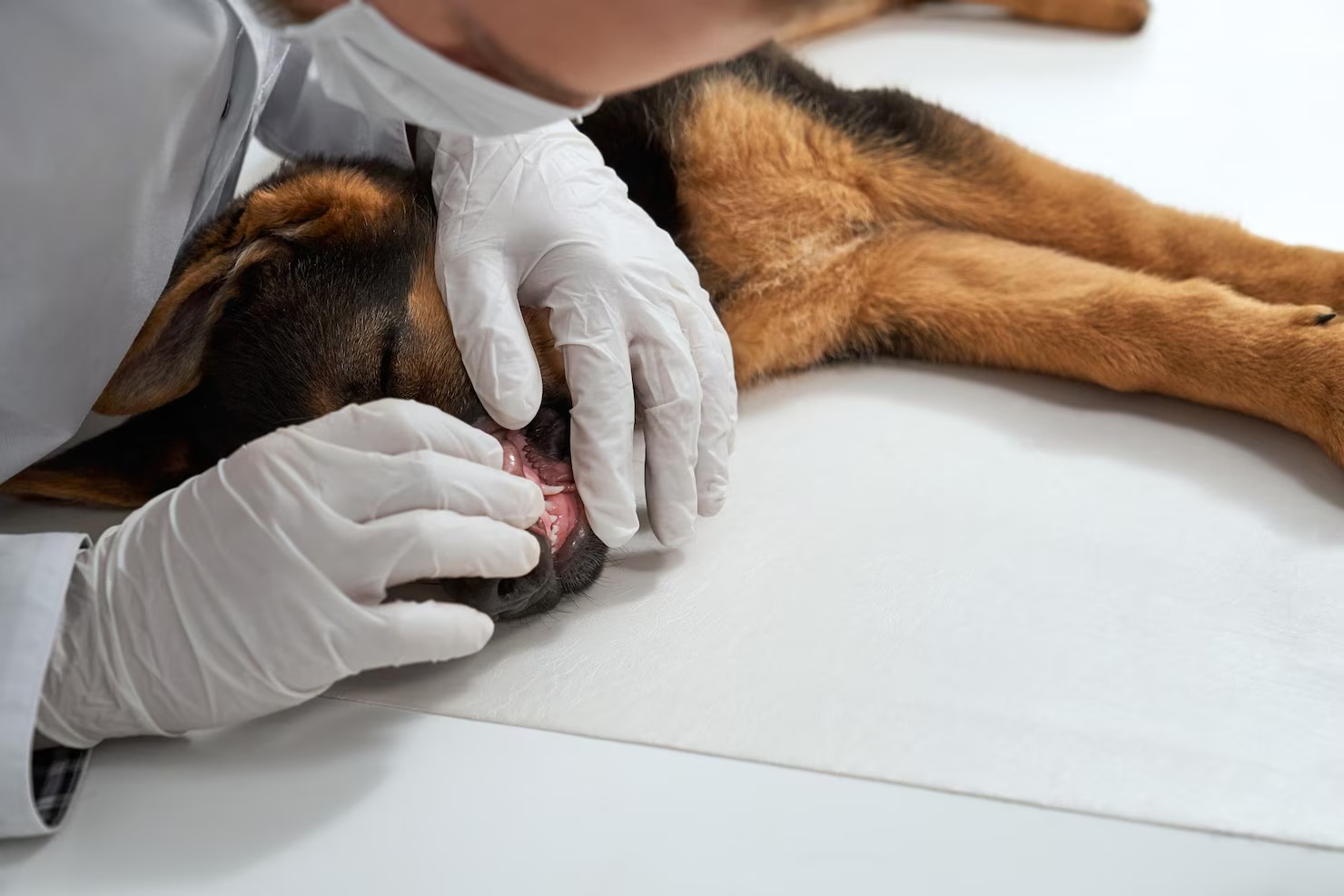
Fine-Needle Aspiration (FNA)
Fine-Needle Aspiration (FNA) is universally considered the foundational, first-line diagnostic modality in veterinary oncology for evaluating almost any palpable cutaneous mass. It is a highly rapid, minimally invasive, highly cost-effective, and exceptionally safe clinical procedure that frequently provides an immediate, highly accurate cellular diagnosis, heavily guiding all subsequent treatment decisions.[10]
- Procedural Technique: The clinician utilizes a standard, sterile hypodermic needle (typically 22 or 25 gauge) attached to a sterile syringe. After thoroughly isolating the mass with their fingers, the needle is aggressively inserted directly into the core of the tumor. The clinician heavily redirects the needle multiple times through different tissue planes within the mass (a technique often called the “woodpecker technique”) to heavily sheer and collect a representative population of cells into the hollow bore of the needle. A syringe can be used to heavily apply negative pressure (suction) to forcefully draw stubborn cells into the needle hub, particularly for firm, highly cohesive epithelial tumors.
- Cytological Slide Preparation: The collected, microscopic cellular material is immediately, forcefully expelled onto a clean glass microscope slide. The clinician carefully uses a second slide to gently but firmly smear the highly fragile cellular material into a very thin, monolayered film. The slide is then heavily air-dried, completely fixed, and stained using specialized, highly differential cytological stains (such as Diff-Quik or Wright-Giemsa).
- Microscopic Cytological Evaluation: The stained slide is heavily evaluated under a high-powered microscope. The clinician (or a board-certified veterinary clinical pathologist) meticulously categorizes the general cell type present (e.g., differentiating between a distinct mesenchymal spindle cell, a highly cohesive epithelial cell cluster, or a discrete, individual round cell). They heavily look for distinct microscopic criteria of malignancy, including massive variation in cell size (anisocytosis), massive variation in nucleus size (anisonucleosis), heavily prominent, multiple nucleoli, highly abnormal, coarse chromatin clumping patterns, and the heavy presence of bizarre, abnormal mitotic figures (cells caught in the act of highly abnormal division).
FNA is incredibly diagnostic for distinctly classifying inflammatory lesions (like abscesses, identifying massive numbers of neutrophils and heavy intracellular bacteria), completely diagnosing benign lipomas (yielding distinct droplets of clear, acellular free lipid), and heavily confirming specific round cell tumors like extremely aggressive mast cell tumors, which shed massive quantities of highly characteristic, deep purple, heavily granulated cells. However, FNA is not infallible. Some heavily fibrotic, heavily scarred, or deeply necrotic tumors simply do not exfoliate or shed enough intact cells into the needle, resulting in a frustratingly non-diagnostic sample. In these complex cases, a formal tissue biopsy becomes absolutely necessary.[11]
Imaging
When a fine-needle aspirate or tissue biopsy definitively confirms the presence of a highly malignant, aggressive skin tumor, the diagnostic focus immediately shifts from identifying the mass to heavily “staging” the entire patient. Clinical staging utilizes highly advanced, specialized diagnostic imaging modalities to meticulously search the entire body for any profound evidence of distant metastasis (cancer spread) or to completely evaluate the deep, localized invasiveness of the primary mass prior to attempting highly complex surgery.[12]
- Thoracic Radiographs (X-rays): The deep pulmonary parenchyma (the lungs) is the single most common, highly frequent site for aggressive distant metastasis from malignant canine skin tumors. Procuring highly detailed, three-view (right lateral, left lateral, and ventrodorsal) thoracic radiographs is absolutely essential. The clinician meticulously scrutinizes the lung fields, searching for the heavy presence of distinct, abnormal soft-tissue nodules, deeply enlarged intrathoracic lymph nodes, or any heavy evidence of malignant pleural effusion (fluid building up around the lungs). Identifying heavy pulmonary metastasis often drastically alters the clinical prognosis from potentially curable to strictly palliative.
- Abdominal Ultrasonography: A highly detailed, comprehensive abdominal ultrasound is frequently mandated, particularly for tumors like high-grade mast cell tumors or malignant melanomas that have a notorious, highly documented propensity to aggressively metastasize to deep internal abdominal organs, specifically the liver, spleen, and the deep, internal sublumbar or mesenteric lymph nodes. Ultrasound provides a heavily detailed, real-time internal view of the architecture of these vital organs and allows the clinician to safely perform heavily targeted, ultrasound-guided fine-needle aspirations of any highly suspicious, abnormal internal lesions discovered.
- Advanced Cross-Sectional Imaging (CT and MRI): Computed Tomography (CT) heavily utilizes advanced X-ray technology to generate highly complex, deeply detailed, 3D cross-sectional slices of the patient’s body. Magnetic Resonance Imaging (MRI) heavily utilizes powerful magnetic fields to provide unparalleled, exquisite detail of soft tissue structures. These highly advanced imaging modalities are absolutely essential for surgical planning when dealing with massive, deeply fixed, or highly infiltrative tumors (such as aggressive soft tissue sarcomas or deeply invasive oral melanomas). A CT scan allows the veterinary surgeon to perfectly visualize exactly how deeply the tumor has violently invaded into the surrounding critical blood vessels, delicate nerve plexuses, and rigid underlying bone, heavily determining if complete, clean surgical removal is physically possible.
Thorough, aggressive clinical staging is fundamentally required to provide the pet owner with an accurate, highly realistic prognosis and to heavily guide the selection of the most profoundly appropriate, effective therapeutic modalities. Without aggressive staging, a surgeon might unknowingly subject a dog to massive, highly painful surgery to remove a primary skin tumor, only to tragically discover weeks later that the cancer had already heavily spread to the lungs.[13]
Blood Tests
While standard, routine hematological tests cannot definitively, microscopically diagnose the exact type of skin tumor, comprehensive blood analysis is a deeply foundational requirement in the exhaustive oncological workup. It provides profoundly vital information regarding the patient’s overall, systemic physiological status, actively uncovers severe, hidden concurrent diseases that heavily complicate anesthesia, and deeply assesses the patient’s biological ability to safely tolerate highly toxic chemotherapy protocols.
- Complete Blood Count (CBC): The CBC heavily quantifies and intimately evaluates the red blood cells, white blood cells, and platelets. It is crucial for detecting severe anemia (which could be directly caused by chronic, heavy hemorrhage from an ulcerated, bleeding tumor or by malignant bone marrow suppression), heavy systemic infection (indicated by a massive elevation in circulating white blood cells), or profound thrombocytopenia (dangerously low platelet counts, which heavily increases the risk of massive, uncontrollable bleeding during surgery). In highly specific cases, such as advanced, systemic cutaneous lymphoma or highly aggressive mast cell disease, the clinician can actually identify actively circulating, highly malignant cancer cells directly within the bloodstream by heavily examining the buffy coat smear under the microscope.
- Serum Biochemistry Profile: This comprehensive blood panel heavily evaluates the deep functional health of the body’s major internal organs, most notably the liver and the kidneys. Severe organ dysfunction heavily dictates the specific choice and precise dosage of potent anesthetic agents and highly toxic chemotherapy drugs, as these organs are primarily responsible for deeply metabolizing and actively clearing these powerful medications from the body. Furthermore, the biochemistry profile is absolutely vital for detecting highly specific paraneoplastic syndromes. The most profound example is hypercalcemia of malignancy, an exceptionally dangerous, life-threatening elevation of systemic blood calcium levels frequently caused by the tumor heavily secreting a specific hormone called parathyroid hormone-related protein (PTHrP).
- Coagulation Profiles: Specific, highly advanced cancers, particularly aggressive hemangiosarcomas or advanced mast cell tumors, can severely disrupt the body’s complex, highly delicate blood-clotting cascade, potentially leading to massive, life-threatening intraoperative hemorrhage. A comprehensive coagulation profile heavily ensures that the patient’s blood can clot normally before any scalpel touches the skin.
Comprehensive bloodwork ensures that the veterinarian possesses a completely holistic, highly detailed understanding of the patient’s internal physiological landscape, heavily maximizing the safety and profound efficacy of the entire subsequent treatment plan.[14]
Treatment for Skin Masses in Dogs

The formulation of a definitive treatment plan for canine skin masses is a highly nuanced, profoundly complex clinical undertaking. The chosen therapeutic strategy is heavily dictated by a synthesis of critical factors: the exact, histologically confirmed type of the tumor, its precise anatomical location and size, its established malignant grade and stage, the patient’s overall physiological health, and the owner’s goals and financial constraints. Treatment heavily ranges from simple surgical cure to complex, multi-modal oncological protocols.
Surgical Treatment of Skin Masses in Dogs
Surgical excision fundamentally remains the absolute cornerstone, heavily relied upon primary treatment modality for the vast majority of solid skin masses in dogs. The primary, highly critical objective of oncological surgery is to completely physically eradicate the entire macroscopic tumor burden along with a meticulously calculated, heavily defined perimeter of completely normal, microscopic healthy tissue—known heavily as the “surgical margin”—to definitively guarantee that absolutely no microscopic, aberrant cancer cells are inadvertently left behind in the tumor bed to cause a devastating future local recurrence.[15]
- Surgical Planning and Anesthetic Protocols: Prior to making any incision, the veterinary surgeon heavily relies on the diagnostic staging (bloodwork, imaging) to meticulously plan the exact surgical approach. General anesthesia is strictly required, utilizing highly tailored, heavily monitored protocols to ensure absolute patient safety and profound, complete pain management throughout the highly invasive procedure.
- Principles of Oncological Surgical Margins: The strict, aggressive width of the normal tissue margin heavily removed along with the tumor is entirely dictated by the tumor’s specific biological behavior. For distinctly benign masses like simple lipomas, a “marginal excision,” where the surgeon dissects precisely closely along the tumor’s true capsule, is highly appropriate and fully curative. However, for deeply invasive, highly aggressive malignancies like fibrosarcomas or high-grade mast cell tumors, the surgeon must execute a “wide margin excision.” This heavily requires removing a massive perimeter of completely normal-appearing skin—often 2 to 3 centimeters completely surrounding the entire tumor in all lateral directions—and, absolutely crucially, removing at least one complete, uninvaded fascial tissue plane deeply underneath the tumor base to heavily ensure complete, definitive microscopic eradication of infiltrating cellular tendrils.
- Reconstructive Surgery Challenges: Creating massive, highly aggressive surgical margins often results in enormous, profoundly gaping tissue defects that cannot simply be pulled closed. Veterinary surgeons must heavily utilize highly complex, advanced reconstructive surgical techniques, utilizing elaborate local skin flaps, massive axial pattern flaps, or highly complex free skin grafts to successfully cover these massive surgical wounds, allowing for primary healing and heavily maintaining the patient’s functional mobility.
- Mandatory Histopathological Margin Evaluation: Following removal, the entire, massive surgical specimen is heavily inked with specialized surgical dyes by the clinician and immediately submitted to a veterinary pathologist. The pathologist meticulously section the tissue to heavily evaluate the microscopic edges of the specimen. If the surgical margins are definitively “clean” (completely devoid of any tumor cells), the surgery is considered highly successful. If the margins are heavily “dirty” or “incomplete” (cancer cells deeply extend to the absolute edge of the cut tissue), it is a clinical certainty that microscopic disease remains actively growing in the patient, strictly necessitating heavily aggressive secondary surgery or immediate, highly targeted radiation therapy to prevent a massive local recurrence.
While heavily invasive and carrying inherent risks of anesthesia, profound post-operative pain, and deep incisional infection, aggressive, properly executed surgery heavily provides the highest possible probability of a complete, definitive clinical cure for solitary, solid skin masses.[16]
Chemotherapy for Skin Masses in Dogs
Chemotherapy is a highly specialized, systemically administered medical treatment that heavily utilizes powerfully toxic pharmacological agents to aggressively target, severely damage, and physically destroy rapidly dividing cancer cells heavily circulating throughout the entire body. In the specific context of canine skin masses, chemotherapy is primarily utilized to aggressively combat highly malignant tumors that possess a profoundly high rate of distant metastasis (such as high-grade mast cell tumors, malignant melanomas, or advanced lymphomas) or as a vital adjunctive “mop-up” therapy following surgery to actively hunt down and destroy any heavily elusive microscopic cancer cells that may have already secretly escaped into the systemic bloodstream.[17]
- Philosophical Objective in Veterinary Medicine: A profound distinction exists between human and veterinary chemotherapy. In veterinary oncology, the absolute primary objective is rarely to push the patient to the absolute brink of toxicity to achieve a total cure at any cost. Instead, the profound goal is maximizing the quality of life while heavily extending survival time. Consequently, veterinary chemotherapy dosages are meticulously calculated to be highly effective against the tumor while purposely generating significantly fewer, much milder systemic side effects than those heavily experienced by human patients.
- Mechanisms of Action and Drug Classes: Chemotherapeutic agents are highly complex toxins that heavily target the intricate cellular mechanisms of actively dividing cells. Different drug classes disrupt the cancer cell life cycle at various critical points. Alkylating agents (like Chlorambucil or Lomustine) heavily cross-link DNA, preventing replication; Vinca alkaloids (like Vincristine) profoundly disrupt the delicate mitotic spindle apparatus, physically stopping cell division in its tracks. A veterinary oncologist will heavily design specific protocols utilizing combinations of these highly potent drugs to maximize tumor cell death while minimizing profound toxicity to normal host tissues.
- Treatment Protocols and Schedules: The chemotherapy protocol is a highly rigorous, tightly scheduled regimen. Treatments, heavily termed “cycles,” are administered at precise, scientifically calculated intervals (often every 1 to 3 weeks). This specific timing is heavily designed to strike the cancer cells when they are most biologically vulnerable while allowing the patient’s normal, healthy tissues (particularly the sensitive bone marrow and gastrointestinal tract lining) a crucial period to physically recover and regenerate.
- Management of Side Effects: While generally well-tolerated, systemic chemotherapy profoundly affects any rapidly dividing cells in the body, including the healthy cells heavily lining the stomach and intestines and the vital white blood cells produced heavily within the bone marrow. Potential side effects include profound, acute nausea, severe, transient inappetence, heavy vomiting, diarrhea, and significant myelosuppression (a dangerous drop in white blood cells, making the dog highly susceptible to overwhelming secondary infections). Remarkably, massive hair loss (alopecia) is heavily uncommon in most dog breeds, heavily excepting breeds with continuously growing hair coats like Poodles, Schnauzers, and Old English Sheepdogs.
- Intensive Clinical Monitoring: A patient undergoing heavy chemotherapy requires exhaustive, continuous clinical monitoring. Prior to every single dose, the veterinarian must perform a rigorous physical exam and run a comprehensive Complete Blood Count (CBC) to definitively ensure the patient possesses an adequate white blood cell count to safely metabolize and survive the highly toxic treatment. If the white blood cell count plunges dangerously low, the chemotherapy must be heavily delayed, or the specific dosage significantly reduced.
Chemotherapy requires a massive, deep commitment from the pet owner regarding intense monitoring, frequent veterinary visits, and significant financial investment. However, for aggressive, systemic skin cancers, it is often the most profoundly effective weapon available to heavily extend a high quality of life.[18]
Radiation Therapy for Skin Masses in Dogs
Radiation therapy (radiotherapy) is a highly sophisticated, technologically advanced oncological modality that heavily utilizes massively high-energy ionizing radiation—typically powerful X-rays or electron beams generated heavily by a massive medical linear accelerator—to physically target and profoundly destroy the DNA specifically within localized cancer cells. When the cancer cell’s DNA is heavily shattered by the radiation beam, the cell utterly loses its physical ability to divide and reproduce, eventually resulting in targeted cellular death. In veterinary dermatology, radiation is a profoundly critical tool for treating skin masses where highly aggressive surgical excision is anatomically impossible without causing unacceptable, severe functional mutilation (such as massive tumors deeply embedded on the face, the distal limbs, or across the delicate joints).[19]
- Definitive vs. Palliative Intent: Radiation therapy is heavily divided into two completely distinct philosophical protocols. Definitive (Curative) Radiation is an aggressive, high-dose protocol heavily designed to completely eradicate all microscopic disease remaining in the tumor bed after an incomplete surgical removal (where the margins were heavily “dirty”). This protocol heavily aims for a long-term, definitive cure. Palliative Radiation utilizes a much lower, less aggressive total dose of radiation over fewer sessions. Its sole, profound objective is not to cure the cancer, but to rapidly shrink a massive, heavily inoperable tumor, thereby massively reducing intense pain, profoundly stopping severe bleeding, and significantly improving the patient’s end-of-life comfort for a matter of months.
- The Procedural Mechanics: Delivering precisely targeted, highly destructive ionizing radiation requires absolute, motionless precision. Therefore, the dog must be heavily anesthetized for every single treatment session. The patient is placed on a specialized treatment couch, and the massive linear accelerator heavily targets the specific tumor bed from multiple complex angles to heavily maximize the destructive dose delivered directly to the tumor while severely sparing the delicate, surrounding healthy tissues from heavy radiation damage.
- Fractionated Treatment Schedules: To heavily protect healthy, normal tissue, the total required dose of heavy radiation is never given all at once. It is meticulously divided into numerous, smaller, highly calculated doses heavily termed “fractions.” A definitive radiation protocol frequently requires the dog to undergo general anesthesia and receive heavy radiation treatments daily, five days a week, for a span of three to four grueling weeks.
- Acute and Late Side Effects: While the radiation beam is completely invisible and entirely painless during administration, it profoundly damages rapidly dividing normal cells in the treatment field. Acute side effects heavily manifest near the end of the strict protocol and deeply include severe, painful radiation dermatitis (profound skin redness, massive hair loss, intense blistering, and moist, painful desquamation mimicking a severe thermal burn). These acute effects are highly painful but heavily expected and typically heal completely within several weeks post-treatment. Late, highly chronic side effects, heavily occurring months or years later, can profoundly include permanent, massive tissue fibrosis, severe skin color changes (leukotrichia), or highly rare secondary radiation-induced tumor formation.
Access to advanced radiation therapy heavily requires referral to a highly specialized, dedicated veterinary oncology center equipped heavily with a linear accelerator, and involves profound financial and significant logistical commitment from the dedicated pet owner.[20]
Cryosurgery for Skin Masses in Dogs
Cryosurgery, or heavily localized clinical cryotherapy, is a highly effective, minimally invasive therapeutic modality that heavily utilizes targeted, extreme freezing temperatures to aggressively induce severe, localized cellular necrosis and subsequent complete destruction of targeted abnormal tissues. In veterinary practice, it is most frequently and heavily utilized for completely eradicating very small, superficial, distinctly benign skin masses such as tiny sebaceous adenomas, heavy viral papillomas, or extremely small, superficial squamous cell carcinomas, particularly in highly delicate, difficult-to-suture anatomical locations like the delicate eyelid margins or within the complex structure of the ear canal.[21]
- The Mechanism of Cryonecrosis: The fundamental biological objective of cryosurgery is to heavily freeze the targeted cells so rapidly and deeply that sharp, destructive intracellular ice crystals violently form, physically shattering the delicate cell membranes and destroying the vital internal cellular organelles. When the tissue is subsequently allowed to slowly, naturally thaw, massive, targeted vascular thrombosis heavily occurs within the microvasculature, completely starving the heavily frozen tissue of vital blood supply, leading to massive, inevitable ischemic necrosis and heavy cellular death.
- Procedural Application: The veterinarian utilizes specialized, highly precision-engineered cryosurgical equipment. Either highly volatile liquid nitrogen (which reaches profoundly frigid temperatures of -196°C) or highly pressurized argon gas is aggressively delivered directly onto or immediately into the targeted mass via a fine, focused spray technique or a highly specialized, freezing contact probe. The clinician meticulously orchestrates a precise, highly calculated rapid freeze followed by a crucial slow, deliberate thaw phase, heavily repeating this grueling cycle two or three distinct times (a double-freeze-thaw cycle) to absolutely maximize the profound destructive tissue effect.
- Anesthesia and Patient Compliance: Because the rapid, deep freezing process profoundly numbs the local sensory nerve endings almost instantly, the procedure is remarkably fast and frequently well-tolerated. Therefore, cryosurgery can often be highly successfully performed utilizing only mild, reversible sedation and highly targeted local anesthetic blocks, completely avoiding the inherent, heavy risks heavily associated with full, deep general anesthesia, making it an extraordinarily attractive, profound option for significantly older, heavily geriatric patients with deep, concurrent systemic health issues.
- Post-Procedural Healing and Monitoring: Immediately following the heavy cryosurgical insult, the heavily treated tissue will typically appear acutely swollen, profoundly hyperemic (red), and may heavily blister or actively ooze serum. Over the subsequent weeks, the targeted mass will slowly turn completely necrotic (blackening), shrivel drastically, and eventually slough off entirely, leaving a highly distinct, circular, completely hairless scar (alopecia) and frequently causing permanent, deep depigmentation (leukotrichia) of any hair that eventually manages to regrow in the heavily treated area.
While profoundly effective for highly selected, very small, benign superficial lesions, cryosurgery is heavily contraindicated and profoundly inappropriate for large, deeply embedded masses, highly infiltrative tumors, or aggressive, confirmed malignancies where heavily guaranteeing complete, clean, microscopically evaluated surgical margins is absolutely, unequivocally vital for preventing a devastating local recurrence.[22]
Immunotherapy for Skin Masses in Dogs
Immunotherapy represents arguably the most exciting, heavily researched, and profoundly innovative frontier in modern veterinary oncology. Historically, cancer treatments heavily relied on exogenous, highly toxic chemicals or massively destructive radiation to kill tumors. Immunotherapy, however, takes a profoundly different approach: it heavily focuses on highly specifically stimulating, artificially reprogramming, and massively boosting the patient’s own native immune system, heavily empowering the body’s own cytotoxic white blood cells to aggressively recognize, actively seek out, and violently destroy highly abnormal, elusive cancerous tumors wherever they attempt to hide throughout the body.[23]
- Overcoming Immune Evasion: Malignant cells are biologically treacherous; they survive and heavily proliferate largely by utilizing highly complex molecular camouflage mechanisms to “hide” completely from the host’s normal immune surveillance, effectively rendering the tumor completely “invisible” to the body’s natural defenses. The profound, primary goal of highly advanced immunotherapy is to violently strip away this molecular camouflage, loudly “tagging” the cancer cells and forcefully provoking a massive, highly specific, systemic immune attack.
- The Oncept Melanoma Vaccine: The absolute shining paradigm of successful veterinary immunotherapy is the Oncept canine melanoma vaccine. This is a highly specialized, profoundly complex xenogeneic DNA vaccine. It heavily utilizes a massively engineered plasmid containing the specific DNA sequence for human tyrosinase (a vital protein heavily found in massive abundance on melanoma cells). When forcefully injected via a highly specialized transdermal device into the dog’s muscle, the dog’s immune system heavily recognizes this human protein as profoundly foreign and rapidly mounts a massive, aggressive immune attack. Crucially, because the human and canine tyrosinase proteins are structurally very similar, the highly agitated canine immune system cross-reacts, unleashing a massive, targeted wave of cytotoxic T-cells that actively hunt down and violently destroy the dog’s own microscopic malignant melanoma cells wherever they have metastasized.
- Monoclonal Antibodies and Checkpoint Inhibitors: The cutting edge of veterinary research is heavily exploring the complex use of specifically engineered Monoclonal Antibodies (mAbs). These are highly specialized, laboratory-produced proteins heavily designed to tightly bind to highly specific, unique receptors heavily located on the surface of malignant tumor cells, essentially marking them heavily for violent immune destruction. While still heavily in deep developmental and clinical trial phases for various canine cancers, these highly targeted therapies offer a profound, incredibly powerful future where tumors can be aggressively attacked with extreme prejudice, leaving completely normal, healthy tissues completely unharmed, vastly minimizing the severe, highly toxic side effects historically associated with traditional chemotherapy.
Immunotherapy is rarely, if ever, utilized as a sole, standalone primary treatment. It is almost exclusively and heavily employed as a profoundly powerful adjunctive, systemic therapy, utilized immediately after the massive, primary tumor bulk has been heavily eradicated via aggressive surgical resection or heavy radiation.[24]
Medication for Treating Skin Masses in Dogs
The vast, complex landscape of modern veterinary pharmacology offers a highly diverse, incredibly potent array of specialized, targeted medications that play a profoundly critical role in the comprehensive management of canine skin masses. While these heavily prescribed oral or systemic medications are occasionally utilized to directly attack and physically shrink the specific tumor volume, they are far more frequently and heavily employed to aggressively manage massive localized inflammation, completely resolve severe, complicating secondary bacterial infections, profoundly alleviate deep tissue pain, or heavily modulate highly specific oncological signaling pathways.[25]
- Tyrosine Kinase Inhibitors (TKIs): The introduction of heavily targeted Tyrosine Kinase Inhibitors (TKIs), most notably the heavily prescribed drug Toceranib phosphate (commercially known as Palladia), has utterly revolutionized veterinary oncology. TKIs are highly advanced, profoundly specific biological drugs heavily engineered to precisely block highly specific, malfunctioning cellular communication receptors (specifically the c-KIT receptor) located heavily on the surface of certain cancer cells, particularly high-grade, aggressive mast cell tumors. When these critical receptors are forcefully blocked by the TKI, the malignant cell essentially loses its primary, critical biological signal to divide, actively grow, and heavily recruit new blood vessels (angiogenesis). This profoundly cuts off the tumor’s vital blood supply and forces the malignant cells to heavily undergo programmed cell death (apoptosis), leading to massive tumor shrinkage and disease stabilization without utilizing traditional chemotherapy.
- Non-Steroidal Anti-Inflammatory Drugs (NSAIDs): While heavily known primarily for their profound ability to powerfully control severe pain and reduce massive acute inflammation, certain specific veterinary NSAIDs (such as Piroxicam, Meloxicam, or Carprofen) exhibit highly remarkable, deeply documented anti-cancer properties. They achieve this primarily by aggressively inhibiting the COX-2 (Cyclooxygenase-2) enzyme, which is frequently and massively overexpressed by numerous highly malignant carcinomas and aggressive sarcomas. Heavy COX-2 inhibition not only deeply manages tumor-associated pain but can also profoundly reduce tumor-associated inflammation and heavily disrupt the tumor’s critical ability to grow new, supportive blood vessels, exerting a deeply meaningful, heavy anti-proliferative effect.
- Corticosteroids: Potent, systemically administered corticosteroids, particularly high-dose Prednisone or Dexamethasone, are heavily utilized in veterinary oncology. They are profoundly effective at rapidly, massively reducing severe peritumoral edema (swelling) and deeply decreasing heavy, localized, painful inflammation heavily surrounding aggressively expanding tumors. Furthermore, corticosteroids are directly, heavily cytotoxic (lethal) to specific, malignant immune cells, making them an absolute foundational, critical component in almost every complex chemotherapy protocol heavily utilized for treating advanced cutaneous lymphomas or aggressive mast cell tumors.
- Antibiotics and Anti-microbials: Because many massive, rapidly growing skin tumors frequently outgrow their delicate blood supply, they inevitably ulcerate, heavily break open, and suffer severe, deep tissue necrosis. These open, highly compromised wounds are instantly and heavily colonized by highly pathogenic environmental and oral bacteria, leading to massive, deep secondary pyoderma. The heavy, systemic administration of precisely targeted, culture-driven oral antibiotics (such as Cephalexin or Clindamycin) is absolutely vital to aggressively resolve the deep infection, massively reduce associated fever and lethargy, deeply improve patient comfort, and completely stabilize the surrounding tissues prior to attempting a highly complex, invasive surgical resection.
Prevention for Skin Tumors in Dogs
Preventing the spontaneous, terrifying emergence of cutaneous neoplasms in dogs is a highly complex, heavily profound clinical challenge. Unlike many infectious pathogens where highly targeted vaccines provide robust, guaranteed immunity, cancer fundamentally arises from deep, complex, highly unpredictable internal genetic malfunctions intimately combined with extensive, lifetime environmental exposures. Therefore, absolute, guaranteed prevention is biologically impossible. However, dedicated pet owners can heavily implement highly strategic, profoundly proactive lifestyle adjustments to massively mitigate known environmental risks and heavily increase the likelihood of life-saving early detection.[26]
- Rigorous Sun Avoidance and UV Protection: For breeds heavily predisposed to deep solar-induced skin damage (such as those with extremely thin, highly sparse, or completely unpigmented white hair coats like Pit Bulls, Dalmatians, and Whippets), profound, absolute protection from intense, direct solar ultraviolet radiation is unequivocally mandatory. Owners must completely restrict prolonged outdoor sunbathing, particularly during peak UV index hours (between 10:00 AM and 4:00 PM). Providing heavily shaded, deep outdoor rest areas is vital. Furthermore, the daily, heavy application of highly specialized, pet-safe, non-toxic zinc-free sunscreens, or physically outfitting the dog with heavily protective UV-blocking specialized clothing (sun shirts), can massively, profoundly reduce the deeply cumulative actinic cellular damage that directly drives the pathogenesis of highly aggressive, lethal squamous cell carcinomas.
- Proactive, Routine Veterinary Surveillance: Establishing a strict, non-negotiable schedule of comprehensive, highly detailed biannual or annual veterinary physical examinations is arguably the most profoundly powerful, highly effective preventive weapon. A highly trained veterinarian possesses the specific tactile expertise to heavily palpate and discover extremely small, deeply hidden, completely asymptomatic lumps that an owner might easily overlook. When an aggressive tumor is discovered while it is still microscopically small and highly localized, before it has heavily invaded deep tissues or metastasized into the lymphatic system, a simple, localized surgical excision is frequently 100% curative.
- Exhaustive Home Grooming and Tactile Screening: Owners must heavily integrate deep, highly methodical “nose-to-tail” tactile massages and rigorous, deep grooming sessions into their weekly routine. Meticulously, deeply running hands heavily through the dog’s coat, specifically feeling the deep axillary (armpit) regions, the sensitive groin, carefully examining the delicate nail beds, and deeply inspecting the hidden mucocutaneous junctions around the mouth and eyes, empowers the owner to instantly detect the smallest, newest abnormality. Any newly discovered bump must be evaluated by a veterinarian immediately; never assume a new lump is “just a fat deposit.”
- Strategic Sterilization Timing: The precise, highly debated timing of spaying (ovariohysterectomy) and neutering (castration) exerts a massive, deeply profound physiological impact on the subsequent, lifetime development of specifically hormonally driven tumors. Spaying a female dog completely prior to her very first estrus cycle massively, profoundly reduces her lifetime statistical risk of developing aggressive malignant mammary gland tumors to less than 0.5%. Similarly, heavily neutering male dogs effectively, completely eliminates the heavy physiological risk of developing systemic testosterone-dependent perianal gland adenomas and completely prevents deadly testicular neoplasia.
- Nutritional Excellence and Immune Support: Feeding a highly premium, rigorously balanced, heavily bio-appropriate diet, exceptionally rich in high-quality, highly digestible proteins, complex vital antioxidants, and heavy essential Omega-3 fatty acids, provides the deep, foundational physiological building blocks required to maintain a highly robust, aggressively vigilant immune system. A powerfully healthy immune system is the dog’s absolute first and strongest line of defense, heavily tasked with constantly patrolling the body to recognize and instantly destroy microscopically small, newly forming aberrant cancer cells before they can establish a massive tumor.
- Comprehensive Pathogen Protection: Ensuring your dog is up-to-date on all recommended vaccines can help prevent certain viral infections that can heavily compromise the immune system or directly induce heavy neoplastic changes (such as managing exposure to the canine papillomavirus). Additionally, maintaining strict, year-round, flawless parasite control (utilizing highly effective flea, tick, and heartworm preventatives) heavily prevents severe, chronic localized tissue inflammation and massive, deep secondary immune hypersensitivity reactions that can deeply complicate dermatological health.
Always consult your veterinarian before making any changes to your pet’s care. Early, heavily aggressive diagnosis and highly targeted, rapid clinical intervention are the absolute, undisputed cornerstones of profoundly successful veterinary oncological medicine. Never ignore a newly discovered skin mass, hoping it will simply vanish. Taking immediate, decisive action ensures your pet receives the highest, most profound standard of compassionate, life-saving care.[27]
Frequently Asked Questions
References
- Merck Veterinary Manual. Cutaneous Neoplasia in Dogs. Merck & Co., 2023.
- Goldschmidt, M. H., & Shofer, F. S. Skin Tumors of the Dog and Cat. Pergamon Press, 1992.
- American Veterinary Medical Association (AVMA). Cancer in Pets: Early Detection. AVMA, 2023.
- Withrow, S. J., & MacEwen, E. G. Small Animal Clinical Oncology. Saunders Elsevier, 2019.
- Dobson, J. M. Breed-predispositions to cancer in pedigree dogs. ISRN Veterinary Science, 2013.
- Kessler, M. Small Animal Oncology. Thieme Medical Publishers, 2010.
- VCA Animal Hospitals. Sebaceous Cysts in Dogs. VCA, 2022.
- Miller, W. H., Griffin, C. E., & Campbell, K. L. Muller and Kirk’s Small Animal Dermatology. Elsevier Mosby, 2013.
- Gross, T. L., et al. Skin Diseases of the Dog and Cat: Clinical and Histopathologic Diagnosis. Blackwell Science, 2005.
- Moore, P. F. A review of histiocytic diseases of dogs and cats. Veterinary Pathology, 2014.
- Affolter, V. K., & Moore, P. F. Canine cutaneous and systemic histiocytosis. American Journal of Veterinary Pathology, 2000.
- Raskin, R. E., & Meyer, D. J. Canine and Feline Cytology: A Color Atlas and Interpretation Guide. Saunders, 2015.
- Welle, M. M., et al. Mast cell tumors in dogs: predispositions, pathology, and clinical relevance. Veterinary Dermatology, 2008.
- Kiupel, M., et al. Proposal of a 2-tier histologic grading system for canine cutaneous mast cell tumors. Veterinary Pathology, 2011.
- London, C. A., et al. Multi-center, placebo-controlled, double-blind, randomized study of oral toceranib phosphate (SU11654). Clinical Cancer Research, 2009.
- Veterinary Information Network (VIN). Aural Hematoma Management. VIN, 2021.
- Fossum, T. W. Small Animal Surgery. Elsevier Mosby, 2018.
- Slatter, D. Textbook of Small Animal Surgery. Saunders, 2003.
- Nicholls, P. K., & Stanley, M. A. The immunology of animal papillomaviruses. Veterinary Immunology and Immunopathology, 2000.
- Goldschmidt, M. H., et al. Tumors of the Skin and Soft Tissues. Tumor Fascicle, Armed Forces Institute of Pathology, 1998.
- Munday, J. S., et al. Papillomaviruses in dogs and cats. Veterinary Journal, 2017.
- Bergman, P. J. Canine oral melanoma. Clinical Techniques in Small Animal Practice, 2007.
- Smith, S. H., et al. A review of the histologic grading of canine cutaneous melanoma. Veterinary Pathology, 2002.
- Bergman, P. J., et al. Long-term survival of dogs with advanced malignant melanoma after DNA vaccination. Clinical Cancer Research, 2006.
- ASPCA. Abscesses and Wound Management in Pets. ASPCA, 2021.
- Greene, C. E. Infectious Diseases of the Dog and Cat. Elsevier Saunders, 2012.
- Sykes, J. E. Canine and Feline Infectious Diseases. Elsevier Saunders, 2014.


