


 March 2, 2023
 Phil Good, DVM
What are Ear Mites in Dogs?
This content was prepared with AI assistance and reviewed by a licensed professional for accuracy.
Introduction
As a veterinarian, I often see pet owners deeply concerned when their beloved canine companions constantly scratch their heads and exhibit extreme discomfort. Often, the culprit behind this agonizing itch is an infestation of ear mites, which are highly contagious, microscopic parasites in dog’s ears. Recognizing the signs of mites early is critical because an untreated ear mite infestation can lead to severe auditory complications, secondary bacterial infections, and permanent tissue damage. Karen, a dedicated pet owner, recently experienced this exact scenario with her lovable Beagle, Buster. For several days, Buster had been persistently scratching at his ears, whimpering in distress, and violently shaking his head. Initially, Karen suspected it might be a minor, transient irritation related to seasonal allergies or a mild ear problem that would resolve on its own. However, when she observed a dark, foul-smelling discharge resembling coffee grounds emanating from Buster’s ear canals, she recognized the urgency of the situation and brought him to our clinic. After a comprehensive otoscopic examination and microscopic cytology, I confirmed that Buster was suffering from a severe case of otodectic mange, a parasitic condition caused by the ear mite Otodectes cynotis.[1]
Ear mites in dogs, scientifically classified as Otodectes cynotis, belong to the family Psoroptidae. These minuscule arthropods are non-burrowing, surface-dwelling parasites that predominantly infest the epithelial lining of the canine ear canal, though in severe cases, they can migrate to the adjacent skin of the head, neck, and tail base.[2] These highly active mites sustain themselves by piercing the outer layer of the skin—the stratum corneum—and feeding on epidermal debris, lymph fluid, tissue exudate, and cerumen (ear wax). The feeding process itself causes significant mechanical irritation, but the primary source of the dog’s intense pruritus (itching) is an allergic hypersensitivity reaction to the mite’s saliva and fecal matter.[3] This hypersensitivity means that even a relatively small population of mites can trigger a massive inflammatory response, leading to the clinical presentation of otodectic mange.[4]
To fully understand why the canine ear is such a hospitable environment for these parasites, we must look at the unique anatomy of a dog’s ear canal. Unlike human ear canals, which are relatively straight and horizontal, canine ear canals are structured in a distinct “L” shape. They consist of a long vertical canal that travels downward before making a sharp, nearly 90-degree turn into a horizontal canal, which ultimately leads to the tympanic membrane (eardrum).[5] This anatomical configuration is excellent for protecting the delicate eardrum from foreign objects and trauma, but it also creates a deep, dark, warm, and poorly ventilated environment that is ideal for trapping moisture, wax, and debris.[6] For an obligate parasite like Otodectes cynotis, this microenvironment provides the perfect incubator for survival and reproduction.[7]
The life cycle of the ear mite is an essential factor in understanding both the pathogenesis of the disease and the rationale behind treatment protocols. The entire life cycle, from egg to adult, takes approximately 21 days and occurs entirely on the host animal.[8] Adult female mites lay individual eggs, cementing them to the surface of the ear canal using a sticky secretion. After an incubation period of about four days, these eggs hatch into six-legged larvae. These larvae feed actively for a few days before molting into eight-legged protonymphs. The protonymphs undergo a resting stage before molting again into deutonymphs.[9] The deutonymph then attaches itself to an adult male mite. If the deutonymph ultimately develops into a female, fertilization occurs during this attachment phase, and the newly formed adult female will soon begin laying eggs, perpetuating the cycle. Because of this continuous, rapid reproductive cycle, a single pregnant female transferred from one animal to another can initiate a massive infestation within a matter of weeks.[10]
What Causes Dog Ear Mites?

The fundamental cause of canine ear mites is exposure to the Otodectes cynotis parasite. However, the dynamics of how these mites spread, survive, and establish infestations involve several clinical and environmental factors. Ear mites are universally recognized as highly contagious among carnivores. They do not spontaneously generate due to poor hygiene; rather, their transmission primarily relies on direct, physical contact with an infected host.[11] When animals sleep closely together, groom each other, or engage in play, mites rapidly transfer from the infected animal’s fur and ears to the new host. This high rate of transmissibility makes ear mites an incredibly common issue in multi-pet households, crowded breeding facilities, animal shelters, dog parks, and boarding kennels.[12]
Cross-species transmission is a major factor in the epidemiology of ear mites. While dogs are frequently affected, cats serve as the primary reservoir for Otodectes cynotis in many domestic environments. The Companion Animal Parasite Council (CAPC) notes that cats are responsible for the vast majority of ear mite cases seen in veterinary medicine, and a single feline host can harbor thousands of mites in a single ear while remaining relatively asymptomatic.[13] When a dog shares a household with an asymptomatically infected cat, the dog is highly likely to contract the parasite. In addition to felines, dogs can also acquire ear mites through interactions with other susceptible mammals. For instance, ferrets are notorious carriers of Otodectes cynotis, and foxes, rabbits, and other small wildlife can also harbor these microscopic pests.[14] Therefore, hunting dogs or pets that spend extensive time roaming in rural or wooded environments face an increased risk of exposure from wildlife reservoirs.[15]
Although direct contact is the overwhelmingly dominant mode of transmission, indirect transmission via fomites (inanimate objects) and the environment is a clinically significant secondary pathway. Otodectes cynotis mites are obligate parasites, meaning they require a living host to complete their life cycle and thrive. However, they are not immediately destroyed upon leaving the host’s body. Depending on environmental conditions, adult mites can survive off the host in the surrounding environment for a limited duration. In a cool, humid climate, mites have been observed to survive in bedding, carpets, and upholstery for up to 12 weeks, although in typical, climate-controlled household environments, their off-host survival time is usually closer to a few days to a couple of weeks.[16] This resilience means that shared sleeping areas, shared grooming equipment like brushes and combs, and shared soft toys can act as vehicles for transmission. If an infested animal sleeps on a dog bed and sheds mites or mite-laden debris, a healthy dog that subsequently uses that same bed is at risk of acquiring the parasites.[17]
Host susceptibility also plays a critical role in the establishment and severity of an ear mite infestation. Age is a significant demographic factor; puppies and young dogs are disproportionately affected compared to mature adults. This increased prevalence in juveniles is largely attributed to their immature and developing immune systems, which are not yet fully capable of mounting a robust defense against parasitic antigens.[18] Furthermore, older dogs that suffer from immunosuppressive conditions, such as endocrine disorders (like hypothyroidism or hyperadrenocorticism), autoimmune diseases, or chronic systemic illnesses, may find themselves highly vulnerable to infestations that a healthy adult immune system would otherwise keep in check. The animal’s local immune response in the ear canal is responsible for managing the microflora; when ear mites disrupt this delicate balance through mechanical damage and the induction of massive inflammation, the local immunity is compromised, opening the door for severe secondary complications.[19]
Signs and Symptoms of Ear Mites in Dogs
The clinical presentation of ear mites in dogs is characterized by a combination of primary signs resulting directly from the mite’s activity and secondary signs stemming from the host’s inflammatory response and subsequent opportunistic infections. Because ear mites provoke both Type I (immediate) and Type III (immune-complex mediated) hypersensitivity reactions, the symptoms are often acute, severe, and highly distressing to the animal.[20] If intervention is delayed, the chronic inflammation alters the microenvironment of the ear canal, leading to changes that can permanently affect the dog’s auditory health and overall quality of life.[21]
The most universal and prominent symptom of an ear mite infestation is intense, unrelenting pruritus. Dogs will frantically scratch at their ears with their hind legs, often traumatizing the delicate pinna (ear flap) and the surrounding skin of the head and neck.[22] This scratching is accompanied by frequent, violent head shaking. The dog may also rub its head vigorously against carpets, furniture, or the ground in a desperate attempt to relieve the deep-seated itch. In veterinary medicine, we often look for a positive “oto-pedal reflex,” which is a highly suggestive clinical sign. When the veterinarian gently manipulates or massages the affected ear canal, the dog will involuntarily begin a scratching motion with its ipsilateral (same-side) hind leg. While this reflex is not exclusively pathognomonic for ear mites—it can occur with any severe otitis externa—its presence is a strong indicator of profound localized pruritus.[23]
Upon visual inspection of the ear canal, the hallmark sign of Otodectes cynotis is the presence of a copious, dark, dry, and crumbly discharge that is universally compared to the appearance of dry coffee grounds.[24] This distinctive exudate is a complex mixture of the dog’s overproduced cerumen (ear wax), dried blood from the mite’s feeding activity and the dog’s self-trauma, cellular debris from the inflamed epithelial lining, and the mites’ own fecal material. In severe infestations, this coffee-ground material can completely occlude the external ear canal, leading to temporary conductive hearing loss simply due to the physical blockage of sound waves reaching the tympanic membrane.[25]
The constant mechanical trauma from the mites and the dog’s self-inflicted scratching invariably leads to severe erythema (redness) and edema (swelling) of the ear canal lining. The skin of the pinna may become excoriated, thickened, and crusty, exhibiting multiple linear abrasions and scabs. One of the most severe secondary complications arising from the violent head shaking is the development of an aural hematoma.[26] An aural hematoma occurs when the centrifugal force of the head shaking causes the delicate blood vessels within the cartilage layers of the ear flap to rupture. The ear flap rapidly fills with blood and serum, transforming into a swollen, painful, fluid-filled balloon. Aural hematomas require prompt surgical intervention or medical drainage; if left to heal on their own, the cartilage will scar and contract, resulting in a permanently deformed “cauliflower ear.”[27]
Furthermore, the inflammation and accumulation of debris destroy the ear canal’s natural protective barrier, leading to secondary opportunistic infections. The damaged, moist tissue becomes an ideal breeding ground for bacteria (such as Staphylococcus pseudintermedius or Pseudomonas aeruginosa) and yeast (such as Malassezia pachydermatis).[28] When a secondary infection takes hold, the character of the ear discharge may change, becoming purulent (pus-like), slimy, or exceptionally malodorous, emitting a distinct foul, sweet, or yeasty smell. If the condition progresses unchecked into chronic otitis, the infection can breach the tympanic membrane, spreading into the middle ear (otitis media) and inner ear (otitis interna). This catastrophic progression can result in permanent deafness, facial nerve paralysis, and severe vestibular signs, such as a severe head tilt, nystagmus (involuntary, rapid eye movements), circling, and an inability to maintain balance.[29]
How to Diagnose Ear Mites in Dogs

Diagnosing ear mites in dogs requires a systematic and comprehensive clinical workup. Because the symptoms of otodectic mange heavily overlap with other dermatological and otic diseases, a definitive diagnosis cannot be made based on clinical signs alone. As a veterinarian, my diagnostic protocol involves a detailed medical history, a thorough physical examination, specialized otoscopic visualization, and microscopic laboratory analysis. Accurate diagnosis is paramount because applying the wrong treatment—such as treating for a bacterial infection when parasites are the root cause—will not only fail to resolve the issue but may also exacerbate the dog’s suffering and lead to antimicrobial resistance.[30]
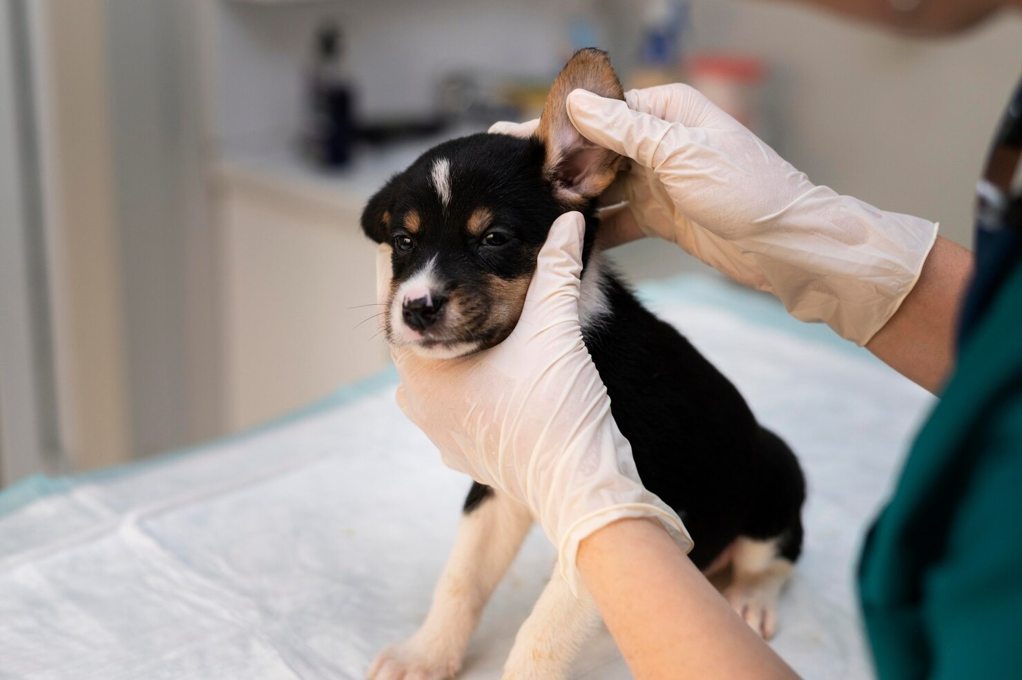
The diagnostic process begins with a comprehensive history taking. We inquire about the onset and duration of the symptoms, the dog’s environment, its travel history, and its exposure to other animals. Specifically, we ask if there are cats in the household and if any of the in-contact animals are exhibiting similar signs of pruritus. Following the history, a general physical examination is conducted to assess the dog’s overall health and to look for evidence of ectopic mites (mites that have migrated out of the ear to the head, neck, or rump). The veterinarian will then closely examine the external pinnae for excoriations, crusting, and signs of hematoma formation. The presence of a positive oto-pedal reflex is noted during palpation of the ear base.[31]
The next critical step is otoscopy. Using a hand-held otoscope or a high-definition video otoscope, the clinician carefully evaluates the vertical and horizontal ear canals. The goal is to assess the degree of inflammation, identify the character of the exudate, look for foreign bodies (such as grass awns or foxtails), and evaluate the integrity of the tympanic membrane. In some cases, the mites themselves can be visualized directly through the otoscope; the intense light and magnification may reveal tiny, white, crab-like specks moving quickly away from the light source across the dark field of cerumen. However, the absence of visible mites during otoscopy does not rule out an infestation, as the parasites often hide deep within the exudate or retreat into the horizontal canal.[32]
The gold standard for definitively diagnosing ear mites is microscopic cytology. The veterinarian uses a specialized cotton-tipped applicator to carefully swab the deep horizontal ear canal to collect a sample of the exudate. This sample is then rolled onto a clean glass microscope slide. To identify live mites, the clinician places a drop of mineral oil over the sample and covers it with a coverslip. The mineral oil breaks down the thick cerumen and immobilizes the mites without killing them immediately, allowing them to be easily identified under low-power magnification (typically the 4x or 10x objective lens). Under the microscope, adult Otodectes cynotis mites are readily identifiable by their oval bodies, short legs, and the presence of suckers (caruncles) on unjointed pedicels. The veterinarian will also look for the large, oval-shaped mite eggs, which confirm active reproduction.[33]
In addition to the mineral oil preparation, a second swab is typically rolled onto another slide, heat-fixed or air-dried, and stained using a Romanowsky-type stain. This stained slide is examined under high-power oil immersion (100x objective) to evaluate the cellular component of the exudate and to identify the presence of secondary invaders. The clinician will look for an overgrowth of Malassezia yeast (which look like dark purple, peanut-shaped organisms) or pathogenic bacteria (such as cocci or rods), as well as an influx of neutrophils, which indicates active bacterial infection.[34] Identifying these secondary pathogens is essential for formulating a comprehensive treatment plan.
Veterinarians must also perform a differential diagnosis to rule out other conditions that present with identical symptoms. If no mites are found on repeated cytology, but the dog exhibits severe ear disease, the clinician must investigate other potential causes. These include atopic dermatitis (environmental allergies), adverse food reactions (food allergies), primary bacterial or yeast otitis, foreign bodies lodged in the canal, endocrinopathies (like hypothyroidism, which alters sebum production), or other parasitic infestations, such as Demodex canis or Sarcoptes scabiei. Skin scrapings of the pinnal margins and surrounding skin may be required to differentiate Otodectes from Sarcoptes, which burrows deeply into the skin. In rare instances where the clinical suspicion is exceptionally high but mites cannot be found—perhaps due to the pet’s hyper-reactive immune response clearing most of the mites while remaining intensely inflamed—a diagnostic therapeutic trial with an anti-parasitic treatment may be initiated to see if symptoms resolve.[35]
Treatment Options for Ear Mites in Dogs

The medical management of a canine ear mite infestation requires a multi-modal approach designed to eliminate the primary parasitic culprit, resolve any secondary bacterial or fungal infections, reduce debilitating inflammation, manage the patient’s pain, and prevent rapid re-infestation. Because Otodectes cynotis has a 21-day life cycle and many traditional topical medications are not ovicidal (they do not kill the eggs), treatment protocols must often span several weeks to ensure that newly hatching nymphs are eradicated before they can reproduce. Modern veterinary pharmacology, however, has provided us with highly effective, long-acting medications that have revolutionized the treatment of this condition.[36]
The first and arguably most critical step in any ear treatment protocol is a thorough ear cleaning. The thick, tarry, coffee-ground exudate creates a physical barrier that prevents topical medications from reaching the epithelial tissue where the mites reside. Furthermore, cellular debris and pus can actually inactivate certain types of antibiotics and anti-parasitics. The veterinary team will carefully flush the ear canal using a gentle, veterinary-grade ceruminolytic cleanser. These specialized solutions often contain specific ceruminolytic ingredients which act to dissolve the waxy, lipid-rich discharge, allowing it to be flushed out or wiped away. This cleaning process must be performed delicately, as the inflamed ear canal is exquisitely painful, and aggressive cleaning can cause further trauma or inadvertently rupture a weakened tympanic membrane. In severe, painful cases, the dog may require heavy sedation or general anesthesia to safely and adequately flush the ears.[37]
Following a thorough cleaning, the specific anti-parasitic therapy is initiated. Historically, the primary treatment involved daily applications of traditional multi-agent topical anti-parasitic drops directly into the ear canal. While effective if used meticulously, these older topical treatments required twice-daily administration for a minimum of 21 to 30 days to cover the parasite’s entire life cycle. This grueling regimen often resulted in poor owner compliance and tremendous stress for the dog, who quickly learns to run and hide when they see the medication bottle.[38]
Today, modern systemic anti-parasitic therapies are considered the standard of care for treating ear mites due to their superior efficacy and ease of administration. Certain long-acting anti-parasitic medications are administered as a topical “spot-on” liquid applied to the skin at the back of the dog’s neck. These drugs are absorbed systemically and distributed to the sebaceous glands and tissues, effectively killing the mites as they feed. A single application is often highly effective, though a second dose 30 days later is generally recommended to ensure complete eradication. Additionally, injectable or oral formulations of systemic anti-parasitic medications may be utilized in specific clinical scenarios under strict veterinary supervision, taking care to avoid use in herding breeds with the MDR1 gene mutation, which makes them highly susceptible to toxicity from specific drug classes.[39]
More recently, advanced classes of systemic parasiticides have shown remarkable efficacy in treating ear mites. These medications, often administered as highly palatable prescription oral chewables, target the parasite’s nervous system, causing fatal paralysis. While primarily indicated for flea and tick prevention, numerous clinical studies have demonstrated that a single dose of these systemic preventatives can rapidly and completely eliminate an Otodectes cynotis infestation, representing a massive leap forward in treatment convenience and patient comfort.[40]
Eradicating the mites is only part of the clinical picture; treating the secondary complications is equally vital. If cytology reveals a secondary Malassezia yeast or bacterial overgrowth, the veterinarian will prescribe appropriate topical or systemic antimicrobial therapy. This may involve prescription otic drops containing antifungal agents and broad-spectrum antibiotics tailored to your pet. To combat the intense pruritus and localized inflammation, glucocorticoids (steroids) are frequently incorporated into the otic drops, or a short, tapering course of oral prescription anti-inflammatory medication may be prescribed to provide rapid relief. Pain management is also a critical consideration; nerve-pain medication prescribed by your veterinarian or prescription non-steroidal anti-inflammatory drugs (NSAIDs) may be utilized to keep the patient comfortable during the initial healing phase.[41]
A fundamental rule of ear mite treatment is that every susceptible animal in the household must be treated simultaneously, regardless of whether they are currently exhibiting clinical signs. Because cats are frequently asymptomatic carriers, treating the symptomatic dog while ignoring the feline housemate guarantees that the dog will be rapidly re-infested as soon as the medication wears off. Environmental management, including washing all pet bedding in hot water (above 60°C or 140°F) and thoroughly vacuuming carpets and upholstery, is also advised to eliminate off-host mites and remove fomites from the home. Follow-up cytological examinations are strongly recommended to verify that the mites have been completely eradicated and that the secondary infections have fully resolved.[42]
How to Prevent Canine Ear Mites
Preventing an ear mite infestation is significantly easier, less stressful, and more cost-effective than enduring the arduous process of diagnosing and treating a severe clinical case. The foundation of prevention lies in routine veterinary care, consistent application of broad-spectrum parasiticides, and proactive environmental management. By integrating simple preventative strategies into your dog’s standard wellness routine, you can effectively shield them from the discomfort and health risks associated with Otodectes cynotis.[43]
The most reliable and effortless method for preventing ear mites is the continuous, year-round administration of modern veterinary parasite preventatives. Many of the prescription medications utilized to protect dogs from fleas and ticks, as well as heartworms and intestinal nematodes, also offer robust prophylactic coverage against ear mites. Topical spot-on prescription preventatives, when applied monthly according to the manufacturer’s directions, establish a systemic barrier that kills mites upon exposure, preventing them from establishing a reproducing population in the ear canal. Similarly, modern prescription oral preventative medications provide sustained systemic protection. It is important to note that Vaccines are typically used to prevent infectious diseases caused by viruses and bacteria; there is no vaccine available for parasitic infestations like ear mites. Therefore, reliance on appropriate pharmacological preventatives is non-negotiable for at-risk pets.[44]
Routine aural hygiene and consistent monitoring are crucial components of preventive care. Pet owners should make it a habit to visually inspect their dog’s ears at least once a week. You should look for any signs of abnormal redness, swelling, or the accumulation of dark, waxy debris. A healthy ear canal should appear light pink, be relatively clean, and be free of any offensive odor. Integrating regular ear cleaning into your dog’s grooming routine using a veterinarian-approved, pH-balanced otic flush can help maintain a healthy microenvironment. This removes excess cerumen and trapped moisture, making the canal less inviting to opportunists and parasites alike. For breeds with heavy, pendulous ears (like Basset Hounds or Cocker Spaniels) or those with excessive hair growth inside the ear canal (like Poodles or Schnauzers), keeping the ear canal dry and carefully managing the hair growth under veterinary guidance is critical.[45]
Managing the dog’s environment and controlling their exposure to potential carriers is the final pillar of prevention. When introducing a new pet into a household—whether it is a newly adopted puppy, a rescue cat, or a small mammal like a ferret—it is imperative to quarantine the new arrival until they have been thoroughly examined by a veterinarian. A simple microscopic ear cytology during the initial wellness exam can identify asymptomatic carriers before they have the opportunity to spread mites to your established pets. Furthermore, if your dog frequents high-risk environments such as boarding kennels, dog daycares, or grooming salons, maintaining their monthly preventative medications is even more critical. In the home, practicing good hygiene by regularly washing dog beds, blankets, and plush toys in hot water, and routinely cleaning grooming implements, will mitigate the risk of fomite transmission and ensure a healthier living space for your canine companion.[46]
Frequently Asked Questions
Can ear mites spread from dogs to humans?
Generally, ear mites (Otodectes cynotis) are highly species-specific, preferring to infest carnivores such as dogs, cats, and ferrets. They do not consider humans to be a suitable host, and they cannot complete their life cycle or reproduce on human skin. However, in rare instances of massive environmental contamination or extremely close physical contact with a heavily infested pet, humans may experience transient, temporary infestations. This condition, sometimes referred to as a “pseudoscabies” or a transient papular dermatitis, manifests as a self-limiting, itchy rash on the arms or torso where the mites have temporarily bitten the skin. The mites will naturally die off quickly on a human host without the need for medical intervention. If you develop persistent skin irritation after handling an infected pet, it is best to consult a human healthcare professional to rule out other dermatological conditions. Always consult your veterinarian before making any changes to your pet’s care to quickly eliminate the source of the mites in your home.[47]
What happens if a dog’s ear mite infestation is left untreated?
If an ear mite infestation is ignored or improperly treated, the consequences for the dog’s health can be severe and permanent. The continuous mechanical trauma from the feeding mites and the massive inflammatory immune response rapidly degrade the protective lining of the ear canal. This inevitably leads to severe secondary bacterial and fungal infections, which significantly amplify the dog’s pain and tissue damage. The violent head shaking and frantic scratching caused by the relentless itching frequently result in aural hematomas—painful, blood-filled swellings of the ear flap that require surgical correction. Left unabated, the chronic inflammation causes the ear canal tissues to thicken and calcify (hyperplastic otitis), permanently narrowing the canal. The infection can also rupture the tympanic membrane (eardrum), spreading deep into the middle and inner ear. This progression to otitis media and interna can cause permanent deafness, severe balance issues (vestibular disease), and chronic facial nerve damage.[48]
Are there effective natural home remedies for treating ear mites in dogs?
While the internet is replete with suggestions for natural remedies—ranging from olive oil and mineral oil to apple cider vinegar and essential oils—veterinary professionals strongly caution against relying on these methods as a primary treatment for ear mites. Applying oils to the ear canal might smother some adult mites, providing temporary relief, but it will not kill the eggs embedded in the ear debris, guaranteeing a rapid relapse within weeks. Furthermore, using acidic solutions like vinegar or highly concentrated essential oils on exquisitely inflamed, excoriated ear tissues can cause excruciating pain, chemical burns, and exacerbate the inflammation. Ear mites are resilient, microscopic parasites that require specific, pharmacological therapies prescribed by your veterinarian to achieve complete eradication. Delaying appropriate medical treatment in favor of unproven home remedies allows secondary bacterial infections and irreversible tissue damage to progress. Always seek a definitive diagnosis and prescription treatment plan from your veterinarian.[49]
Concerned About Your Dog’s Ears?
If your dog is exhibiting signs of intense scratching, head shaking, or unusual ear discharge, do not wait. Schedule an appointment with a veterinarian today for a definitive diagnosis and a safe, effective, tailored treatment plan.
References
- Merck Veterinary Manual. Ear Mites in Dogs. Merck & Co., Inc., 2023.
- Bowman DD. Georgis’ Parasitology for Veterinarians. 10th ed. Elsevier, 2014.
- Companion Animal Parasite Council (CAPC). Otodectes cynotis Guidelines. CAPC, 2022.
- Miller WH, Griffin CE, Campbell KL. Muller and Kirk’s Small Animal Dermatology. 7th ed. Elsevier, 2013.
- Harvey RG, Harari J, Delauche AJ. Ear Diseases of the Dog and Cat. Manson Publishing, 2001.
- Gotthelf LN. Small Animal Ear Diseases: An Illustrated Guide. 2nd ed. Elsevier, 2004.
- American Veterinary Medical Association (AVMA). Ear Infections and Ear Mites. AVMA, 2023.
- Paterson S. Manual of Skin Diseases of the Dog and Cat. 2nd ed. Wiley-Blackwell, 2008.
- Merck Veterinary Manual. Otodectic Mange. Merck & Co., Inc., 2022.
- Curtis CF. Current trends in the treatment of Sarcoptes, Cheyletiella and Otodectes mite infestations in dogs and cats. Vet Dermatol, 2004.
- VCA Animal Hospitals. Ear Mites in Dogs. VCA Hospitals, 2023.
- ESCCAP. Control of Ectoparasites in Dogs and Cats. European Scientific Counsel Companion Animal Parasites, 2018.
- Ginel PJ, et al. A survey of the prevalence of Otodectes cynotis in domestic dogs and cats. Vet Parasitol, 2015.
- Nuttall T, Harvey RG, McKeever PJ. Skin Diseases of the Dog and Cat. Manson Publishing, 2009.
- Lecru LA, et al. Prevalence of ear mites in dogs from rural environments. Vet Parasitol, 2015.
- Otranto D, Wall R. Biology and control of non-burrowing mange mites. Vet Parasitol, 2008.
- Zajac AM, Conboy GA. Veterinary Clinical Parasitology. 8th ed. Wiley-Blackwell, 2012.
- Kwochka KW. Mites and related diseases. Vet Clin North Am Small Anim Pract, 1987.
- ASPCA. Ear Mites and Common Dog Behavior Issues. ASPCA, 2023.
- Griffin CE. Otitis externa and media. In: Small Animal Dermatology. Elsevier, 2013.
- Rosychuk RA. Management of otitis externa. Vet Clin North Am Small Anim Pract, 1994.
- Scott DW, Miller WH, Griffin CE. Muller and Kirk’s Small Animal Dermatology. 6th ed. W.B. Saunders, 2001.
- Cole LK. Otoscopic evaluation of the ear canal. Vet Clin North Am Small Anim Pract, 2004.
- Harvey RG. Ear Diseases of the Dog and Cat. CRC Press, 2005.
- Gotthelf LN. Diagnosis and treatment of otitis media in dogs and cats. Vet Clin North Am Small Anim Pract, 2004.
- Fossum TW. Small Animal Surgery. 4th ed. Mosby, 2012.
- Henderson MacCallum A. Aural hematomas in dogs and cats. Vet Rec, 2013.
- Morris DO. Medical therapy of otitis externa and otitis media. Vet Clin North Am Small Anim Pract, 2004.
- Strain GM. Deafness in Dogs and Cats. CABI, 2011.
- Hillier A, et al. Guidelines for the diagnosis and antimicrobial therapy of canine superficial bacterial folliculitis. Vet Dermatol, 2014.
- Merck Veterinary Manual. Physical Examination of the Skin. Merck & Co., Inc., 2022.
- Angus JC. Otic cytology in health and disease. Vet Clin North Am Small Anim Pract, 2004.
- Tater KC, Patterson AP. Canine and feline otitis externa. Vet Med, 2008.
- Mendelsohn C, et al. Practical cytology for the veterinary clinic. Compend Contin Educ Vet, 2006.
- Logas DB. Diseases of the ear canal. Vet Clin North Am Small Anim Pract, 1994.
- Gatellet M, et al. Efficacy of a systemic acaricide against Otodectes cynotis in dogs. Vet Dermatol, 2014.
- Mansfield PD. Preventive ear care for dogs and cats. Vet Clin North Am Small Anim Pract, 2004.
- Sivajothi S, et al. Comparative efficacy of topical acaricides. J Vet Parasitol, 2015.
- Six RH, et al. Efficacy and safety of topical anti-parasitic medication against Sarcoptes scabiei on dogs and Otodectes cynotis on dogs and cats. Vet Parasitol, 2000.
- Taenzler J, et al. Efficacy of a systemic parasiticide against Otodectes cynotis infestations in dogs and cats. Parasit Vectors, 2015.
- Hnilica KA. Small Animal Dermatology: A Color Atlas and Therapeutic Guide. 3rd ed. Saunders, 2011.
- Blagburn BL, Dryden MW. Biology, treatment, and control of flea and tick infestations. Vet Clin North Am Small Anim Pract, 2009.
- CDC. Healthy Pets, Healthy People: Dogs. Centers for Disease Control and Prevention, 2023.
- World Health Organization. Zoonoses. WHO, 2020.
- Rougier S, et al. Comparative efficacy of different ear cleansers in dogs. Vet Rec, 2005.
- Dryden MW. Flea and tick control in the 21st century. Vet Clin North Am Small Anim Pract, 2009.
- Hewitt M, et al. Cheyletiella and Otodectes species causing human dermatoses. Br J Dermatol, 1973.
- Pye C. Otitis externa in dogs. Can Vet J, 2018.
- Ebner R. Natural remedies in veterinary medicine: risks and benefits. Vet Med Res, 2016.


