


 March 4, 2023
 Phil Good, DVM
What is Ear Infection in Dogs?
This content was prepared with AI assistance and reviewed by a licensed professional for accuracy.
Introduction
When an ordinarily happy, energetic dog begins relentlessly shaking their head, pawing at their face, or whimpering when petted, an underlying medical issue is often to blame. Among the most frequent reasons a dog is brought into a veterinary clinic is to address an ear infection, a condition that stems from the overgrowth of microscopic organisms leading to severe inflammation. Specifically, bacterial contamination of the outer ear canal, along with the proliferation of opportunistic yeast, can cause a cascade of painful symptoms. The various causes of ear infections in dogs range from anatomical predispositions to underlying allergic diseases, making this a highly complex dermatological and otological issue. [1] Understanding the anatomy of the ear, the pathophysiology of the disease, and the myriad Symptoms of Ear Infection in dogs is essential for any responsible pet owner looking to manage and prevent these frustrating ear problems.
To truly comprehend why ear issues are so widespread, it is necessary to examine the unique anatomy of the canine ear. Unlike the human ear canal, which is relatively short and horizontal, a dog’s ear canal is shaped like the letter “L.” It consists of a long vertical portion that travels downward before making a sharp nearly 90-degree turn into a horizontal portion, ending at the tympanic membrane, or eardrum. [2] This distinct anatomical structure is an evolutionary adaptation for enhanced hearing, but it also creates a deep, dark, and potentially warm and moist environment that is exceptionally prone to trapping debris and fluid. [3] When the delicate microenvironment of the ear canal is disrupted, the normal protective mechanisms—such as the epithelial migration that naturally sweeps wax and debris outward—begin to fail. [4]
Current veterinary epidemiological data suggests that up to 20 percent of the general canine population will suffer from some form of ear disease during their lifetime. [5] Furthermore, in dogs that suffer from allergic skin disease, the incidence of ear infections skyrockets to over 80 percent. [6] These statistics underscore the fact that an ear infection is rarely an isolated, random event; rather, it is usually a secondary symptom of a primary underlying condition. [7] If these early warning signs are ignored or treated improperly with over-the-counter remedies, the localized inflammation can progress to chronic, irreversible tissue changes. Over time, the soft, pliable cartilage of the ear canal can become thickened, scarred, and even calcified, turning into bone-like tissue that permanently narrows the canal and traps even more bacteria. [8]
Types of Dog Ear Infections
Veterinary professionals categorize ear infections based on the specific anatomical segment of the ear that is affected. The ear is divided into three distinct zones: the outer ear, the middle ear, and the inner ear. Determining exactly which parts of the ear are compromised is a critical step in establishing a safe and effective treatment protocol, as medications that are safe for the outer ear can be highly toxic if they reach the delicate structures of the inner ear. [9]
Otitis Externa
Otitis externa is defined as the inflammation of the external ear canal, which includes the vertical and horizontal canals leading up to the tympanic membrane. This is the most frequently diagnosed form of ear disease and the infection most commonly affects dogs of all ages, though it is particularly prevalent in adult dogs suffering from underlying allergies. [10] The external ear canal is lined with specialized skin that contains sebaceous and ceruminous glands, which produce earwax (cerumen). In a healthy dog, this wax coats the lining of the ear, trapping dust and microbes, while the skin cells naturally migrate outward to expel the debris. However, when inflammation strikes, these glands become hyperactive and enlarged (hyperplastic), producing excessive amounts of abnormal, watery, or highly thickened wax. [11] This creates a nutrient-rich breeding ground for the normal flora (bacteria and yeast that naturally live on the skin in small numbers) to multiply out of control. If otitis externa is caught early, it is generally highly manageable. If neglected, the chronic swelling forces the canal to narrow, trapping the infection deep against the eardrum and setting the stage for more severe disease. [12]
Otitis Media
When the infection breaches the eardrum, it enters the middle ear, resulting in a much more serious condition known as otitis media. The middle ear is a hollow, air-filled bony cavity called the tympanic bulla, which houses the three tiny auditory ossicles (bones of hearing) that transmit sound waves to the inner ear. [13] Otitis media almost always occurs as a direct extension of chronic, unresolved otitis externa. The sheer volume of infected debris and the enzymatic toxins produced by bacteria weaken the pars tensa (the largest portion of the eardrum), eventually causing it to rupture. [14] Once the bacteria flood into the tympanic bulla, the infection becomes deeply seated and incredibly difficult to eradicate with standard topical treatments. In addition to severe pain, dogs with otitis media may exhibit signs of neurological impairment, because both the facial nerve and the sympathetic nerve fibers travel directly through or adjacent to the middle ear. [15] Damage to these nerves can lead to Horner’s syndrome (characterized by a drooping eyelid, sunken eye, and elevated third eyelid) or facial nerve paralysis, resulting in a drooping lip or inability to blink on the affected side. [16]
Otitis Interna
The deepest and most severe form of an ear infection is otitis interna, which involves the inner ear structures. The inner ear is encased in dense bone and contains the cochlea (responsible for hearing) and the vestibular apparatus (responsible for maintaining balance and spatial orientation). [17] When a bacterial infection penetrates from the middle ear into the inner ear, it causes profound inflammation of these delicate neuro-sensory organs. This condition is a true medical emergency. Dogs suffering from otitis interna frequently display dramatic signs of peripheral vestibular disease. [18] An owner may observe a sudden, severe head tilt, uncontrolled rhythmic flicking of the eyes (nystagmus), staggering or falling to one side (ataxia), walking in tight circles, and profound nausea or vomiting due to the extreme vertigo they are experiencing. In many cases, otitis interna results in permanent, irreversible deafness in the affected ear, and the dog may maintain a slight head tilt for the remainder of their life even after the active infection has been cured. [19]
What are the Causes of Ear Infections in Dogs?

Veterinary dermatologists utilize a highly specific diagnostic framework known as the PSPP system to categorize the causes of ear infections in dogs. PSPP stands for Primary, Secondary, Predisposing, and Perpetuating factors. Understanding this system is crucial, as treating the infection without addressing all four categories guarantees that the ear problems will return. [20] Primary factors are the actual root causes that initiate the inflammation in a previously healthy ear canal. Secondary factors are the microscopic organisms that cause the active infection. Predisposing factors are characteristics that increase the risk of disease, and perpetuating factors are the pathological changes to the ear tissue that prevent healing. [21]
By far, the most common primary cause of ear disease in dogs is hypersensitivity disorders. Canines prone to food-related or environmental allergies almost universally suffer from inflamed ears. In fact, in some dogs with cutaneous adverse food reactions (food allergies), recurrent bilateral ear infections are the only clinical sign they ever display. [22] Atopic dermatitis (environmental allergies to pollen, dust mites, and molds) causes systemic inflammation that alters the epidermal barrier of the skin, including the skin lining the ear canal. This barrier dysfunction allows moisture to escape and irritants to enter, changing the local microclimate and paving the way for bacterial contamination of the outer ear canal. [23]
Another major primary cause is Parasites: Infestations of ear mites or other parasites. The ear mite, known scientifically as Otodectes cynotis, is a microscopic, spider-like parasite that lives on the surface of the ear canal skin. They feed on epidermal debris and tissue fluids, causing intense, hypersensitive pruritus (itching). [24] While ear mites are incredibly common in puppies and shelter dogs, they are less common in adult dogs that receive regular parasite preventatives. Endocrine diseases, such as hypothyroidism and hyperadrenocorticism (Cushing’s disease), are also primary causes. These hormonal imbalances alter the dog’s immune function and lipid metabolism, often resulting in seborrhea, a condition where the skin overproduces thick, rancid sebum that accumulates in the ear canal. [1]
Predisposing factors do not directly cause inflammation, but they make an ear much more likely to become infected if a primary cause is present. Anatomy plays a massive role here. Breeds with long, heavy, pendulous pinnae (ear flaps), such as Basset Hounds and Bloodhounds, suffer from poor ventilation in the ear canal. [3] This traps heat and humidity, creating a tropical environment perfect for microbial growth. Breeds like Poodles and Schnauzers grow excessive amounts of hair deep within the vertical canal, which acts like a net, trapping wax and debris. Swimming or bathing can also be a predisposing factor; when water becomes trapped in the lower horizontal canal, the resulting maceration of the skin cells destroys their protective barrier function. [8]
Secondary causes are the actual pathogenic microbes that capitalize on the inflamed, moist environment. The normal canine ear canal contains tiny numbers of commensal organisms. However, when the environment shifts, these organisms undergo massive proliferation. The yeast Malassezia pachydermatis is a frequent culprit, producing a characteristic dark brown, sweet-smelling discharge. [9] On the bacterial side, Staphylococcus pseudintermedius (a spherical bacterium) and Pseudomonas aeruginosa (a rod-shaped bacterium) are the most common offenders. Pseudomonas is particularly dangerous, as it produces powerful enzymes that rapidly ulcerate the skin and destroy the eardrum, and it is naturally resistant to many common classes of antibiotics. [10]
Finally, perpetuating factors are the chronic structural changes that occur when an ear infection is left untreated. Ongoing inflammation causes the skin lining the canal to thicken and fold (hyperplasia), and the ceruminous glands to balloon in size. Eventually, the underlying cartilage undergoes mineralization and turns into bone. [11] These irreversible changes physically obstruct the canal, making it impossible for medications to reach the deeper structures and preventing the ear from ever cleaning itself properly. [12]
Symptoms of Ear Infection in Dogs

Recognizing the Symptoms of Ear Infection in dogs early is vital to preventing chronic, irreversible tissue damage. The clinical presentation of otitis can vary dramatically depending on the primary cause, the specific organism involved, and the depth of the infection. [13] However, the most universally observed symptom is intense, unrelenting pruritus (itching). A dog will repeatedly scratch at the base of their ears with their hind legs, rub the sides of their head against furniture or the carpet, and violently shake their head. This vigorous head shaking can be so forceful that the fragile blood vessels inside the ear flap rupture, filling the pinna with blood to create a large, swollen mass known as an aural hematoma, which often requires surgical correction. [14]
Upon closer physical inspection, owners will typically notice marked erythema (redness) and swelling of the ear flap and the visible opening of the canal. The skin may feel hot to the touch and appear thickened or corrugated. Discharge is almost always present, and its characteristics can provide clues about the secondary infection. [15] A dry, crumbly, dark brown discharge resembling coffee grounds is classically associated with ear mites. A greasy, waxy, dark brown discharge with an intensely sweet or musty odor strongly suggests an overgrowth of Malassezia yeast. Conversely, a thick, pale yellow, or greenish purulent discharge (pus) with a foul, putrid odor is highly indicative of a severe bacterial infection, such as Pseudomonas. [16]
Pain is another hallmark symptom. Dogs that are normally highly affectionate may become head-shy, pulling away, vocalizing, or even snapping when someone attempts to pet their head or examine their ears. [17] If the infection has ruptured the eardrum and progressed into the middle or inner ear, owners may notice profound lethargy, a loss of appetite due to pain when chewing (since the temporomandibular joint is located right next to the ear canal), and alarming neurological deficits. These can include a severe head tilt, stumbling, walking in circles, and involuntary darting of the eyes. Immediate veterinary intervention is required when any of these advanced neurological signs are present. [18]
Diagnosis of Canine Ear Infections

To successfully treat and manage otitis, a veterinarian must execute a systematic diagnostic workup. Because an ear infection is a multifactorial disease, simply looking at the ear and handing out medication is considered substandard care. The diagnostic process aims to identify the specific microbes present, assess the structural integrity of the ear canal and eardrum, and uncover the primary underlying disease. [19]
Thorough Examination
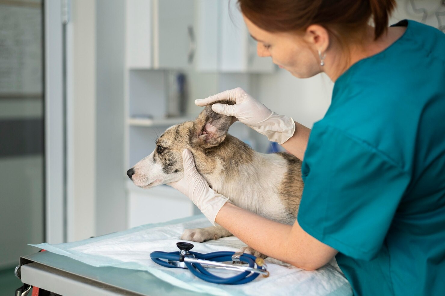
The diagnostic process begins with a comprehensive physical and dermatological examination. The veterinarian will palpate the base of the ear to assess the degree of calcification in the cartilage and to check for pain. [20] They will also examine the rest of the dog’s body, looking for signs of systemic allergies, such as redness between the toes, hair loss on the ventrum (belly), or a dull, flaky hair coat indicative of an endocrine disorder. [21] This holistic approach is crucial for establishing long-term management.
Utilizing an Otoscope
Otoscopy is the visual examination of the internal ear structures using a specialized, lighted instrument called an otoscope. To perform this correctly, the veterinarian must gently grasp the pinna and pull it outward and downward to straighten the L-shaped bend, allowing visualization of the horizontal canal and the tympanic membrane. [2] The otoscope allows the clinician to identify the presence of masses, polyps, or foreign bodies like plant awns (foxtails) that may have migrated deep into the canal and pierced the tissue. Many modern clinics now use video otoscopy, which utilizes a small fiber-optic camera to project a highly magnified image of the eardrum onto a screen, enabling precise assessment of the pars tensa and pars flaccida for ruptures or bulging. [4]
Ear Cytology
Otic cytology is arguably the most critical diagnostic test in the management of ear disease. It is impossible to accurately identify the specific type of bacteria or yeast with the naked eye. The veterinarian will collect a sample of the ear exudate using a cotton-tipped applicator, roll the material onto a glass microscope slide, and use a lighter or heat source to gently heat-fix the sample. [6] The slide is then dipped sequentially through a three-step differential stain (such as Diff-Quik) to dye the cellular structures and microorganisms. Under a high-powered microscope utilizing a 100x oil-immersion objective lens, the clinician evaluates the slide. They look for the presence of budding, peanut-shaped Malassezia yeast, spherical Staphylococcus bacteria (cocci), or elongated, pill-shaped Pseudomonas bacteria (rods). [7] The presence of toxic neutrophils (white blood cells) indicates a severe, active immune response to a bacterial invader.
Culture Testing
While cytology is mandatory for every ear infection, a bacterial culture and susceptibility test is reserved for specific scenarios. [12] If the cytology reveals rod-shaped bacteria, if the infection is chronic and unresponsive to first-line therapies, or if systemic oral antibiotics are required (such as in cases of otitis media), a sterile swab must be sent to an outside laboratory. The lab will isolate the exact species of bacteria and expose it to a panel of different antibiotics to determine the Minimum Inhibitory Concentration (MIC). This tells the veterinarian exactly which antibiotic will successfully kill the bacteria and which ones the bacteria are resistant to, preventing the dangerous misuse of antimicrobial drugs. [10]
Allergy Screening
If a dog has known allergies or presents with recurrent bilateral ear infections, the primary trigger must be identified. To identify any early signs of infection or underlying conditions, the veterinarian may recommend an 8 to 12-week strict dietary elimination trial using a prescription hydrolyzed-protein diet to rule out food allergies. [22] For environmental allergies (atopy), intradermal skin testing or serum IgE blood testing can be performed to identify the specific pollens, molds, or dust mites causing the hyperactive immune response, which paves the way for allergen-specific immunotherapy. [23]
Treatment of Canine Ear Infection

Because the causes and severity of ear infections vary so widely, there is no “one-size-fits-all” treatment. The therapeutic protocol must be custom-tailored based on the results of the otoscopy, cytology, and underlying disease assessment. The overarching goals of treatment are to eliminate the microscopic organisms, reduce the severe tissue inflammation, resolve the patient’s pain, and restore the normal epithelial migration of the ear canal. [24]
Cleaning and Topical Medication
The foundation of treating otitis externa is thorough ear cleaning followed by the application of targeted topical medications. Thick wax and purulent discharge physically block medications from reaching the skin and can chemically inactivate certain classes of antibiotics, such as aminoglycosides and polymyxin B. [11] The veterinarian will typically prescribe a specific ceruminolytic (wax-dissolving) ear flush containing ingredients like salicylic acid or squalene. In severe cases of Pseudomonas, a flush containing Tris-EDTA may be utilized. Tris-EDTA is a chemical that actively breaks down the protective biofilm produced by the bacteria and compromises their cell walls, making them highly susceptible to the subsequent antibiotic drops. [15] After cleaning, a multimodal topical ointment or liquid drop is applied. These topicals generally combine a potent antimicrobial (like gentamicin or florfenicol), a broad-spectrum antifungal (like clotrimazole or miconazole), and a high-potency glucocorticoid steroid (like betamethasone or dexamethasone). The steroid is absolutely critical; it rapidly reduces the intense swelling, alleviates the pet’s pain, and opens the narrowed ear canal so the antimicrobial agents can penetrate deeply. [20]
Oral Medications for Chronic or Recurrent Infections
While topical therapy is highly effective for otitis externa, cases that are severe, chronic, or involve the middle ear (otitis media) require systemic oral medications. [13] If the eardrum is ruptured, many common topical medications are contraindicated because they are ototoxic and can cause permanent deafness if they pool in the inner ear. In these scenarios, oral or injectable antibiotics (like cephalexin, clindamycin, or a fluoroquinolone based on culture results) are prescribed for an extended duration, often 4 to 8 weeks. It is important to note that prolonged use of systemic antibiotics alters the normal flora of the gastrointestinal tract, which can sometimes lead dogs to cause diarrhea. [21] Additionally, a short, tapering course of an oral corticosteroid like prednisone may be utilized to quickly break the cycle of severe inflammation and hyperplasia. To control the intense pruritus and prevent self-mutilation, veterinarians may also prescribe Janus kinase inhibitors (like oclacitinib) or monoclonal antibody therapies (like lokivetmab). [23]
Surgical Intervention in Severe Cases
Medical management eventually fails in ears that have suffered years of chronic, unresolved inflammation. When the cartilaginous canal becomes completely occluded by hyperplastic scar tissue and calcification, it forms an impenetrable, infected biological tube. At this end-stage of disease, surgical intervention is the only humane option. [8] A Lateral Ear Resection (LER) may be performed in mild cases to open the vertical canal and improve airflow. However, the most definitive salvage procedure is a Total Ear Canal Ablation with Bulla Osteotomy (TECA-BO). [16] In a TECA-BO, the surgeon completely excises the diseased vertical and horizontal ear canals and opens the tympanic bulla (middle ear) to thoroughly curette and flush out all infected bone and tissue. The skin is then sutured closed over the site. While the dog will be deaf in that ear, the surgery provides an immediate, permanent cure for the agonizing pain and recurring infections. Surgery may also be required if the ear has sustained severe traumatic injuries that have permanently distorted the ear canal architecture. [14]
Treating the Underlying Cause to Prevent Recurring Infections
Curing the current infection is only half the battle; preventing the next one requires managing the primary underlying cause. [22] If cytology and testing reveal a primary hypothyroidism, the dog must be placed on lifelong synthetic levothyroxine. If the primary cause is atopic dermatitis, long-term management strategies, such as strict adherence to a hypoallergenic prescription diet, monthly administration of systemic anti-itch medications, or allergen-specific immunotherapy (allergy drops or injections), must be implemented. Without this crucial step, the ear infections will inevitably return the moment the topical antibiotics are discontinued. [1]
Home Remedy
When a dog begins showing signs of ear discomfort, owners often turn to the internet for home remedies. This can be a dangerous and counterproductive decision. The microenvironment of the ear canal relies on a highly specific pH balance to maintain a healthy epithelial barrier. [5] Introducing harsh household chemicals like hydrogen peroxide, rubbing alcohol, or undiluted apple cider vinegar into an actively inflamed and ulcerated ear canal will cause excruciating pain and further destroy the delicate epithelial cells, worsening the disease. Hydrogen peroxide, in particular, causes oxidative damage to the tissue and leaves behind water, creating the exact moist environment that yeast and bacteria need to thrive. [19] Always consult your veterinarian before making any changes to your pet’s care, and only use veterinary-approved, pH-balanced ear cleansers that are specifically formulated to gently break down cerumen without damaging the tissue. [24]
Prevention of Canine Ear Infections
Preventative care is essential, particularly for dogs that have suffered from otitis in the past or belong to high-risk groups. Certain dog breeds are more prone to ear infections, requiring owners to be exceptionally diligent. The cornerstone of prevention is maintaining a dry, well-ventilated ear canal. [3] After a dog bathes, swims in a lake, or plays in heavy rain, owners should immediately use a soft cotton ball or a veterinary-approved drying agent containing salicylic acid to absorb any trapped moisture in the vertical canal. Avoid inserting cotton swabs (Q-tips) deep into the ear, as this will merely pack the debris further down against the eardrum and risk rupturing it. [11]
Routine cleaning is recommended, but the frequency must be tailored to the individual dog. Over-cleaning a healthy ear can strip away the normal, protective cerumen and cause maceration of the tissue, leading to the very infection you are trying to prevent. Generally, a prophylactic cleaning once every one to two weeks is sufficient for breeds predisposed to wax buildup. [15] For dogs with excessive hair growth inside the canal, such as Poodles and Bichon Frises, veterinary dermatologists debate the necessity of routine hair plucking. If the ear is healthy, it is often best to leave the hair alone, as plucking causes micro-trauma to the hair follicles, triggering inflammation. However, if the hair is thick enough to block airflow and form a matted plug of wax, careful plucking by a veterinary professional is warranted to prevent the trapping of debris. Finally, remaining vigilant for signs of other systemic illnesses is crucial. While rare, upper respiratory infections involving the sinuses can occasionally travel down the Eustachian tube into the middle ear, highlighting the need for prompt medical attention for any illness. [17]
Frequently Asked Questions
How long does it take for a dog ear infection to go away?
The timeline for resolving a canine ear infection depends entirely on the severity of the tissue inflammation and the type of microorganism causing the disease. For a mild, acute case of otitis externa caused by an overgrowth of Malassezia yeast or Staphylococcus bacteria, you will typically see a dramatic improvement in the dog’s comfort within 48 to 72 hours of starting prescription topical drops. However, the full course of treatment usually lasts between 7 and 14 days. It is absolutely critical that owners complete the entire prescribed course of medication, even if the ear looks completely normal after a few days. Stopping treatment prematurely allows the hardiest, surviving microbes to multiply, leading to a rapid relapse and the development of antibiotic resistance. If the infection involves the middle ear (otitis media) or is caused by resistant Pseudomonas bacteria, treatment with systemic and topical medications may take 4 to 8 weeks, or longer, to fully resolve the deep-seated infection and allow the ruptured eardrum to heal.
What happens if a dog’s ear infection is left untreated?
Leaving an ear infection untreated is a severe welfare issue that leads to chronic agony and permanent anatomical damage. Initially, the local inflammation causes the ceruminous glands to swell and produce excessive, abnormal wax. Over time, the skin lining the ear canal thickens and folds (hyperplasia), and the flexible cartilage begins to calcify, turning into rigid bone. This permanently narrows the canal (stenosis), making it impossible for the ear to naturally drain debris or for topical medications to reach the infection. The trapped bacteria and powerful enzymes they secrete will eventually eat through the tympanic membrane (eardrum), spilling the infection into the middle and inner ear. Once this occurs, the dog may suffer from permanent hearing loss, excruciating facial pain, facial nerve paralysis, and severe vestibular signs like an inability to stand, walking in tight circles, and a permanent head tilt. At this end-stage, the only viable option to relieve the dog’s suffering is a complex and highly invasive salvage surgery to completely remove the diseased ear canal.
Can a dog recover from an ear infection without antibiotics?
Whether a dog can recover without antibiotics depends entirely on the specific organism responsible for the infection, which can only be determined through microscopic cytology performed by a veterinarian. If the cytology reveals that the infection is solely caused by an overgrowth of yeast (Malassezia) or ear mites (Otodectes cynotis), antibiotics are entirely unnecessary and inappropriate. In those cases, the condition is treated with antifungal medications, parasiticides, and anti-inflammatory steroids to calm the tissue. However, if the cytology reveals the presence of pathogenic bacteria—such as Staphylococcus cocci or Pseudomonas rods—antibiotics are strictly required. Attempting to manage a true bacterial infection with over-the-counter cleaners, home remedies, or natural supplements will not eradicate the bacteria. Instead, it allows the infection to dive deeper into the tissue, potentially breaching the eardrum and causing catastrophic damage to the middle ear. Precise diagnosis via cytology is the only way to know exactly which class of medication is required.
References
- Merck Veterinary Manual. Ear Infections and Otitis Externa in Dogs. Merck & Co., 2022.
- Cole, L.K. Anatomy and physiology of the canine ear. Veterinary Dermatology, 2009.
- VCA Animal Hospitals. Ear Infections in Dogs – Otitis Externa. VCA, 2023.
- Griffin, C.E. Otitis externa and media. Veterinary Clinics of North America: Small Animal Practice, 2004.
- American Veterinary Medical Association. Ear infections in dogs. AVMA, 2023.
- Tater, K.C., et al. The cytology of the normal canine ear. Veterinary Dermatology, 2003.
- Ginel, P.J., et al. A semi-quantitative cytological evaluation of normal and pathological samples from the external ear canal of dogs. Veterinary Dermatology, 2002.
- Smeak, D.D. Total ear canal ablation and ventral bulla osteotomy. Veterinary Clinics of North America: Small Animal Practice, 2004.
- Paterson, S. Discovering the causes of otitis externa. In Practice, 2016.
- Pye, C. Pseudomonas otitis in dogs. The Canadian Veterinary Journal, 2018.
- Morris, D.O. Medical therapy of otitis externa and otitis media. Veterinary Clinics of North America: Small Animal Practice, 2004.
- Bajwa, J. Canine otitis externa – Treatment and complications. The Canadian Veterinary Journal, 2019.
- Gotthelf, L.N. Diagnosis and treatment of otitis media in dogs and cats. Veterinary Clinics of North America: Small Animal Practice, 2004.
- Jacobson, L.S. Diagnosis and medical treatment of otitis externa. Journal of the South African Veterinary Association, 2002.
- Angus, J.C. Otoscopy and biopsy of the ear canal. Veterinary Clinics of North America: Small Animal Practice, 2004.
- Garosi, L.S., et al. Neurological signs associated with otitis media/interna in dogs. Journal of Small Animal Practice, 2001.
- Rossignol, H., et al. Peripheral vestibular syndrome in dogs. Veterinary Journal, 2012.
- Veterinary Information Network. Vestibular Disease in Dogs and Cats. VIN, 2021.
- Nuttall, T. Successful management of otitis externa. In Practice, 2016.
- Rosser, E.J. Causes of otitis externa. Veterinary Clinics of North America: Small Animal Practice, 2004.
- Hill, P.B., et al. Survey of the prevalence, diagnosis and treatment of dermatological conditions in small animals in general practice. Veterinary Record, 2006.
- Carlotti, D.N., et al. Food allergy in dogs and cats. A review. Veterinary Dermatology, 1990.
- Hensel, P., et al. Canine atopic dermatitis: detailed guidelines for diagnosis and allergen identification. BMC Veterinary Research, 2015.
- Sousa, C.A. Ear mites (Otodectes cynotis). Veterinary Clinics of North America: Small Animal Practice, 2004.


