


 March 2, 2023
 Phil Good, DVM
Fractured Canine Teeth: Causes, Symptoms, and Treatment Explained
This content was prepared with AI assistance and reviewed by a licensed professional for accuracy.
Introduction
When it comes to maintaining your pet’s overall well-being, addressing fractured teeth, broken teeth, and teeth problems is an absolutely critical, yet frequently overlooked, aspect of veterinary care. Dogs utilize their mouths for far more than just eating; their mouths act as their hands, their primary tool for exploring the world, and their primary means of playing and self-soothing. Because dogs interact with their environment so physically, their teeth are subjected to immense forces daily. A healthy adult dog has forty-two permanent teeth, all of which play a specific role in grasping, tearing, and grinding food. When a traumatic event or excessive chewing force overwhelms the structural integrity of these teeth, fractures occur. Dental trauma is exceptionally common in veterinary medicine, representing one of the most frequently diagnosed oral pathologies in canine patients.[1]
To fully understand the severity of tooth fractures, one must first understand canine dental anatomy. A tooth is composed of several distinct layers. The outermost layer of the visible portion (the crown) is the enamel. Enamel is the hardest substance in the mammalian body, designed to withstand the abrasive forces of chewing. Beneath the enamel lies the dentin, a porous, bone-like layer that makes up the bulk of the tooth. Finally, at the very center of the tooth lies the pulp cavity. The pulp is the living tissue of the tooth, containing a rich network of blood vessels, sensitive nerve endings, and connective tissue. Because the nerves inside the tooth are encased in rigid walls of dentin, any inflammation or injury to the pulp causes immense pressure and agonizing pain.[2]
When a dog experiences a broken tooth, the consequences extend far beyond mere cosmetic imperfection. Unlike human enamel, which can be up to 2.5 millimeters thick, canine enamel is surprisingly thin—often less than 0.5 millimeters thick even on their largest teeth. This structural difference makes their teeth highly susceptible to damage when they chew on materials harder than the teeth themselves. From minor chips that only scrape the surface layer to catastrophic breaks that expose the internal nerve tissue, every type of dental fracture requires professional evaluation. Ignoring a broken tooth forces a dog to live in silent, chronic pain and opens the door to severe, systemic infections that can dramatically shorten their lifespan.[3]
Types and Causes of Tooth Fractures

The American Veterinary Dental College (AVDC) has established a standardized classification system for dental trauma in animals. This precise categorization helps veterinary professionals communicate the exact nature of the injury and determine the most appropriate medical or surgical intervention. Fractures are generally classified based on which anatomical layers of the tooth are involved and whether the fracture line extends below the gumline.[4] The progression of severity directly correlates with the amount of sensitive tissue exposed to the oral cavity.
Enamel Fractures
Enamel fractures (often abbreviated in veterinary charting as EF) represent the mildest form of dental trauma. In these instances, the damage is completely restricted to the outermost layer of the tooth. The enamel is chipped or cracked, but the underlying dentin remains entirely protected. Because enamel does not contain any nerve endings or blood vessels, an isolated enamel fracture is not inherently painful for the dog. The protective seal of the tooth has been compromised, but the internal living structures remain safe from bacterial contamination.[5]
Despite being the least severe, enamel fractures should never be ignored. First, they create a rough surface on the tooth that acts as a magnet for plaque and tartar accumulation, accelerating the progression of periodontal disease. Second, a fracture that appears to only involve the enamel to the naked eye may actually possess microscopic stress fractures that penetrate deeper into the tooth. Enamel fractures are most frequently caused by abrasive chewing over long periods, such as aggressively gnawing on tennis balls, which act like sandpaper against the thin canine enamel, or from minor bumps during roughhouse play.[6]
Uncomplicated Crown Fractures
An uncomplicated crown fracture (UCF) occurs when the break extends through the outer enamel and exposes the underlying dentin, but stops short of exposing the pulp cavity. While the term “uncomplicated” might sound reassuring, this type of fracture is anything but benign. Dentin is composed of millions of microscopic channels called dentinal tubules. These tubules run directly from the outside of the tooth straight into the central nerve pulp. When dentin is exposed to the oral cavity, these open tubules act as direct highways for bacteria and sensory stimuli.[7]
Dogs with uncomplicated crown fractures often experience significant pain due to the “hydrodynamic theory” of dentin sensitivity. When the exposed dentin contacts hot, cold, sweet, or acidic substances—or when physical pressure is applied—the fluid inside the microscopic tubules shifts. This fluid movement stimulates the nerve endings in the pulp, triggering sharp, sudden pain. Over time, bacteria migrating down these tubules can cause slow, chronic inflammation of the pulp. Common causes of uncomplicated crown fractures include chewing on hard plastic toys, ice cubes, or catching hard flying discs at awkward angles.[8]
Complicated Crown Fractures
A complicated crown fracture (CCF) is an absolute veterinary emergency. In this scenario, the traumatic event shears away the enamel and dentin, directly exposing the pulp cavity to the outside world. This is immediately visible as a pink or red spot (if the fracture is fresh and actively bleeding) or a black hole (if the fracture is older and the exposed pulp tissue has become necrotic) in the center of the broken tooth. Complicated crown fractures cause immediate, excruciating, and debilitating pain as the raw nerve is exposed to air, food, and saliva.[9]
Once the pulp is exposed, it is instantaneously contaminated by the billions of bacteria residing in the dog’s mouth. This bacterial invasion triggers acute pulpitis (inflammation of the nerve). Because the pulp is trapped within the unyielding dentin walls, the inflammatory swelling cuts off its own blood supply, leading to ischemic necrosis—meaning the tooth dies from the inside out. The bacteria then travel down the root canal and exit the tip of the root deep within the jawbone, creating a massive periapical abscess. Complicated crown fractures are most often seen in the large carnassial teeth (upper fourth premolars) and canine teeth, usually as a result of chewing on marrow bones, deer antlers, or rocks.[10]
Root Fractures
Root fractures are insidious because they occur entirely below the gumline and are completely invisible during a standard awake oral examination. These fractures involve the cementum (the tissue covering the root), the dentin, and the pulp, all hidden beneath the jaw’s mucosal tissue. A root fracture typically results from acute, massive blunt force trauma to the face rather than simple chewing forces. When the crown of the tooth is struck violently, the energy is transferred down the tooth, causing the root to snap within the alveolar bone socket.[11]
Depending on the angle and location of the break, root fractures can be classified as horizontal, vertical, or oblique. The prognosis for a tooth with a root fracture is generally poor. The fracture creates an unsealable gap within the periodontal space, leading to rapid, deep-seated infection, mobility of the tooth crown, and severe pain every time the dog attempts to bite down. Because these injuries cannot be seen with the naked eye, they underscore the absolute necessity of advanced diagnostic imaging in veterinary dentistry.[12]
Crown-Root Fractures
Crown-root fractures are devastating injuries where the fracture line begins on the visible crown of the tooth and extends diagonally downward beneath the gumline, splitting the root. These fractures can be either “uncomplicated” (not exposing the pulp) or “complicated” (exposing the pulp), but the subgingival (below the gum) extension makes them incredibly difficult to manage. Often, a large “slab” of the tooth will shear off, a condition known as a slab fracture, which is incredibly common in the upper fourth premolars of large breed dogs.[13]
The primary issue with crown-root fractures is the disruption of the “biologic width”—the natural, healthy attachment of the gum tissue to the tooth. Because the fracture extends under the gum, it creates a permanent pocket where food, debris, and bacteria accumulate. This inevitably leads to localized periodontal disease, bone loss, and chronic pain. Repairing these teeth is mathematically and surgically complex, as a veterinarian must somehow restore the tooth’s structure well below the bleeding gumline while maintaining a healthy seal. Therefore, many of these cases ultimately require advanced surgical intervention.[14]
Complete Fractures
Complete fractures, sometimes referred to as avulsion injuries or total luxations, occur when the entire tooth is completely displaced or knocked entirely out of its alveolar socket. This catastrophic level of trauma severs the periodontal ligament, the blood supply, and the nerve supply simultaneously. Complete fractures are almost never caused by chewing; rather, they are the result of severe, high-impact external trauma. For example, a complete avulsion is a common secondary injury when a dog is hit by a car or experiencing a severe fall.[15]
In human dentistry, a knocked-out tooth can sometimes be replanted if the patient reaches a dentist within an hour. In veterinary medicine, replantation is incredibly rare and difficult because dogs will not tolerate the restrictive splints required to stabilize the replanted tooth during the healing process, nor will they restrict their chewing activities. Consequently, complete fractures usually result in the permanent loss of the tooth, and the primary medical focus shifts to treating the massive soft tissue and bone damage left behind in the oral cavity. Immediate surgical debridement and closure of the empty socket are required to prevent necrotic bone infections.[16]
Symptoms of Fractured Canine Teeth
One of the greatest challenges in veterinary dentistry is that dogs are evolutionary masters at hiding pain. In the wild, showing vulnerability or oral pain makes an animal a target. Consequently, domestic dogs will often continue to eat, play, and act relatively normal even while enduring the agonizing pain of an exposed nerve. It is a dangerous misconception that a dog who is still eating cannot possibly have a painful dental issue. Dogs will swallow their kibble whole or chew exclusively on the opposite side of their mouth rather than starve. Recognizing a fractured tooth requires a highly observant owner looking for subtle, secondary clues.[17]
Behavioral changes are often the first indicator of oral discomfort. You may notice your dog dropping food from their mouth, demonstrating a sudden reluctance to play with their favorite chew toys, or exhibiting head shyness when you attempt to pet their face. Some dogs will constantly lick their lips, drool excessively (ptyalism), or paw frantically at their muzzle. You might also notice a dog aggressively rubbing the side of their face against the carpet or furniture in an attempt to soothe a deep, throbbing ache within the jawbone. Furthermore, uncharacteristic aggression or irritability in a normally sweet-tempered dog is a classic hallmark of hidden chronic pain.[18]
Physical signs of a fractured tooth become more apparent as secondary infections set in. If a dog is avoiding chewing on one side of their mouth due to a fracture, that side will rapidly accumulate heavy, thick brown tartar and calculus, creating a visible asymmetry when comparing the left and right sides of the mouth. The breath will become incredibly foul (halitosis) as the internal pulp tissue dies and begins to rot. In cases where a periapical abscess has formed, you may observe profound facial swelling. Specifically, a fractured upper fourth premolar often causes a painful, fluid-filled swelling located just below the dog’s eye. If left untreated, this swelling will burst through the skin, creating an open, draining wound known as an infraorbital fistula.[19]
The consequences of ignoring these symptoms extend far beyond the mouth. The oral cavity is highly vascular. When a broken tooth harbors a deep infection, that bacteria does not stay localized. Every time the dog chews, bacteria are pumped directly into the bloodstream in a process known as bacteremia. These circulating bacteria cause chronic systemic inflammation and can colonize major organs. Long-term, untreated dental infections are strongly linked to the development of endocarditis (infection of the heart valves) and can severely exacerbate kidney disease by overloading the renal filtration system with inflammatory complexes. Timely intervention is not just about saving a tooth; it is about preserving the dog’s overall health and longevity.[20]
Diagnosis of Fractured Teeth in Dogs

Diagnosing a fractured tooth and determining the exact extent of the damage is a multi-step process that requires both awake assessments and advanced evaluations performed under general anesthesia. A cursory glance at a conscious dog’s mouth in the exam room is merely the tip of the iceberg and is never sufficient to formulate a definitive treatment plan. Comprehensive dental diagnostics ensure that the hidden structures of the tooth and the surrounding jawbone are accurately assessed.[21]
Oral Examination
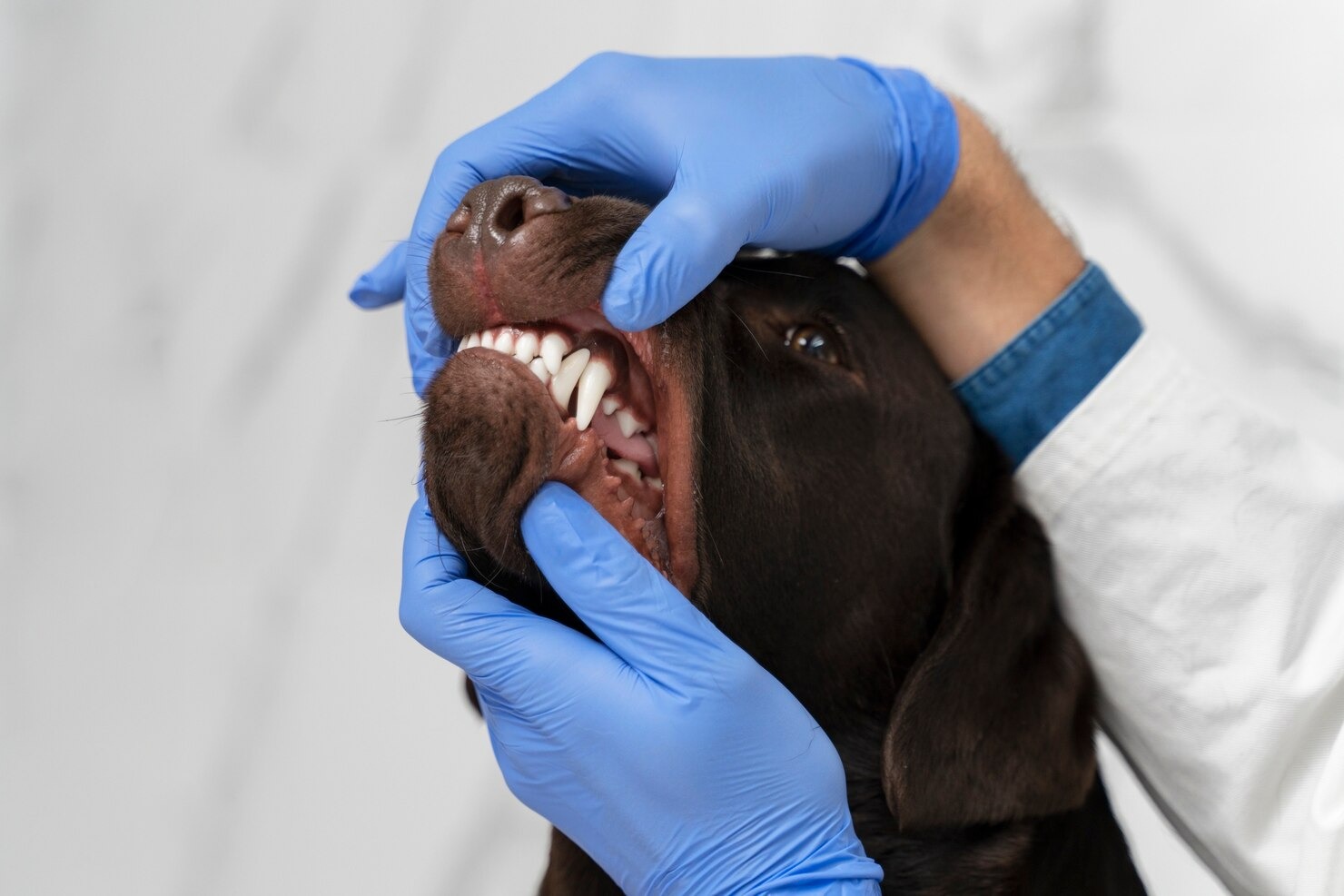
The diagnostic process begins with a conscious oral examination. During this initial step, the veterinarian lifts the dog’s lips to look for gross abnormalities, such as missing crowns, visible pulp exposure, asymmetrical tartar buildup, or draining tracts on the gums. However, because a dog will naturally resist having a painful tooth prodded while awake, the true, detailed examination must be performed after the pet is safely anesthetized and a cuffed endotracheal tube is placed to protect their airway. Under anesthesia, the veterinarian will systematically evaluate all 42 teeth. Using a specialized tool called a dental explorer, the vet will carefully glide the sharp tip over the surface of the fractured tooth. If the explorer tip catches in a hole or drops into an open pulp chamber, a complicated fracture is confirmed. This step is critical for differentiating between an uncomplicated fracture (which feels smooth and hard) and a complicated fracture (which feels soft or hollow).[22]
Palpation
Palpation involves the tactical assessment of the teeth, gums, and surrounding jaw structures. While the dog is anesthetized, the veterinarian will apply gentle but firm pressure to the fractured tooth to assess its mobility. Teeth are naturally held tightly in their sockets by the periodontal ligament; abnormal wiggling or shifting indicates severe root damage, bone loss, or a complete root fracture beneath the gumline. Additionally, the veterinarian will palpate the regional lymph nodes, specifically the submandibular nodes located just under the jaw. Enlarged, firm, or reactive lymph nodes are a strong physiological indicator that the body is actively fighting a significant localized infection stemming from the broken tooth. The gums surrounding the fractured tooth are also palpated for fluctuant swellings that might indicate a developing abscess or a subgingival cyst.[23]
Dental Radiographs (Dental X-rays)
No dental evaluation is complete without intraoral dental radiographs. Standard, whole-body X-rays are useless for dentistry due to the superimposition of the skull bones. Intraoral radiography utilizes high-resolution digital sensors placed directly inside the dog’s mouth, capturing the specific tooth root and the surrounding alveolar bone in microscopic detail. Radiographs are the only way to see the 60% of the tooth that lies hidden beneath the gumline. On an X-ray, the veterinarian will examine the width of the pulp cavity. If a tooth died months ago from a fracture, its pulp cavity will appear much wider than the surrounding healthy teeth because it stopped maturing. Furthermore, radiographs reveal periapical lucencies—dark halos in the bone around the root tip that indicate the bone has been dissolved by an actively expanding abscess. Without radiographs, treating dental fractures is essentially guesswork.[24]
Vitality Testing
In advanced veterinary dental practices, vitality testing may be employed to determine if the nerve inside a fractured tooth is still alive. While highly common in human dentistry, it is somewhat challenging in veterinary medicine because the patient cannot verbally report what they feel. However, transillumination is frequently used. A high-intensity fiber-optic light is placed against the base of the tooth. A healthy, vital tooth will transmit the light and appear a bright, glowing pink. A tooth with a dead, necrotic pulp will appear opaque, dark, or grayish-brown because the degraded blood products block the light transmission. While thermal and electrical pulp testers exist in veterinary medicine, they must be used extremely carefully on lightly sedated animals to gauge autonomic responses (like a slight heart rate increase), though this is largely reserved for specialized, board-certified veterinary dentists.[25]
Treatment Options for Fractured Teeth in Dogs

Once a definitive diagnosis is reached, the veterinarian will outline a specific treatment plan. The “wait and see” approach is never an acceptable medical strategy for a fractured tooth. Leaving a broken tooth untreated is tantamount to veterinary malpractice, as it guarantees chronic pain and progressive infection. The chosen treatment modality depends entirely on whether the pulp is exposed, the age of the dog, the specific tooth involved, and the owner’s commitment to post-operative care. Modern veterinary dentistry offers several sophisticated treatment avenues.[26]
Dental Fillings or Bonding
For uncomplicated crown fractures where the pulp is safely sealed away, the treatment of choice is the application of a bonded dental sealant or filling. Leaving exposed dentin untreated allows bacteria to migrate through the dentinal tubules and slowly kill the tooth over time. To prevent this, the tooth is meticulously cleaned and smoothed using a high-speed diamond bur—a process known as odontoplasty. The surface is then treated with a mild phosphoric acid etch, which creates microscopic pores in the tooth structure. A light-cured dentin bonding agent is applied to seal the microscopic tubules, completely eliminating the dog’s pain and blocking bacterial ingress. Finally, a composite resin may be layered over the defect to restore the tooth’s smooth contour, preventing plaque accumulation. This non-invasive procedure saves the tooth’s vitality and structural integrity.[27]
Vital Pulp Therapy or Root Canal Therapy
When a complicated fracture occurs and the pulp is exposed, the tooth must either be extracted or saved via endodontic treatment. Vital Pulp Therapy (VPT) is a highly specialized procedure used on very young dogs (usually under 18 months of age) who have suffered a complicated fracture within the last 48 hours. Because young dogs have very thin, immature tooth walls, extracting the pulp completely would leave the tooth fatally fragile. In VPT, only the topmost contaminated layer of the pulp is removed. A bioceramic material is placed directly on the bleeding nerve to stimulate the tooth to lay down a protective layer of “tertiary dentin,” allowing the tooth to stay alive and continue thickening its walls.[28]
For adult dogs with complicated fractures, standard Root Canal Therapy (RCT) is the gold standard for saving the tooth. Root canals are highly preferable for the structurally crucial “strategic teeth”—the canine teeth (fangs) and the major chewing teeth (maxillary fourth premolars and mandibular first molars). During an RCT, the veterinarian uses specialized endodontic files to meticulously scrape out the entire dead or infected nerve and blood supply. The hollow canal is then heavily disinfected with sodium hypochlorite solutions to destroy all remaining bacteria. Once sterile, the empty canal is completely filled with an inert, rubber-like material called gutta-percha, mixed with an antimicrobial sealer. The access hole is then sealed with a composite filling. RCT boasts a success rate of over 95%, permanently eliminating the pain while allowing the dog to keep their natural tooth for chewing.[29]
Dental Crowns
Following a root canal, the tooth is essentially hollowed out and dead, making it slightly more brittle over time. If the dog is a highly active working dog (such as a police K9 or a protection dog) or a relentless, aggressive chewer, the veterinarian may recommend installing a prosthodontic crown over the treated tooth. After the root canal is finished, the tooth is filed down to a specific geometric stump. A highly accurate physical impression or 3D digital scan is taken of the prepared tooth and sent to a specialized veterinary dental laboratory. The lab fabricates a custom-fitted crown, usually out of surgical-grade titanium or zirconium, as porcelain crowns used in humans are not strong enough to withstand a dog’s bite force. The metallic crown is then cemented onto the tooth with permanent resin cement, providing impenetrable armor that prevents the brittle tooth from shattering in the future.[30]
Tooth Extraction or Oral Surgery
If the fractured tooth is severely diseased, if the root itself is fractured, if severe bone loss has already occurred, or if an owner declines the financial investment of a root canal, surgical extraction is the necessary treatment. Extracting a large, solid canine tooth or a three-rooted molar is a major oral surgery, not a simple “pulling” of a tooth. Because dog roots are incredibly long and curved, attempting to forcefully yank a tooth will fracture the patient’s jawbone.[31]
Surgical extraction involves first administering a regional nerve block to completely numb the jaw. The surgeon then creates a mucoperiosteal flap, cutting and peeling back the gum tissue to expose the jawbone. A high-speed surgical drill is used to carefully shave away the alveolar bone overlying the roots. For multi-rooted teeth, the crown is physically sectioned into multiple pieces so each root can be gently elevated out of its socket individually. Once the diseased roots are removed, the empty socket is flushed with sterile saline, the sharp bone edges are filed smooth (alveoloplasty), and the gum flap is sutured closed over the hole using absorbable stitches. This tension-free closure ensures the surgical site heals cleanly and rapidly, preventing food impaction.[32]
Pain Management and Antibiotics
Comprehensive pain management is the ethical cornerstone of veterinary dentistry. Dental procedures are painful, and controlling that pain requires a multi-modal approach. As mentioned, local anesthetic nerve blocks are administered during the procedure. This ensures the dog wakes up completely pain-free, dramatically reducing their need for heavy, systemic anesthesia gases during surgery. Post-operatively, patients are sent home with a robust regimen of prescription non-steroidal anti-inflammatory medications (NSAIDs), and frequently a secondary nerve-pain medication prescribed by your veterinarian to target neuropathic pain.[33]
Historically, veterinarians prescribed antibiotics for nearly every dental procedure. Today, strict antibiotic stewardship protocols dictate a more conservative approach. Antibiotics do not cure a fractured tooth; they only temporarily suppress the surrounding soft tissue infection. The only way to cure the infection is to remove the source—either by extracting the tooth or performing a root canal. Therefore, oral antibiotics are generally only prescribed if the dog has a massive, acute facial swelling, severe systemic illness, or is immunocompromised. Routine extractions in healthy dogs generally heal perfectly with proper surgical technique and pain management alone.[34]
Prevention of Fractured Teeth in Dogs
While accidents happen, the vast majority of dental fractures are entirely preventable through mindful pet ownership. The primary culprit behind broken teeth in dogs is inappropriate chew toys. Dogs possess incredible jaw strength, capable of generating hundreds of pounds of pressure per square inch. When they bite down on an unyielding object, that immense force is reflected directly back into the thin enamel of their teeth, causing catastrophic shear fractures. Understanding the mechanical limits of a dog’s tooth is the first step in prevention.[35]
- The “Kneecap Rule” for Chew Toy Selection: Veterinary dentists employ a simple rule of thumb: if you hit yourself hard in the kneecap with a dog toy and it hurts you, it is too hard for your dog’s teeth. You should be able to indent the surface of a toy with your thumbnail. Hard nylon toys, animal hooves, dried marrow bones, thick raw bones, and shed deer antlers are notorious tooth-breakers and should be strictly banned from your home. Always choose durable rubber toys, heavy-duty rope toys, or products that have been explicitly accepted by the Veterinary Oral Health Council (VOHC).
- Routine Veterinary Check-Ups: Preventative care is paramount. Your dog should have a comprehensive physical examination every six to twelve months, which includes a conscious oral exam to catch minor enamel chips or hidden uncomplicated fractures before they progress into massive pulp infections. Annual professional dental cleanings under anesthesia allow for full-mouth radiographs, catching subgingival damage early.
- Daily Dental Hygiene: Plaque forms on the teeth within 24 hours. Brushing your dog’s teeth daily using a pet-safe enzymatic toothpaste (never human toothpaste, which contains toxic xylitol and foaming agents) prevents periodontal disease. Healthy gums and strong alveolar bone provide a better, more resilient foundation for the teeth, making them slightly less prone to traumatic avulsion.
- Training and Behavior Modification: Many dogs fracture their teeth by biting at cage bars out of confinement anxiety or catching rocks thrown by well-meaning owners. Crate training your dog properly to reduce cage-biting, and exclusively using soft, flexible discs or rubber balls for fetch games, eliminates these specific environmental hazards.
- Nutritional Support: Feeding a high-quality, scientifically formulated diet ensures your dog has the necessary calcium, phosphorus, and trace minerals required to maintain bone density and structural integrity throughout their skeletal and dental systems.
Protecting your dog’s mouth requires vigilance. Always ensure you consult your veterinarian before making any changes to your pet’s care, diet, or chew toy regimen to ensure you are selecting options appropriate for your dog’s specific age, breed, and health status.
Frequently Asked Questions
Can a dog’s fractured tooth heal on its own?
No, a fractured tooth cannot heal itself. Unlike skin or bone, the enamel and dentin layers of a dog’s tooth do not contain the cellular machinery necessary to regenerate or repair tissue. Once the structural integrity of the tooth is compromised, it is permanently damaged. If the inner pulp is exposed, the nerve tissue will eventually die, leading to a chronic, painful abscess that slowly destroys the surrounding jaw bone. Immediate veterinary intervention is required to either seal the fracture, perform a root canal to save the tooth structure, or extract the tooth to eliminate the source of infection.
Is a cracked tooth considered a veterinary emergency?
Yes, a cracked or fractured tooth should be treated as an urgent veterinary issue, and in some cases, a true emergency. If the fracture is complicated (meaning the red or black pulp is visibly exposed in the center of the break), the dog is experiencing acute, excruciating nerve pain and is at immediate risk for a systemic bacterial infection. Even if the fracture appears uncomplicated, the exposed dentin causes significant hydrodynamic pain. While you may not need to rush to a 24-hour emergency clinic at 3:00 AM unless there is massive facial swelling or bleeding, you should secure the earliest possible appointment with your primary care veterinarian or a veterinary dental specialist.
How much does it cost to treat a broken dog tooth?
The cost of treating a fractured tooth varies dramatically based on the severity of the fracture, the specific tooth involved, the geographical location of the clinic, and whether a general practitioner or a board-certified veterinary dentist performs the procedure. Basic treatments for minor chips, such as applying a bonded sealant, may range from $200 to $500, inclusive of anesthesia. Surgical extractions of large, multi-rooted teeth like molars or canines often range from $800 to $1,500 due to the required surgical time, nerve blocks, and specialized equipment. Advanced endodontic procedures, such as a root canal followed by a custom titanium crown, are significant investments that typically range from $2,500 to $4,500 or more. A detailed consultation with your veterinarian is necessary to receive an accurate, customized estimate.
Schedule a Veterinary Appointment
If you suspect your dog has suffered a fractured tooth or is experiencing any oral discomfort, do not wait for the symptoms to worsen. Early diagnosis and treatment are crucial for preventing infection and relieving pain. Contact your veterinarian today to schedule an appointment and protect your pet’s long-term health and well-being.
References
- American Veterinary Medical Association (AVMA). Pet Dental Care. AVMA, 2024.
- Merck Veterinary Manual. Dental Anatomy and Physiology in Pets. Merck & Co., 2023.
- VCA Animal Hospitals. Fractured Teeth in Dogs. VCA, 2022.
- American Veterinary Dental College (AVDC). AVDC Nomenclature and Classification. AVDC, 2023.
- Journal of Veterinary Dentistry. Enamel Microfractures in the Canine Patient. JVD, 2017.
- Cornell University College of Veterinary Medicine. Canine Dental Health. Cornell University, 2022.
- Merck Veterinary Manual. Dental Trauma in Small Animals. Merck & Co., 2023.
- Journal of Veterinary Dentistry. Hydrodynamic Theory and Canine Dentin Hypersensitivity. JVD, 2018.
- American Animal Hospital Association (AAHA). Dental Care Guidelines for Dogs and Cats. AAHA, 2019.
- Journal of Veterinary Dentistry. Endodontic Disease and Periapical Abscess Formation in Dogs. JVD, 2015.
- Veterinary Information Network (VIN). Root Fractures and Alveolar Trauma. VIN, 2020.
- VCA Animal Hospitals. Dental X-Rays in Dogs. VCA, 2021.
- Journal of Veterinary Dentistry. Surgical Management of Slab Fractures in the Maxillary Fourth Premolar. JVD, 2014.
- Merck Veterinary Manual. Periodontal Disease in Small Animals. Merck & Co., 2023.
- Journal of Veterinary Dentistry. Dental Traumatology and Maxillofacial Injuries in Dogs. JVD, 2012.
- American Veterinary Medical Association (AVMA). First Aid Basics for Pets. AVMA, 2023.
- Journal of Veterinary Behavior. Behavioral Manifestations of Chronic Dental Pain in Dogs. JVB, 2019.
- World Small Animal Veterinary Association (WSAVA). Guidelines for the Recognition, Assessment and Treatment of Pain. WSAVA, 2014.
- VCA Animal Hospitals. Tooth Root Abscess in Dogs. VCA, 2022.
- Journal of Veterinary Internal Medicine. Systemic Consequences of Periodontal and Endodontic Disease. JVIM, 2014.
- American Veterinary Medical Association (AVMA). The Importance of General Anesthesia for Veterinary Dental Procedures. AVMA, 2023.
- American Animal Hospital Association (AAHA). Oral Examination and Diagnostics. AAHA, 2019.
- Merck Veterinary Manual. Oral Examination in Small Animals. Merck & Co., 2023.
- Journal of Veterinary Dentistry. The Diagnostic Yield of Full-Mouth Radiographs in Canine Dentistry. JVD, 2017.
- Journal of Veterinary Dentistry. Pulp Vitality Testing in the Canine Patient. JVD, 2004.
- VCA Animal Hospitals. Dentistry for Dogs. VCA, 2022.
- Journal of Veterinary Dentistry. Restorative Dentistry: Bonded Sealants for Uncomplicated Crown Fractures. JVD, 2009.
- Journal of Veterinary Dentistry. Vital Pulp Therapy in Immature Dogs. JVD, 2016.
- Journal of Veterinary Dentistry. Long-term Success of Root Canal Therapy in Dogs. JVD, 2013.
- Veterinary Information Network (VIN). Prosthodontics: Crown Placement in Working Dogs. VIN, 2018.
- Merck Veterinary Manual. Exodontics in Small Animals. Merck & Co., 2023.
- Journal of Veterinary Dentistry. Surgical Extraction Techniques and Complications. JVD, 2015.
- American Animal Hospital Association (AAHA). Pain Management Guidelines for Dogs and Cats. AAHA, 2015.
- Centers for Disease Control and Prevention (CDC). Antimicrobial Stewardship in Veterinary Practice. CDC, 2023.
- Veterinary Oral Health Council (VOHC). VOHC Accepted Products for Dogs. VOHC, 2024.


