


 March 2, 2023
 Phil Good, DVM
What is Intervertebral Disc Disease in Dogs?
This content was prepared with AI assistance and reviewed by a licensed professional for accuracy.
A sudden diagnosis of Intervertebral Disc Disease in Dogs (frequently referred to as IVDD) represents one of the most significant, emotionally demanding, and clinically complex medical challenges a pet owner can ever encounter. The spine is the core structural support of the canine body, acting as a flexible protective tunnel for the spinal cord—the crucial information superhighway that transmits motor commands from the brain to the limbs and relays sensory information back to the brain. Between each of the bony vertebrae that make up the spine lies a specialized, cartilaginous cushion known as an intervertebral disc. These discs function as highly efficient shock absorbers, allowing your dog to run, jump, twist, and play without bone grinding against bone. When these vital structures fail, degenerate, or rupture, it leads to devastating neurological consequences. Understanding the sophisticated mechanics of your dog’s spine, the pathophysiology of degenerative disc disease, and the rapid interventions required is absolutely essential for safeguarding your pet’s long-term mobility and quality of life.[1]
Introduction
Consider the harrowing but unfortunately common scenario experienced by Lisa, a devoted pet owner who was watching her cherished Dachshund, Frankie, trot across the living room. Without warning or any obvious traumatic event, Frankie suddenly cried out in agonizing pain, arched his back intensely, and within minutes, completely lost the ability to move his hind legs. Paralyzed and visibly distressed, Frankie was rushed to an emergency veterinary hospital for a comprehensive neurological examination. After a battery of urgent diagnostics, the attending veterinarian diagnosed Frankie with acute intervertebral disc disease (IVDD), a severe spinal condition that disproportionately affects breeds with long backs and short legs, though it can strike virtually any dog at any time.[2]
Intervertebral Disc Disease (IVDD) in dogs is a highly prevalent, debilitating neurological ailment characterized by the progressive deterioration, bulging, or explosive herniation of the intervertebral discs within the spinal column. To fully grasp this condition, one must understand the anatomy of a healthy disc, which is often compared to a jelly donut. The outer layer, called the annulus fibrosus, is a thick, tough, fibrous band that holds everything in place (the dough). The inner layer, known as the nucleus pulposus, is a gel-like, highly hydrated substance that provides the shock-absorbing properties (the jelly). As dogs age or succumb to genetic predispositions, these discs undergo pathological changes. They dehydrate, become brittle, and lose their shock-absorbing capabilities. When the outer annulus weakens, the inner nucleus can either bulge outward (protrusion) or burst completely through the fibrous ring (extrusion). In either scenario, the displaced disc material forcefully invades the spinal canal, placing immense mechanical pressure on the delicate spinal cord and sensitive nerve roots. This compression leads to rapid-onset inflammation, severe pain, localized ischemia (loss of blood flow to the spinal cord tissues), neurological deficits, and in the most severe cases, permanent, irreversible paralysis.[3]
Types of IVDD in Dogs
Veterinary medicine categorizes Intervertebral Disc Disease into distinct classifications based on the underlying microscopic cellular changes within the disc, the timeline of the degeneration, and the clinical presentation of the patient. The disease typically manifests in two primary, well-documented forms, traditionally classified by Dr. H.J. Hansen in the 1950s:
Hansen Type I IVDD
Hansen Type I IVDD, often synonymous with acute disc extrusion, is defined by the sudden, rapid rupture of the annulus fibrosus, allowing the inner nucleus pulposus to extrude violently into the spinal canal, causing immediate and severe spinal cord compression. This specific manifestation of the disease is predominantly, though not exclusively, observed in chondrodystrophic dog breeds—canines selectively bred for a specific physical appearance characterized by shortened, bowed limbs and relatively long backs. Classic examples include Dachshunds, French Bulldogs, Beagles, Pembroke Welsh Corgis, Shih Tzus, and Pekingese.[4]
The underlying pathology of Hansen Type I IVDD is deeply rooted in canine genetics. In these predisposed breeds, the intervertebral discs undergo a premature, abnormal aging process known as chondroid metaplasia. During the first year or two of the dog’s life, the healthy, water-rich, gel-like nucleus pulposus is pathologically replaced by dry, brittle, calcified cartilage. Because the disc has lost its hydration, it can no longer absorb the mechanical shock of daily activities like jumping off a couch or climbing stairs. Eventually, normal biomechanical stress causes the outer fibrous layer to tear. The calcified, hard inner material shoots upward into the spinal canal, striking the spinal cord with concussive force and wedging itself against the nerves. Due to the explosive nature of this rupture, clinical signs of Hansen Type I IVDD develop incredibly rapidly, often over the course of a few hours to a couple of days, presenting as an acute veterinary emergency requiring immediate medical or surgical intervention to prevent permanent paralysis.[5]
Hansen Type II IVDD
Conversely, Hansen Type II IVDD involves a much slower, chronic, and progressively degenerative process affecting the intervertebral discs. This type contrasts sharply with Type I, as it does not typically involve an explosive rupture of the inner gel. Instead, it is characterized by fibroid metaplasia, a gradual thickening, hypertrophy, and bulging of the annulus fibrosus (the outer layer) itself. Over months or years, this weakened outer layer begins to slowly bulge or protrude upward into the spinal canal, leading to gradual spinal cord compression.[6]
Type II IVDD most commonly affects non-chondrodystrophic breeds, particularly medium to large-breed dogs such as German Shepherds, Labrador Retrievers, Golden Retrievers, Doberman Pinschers, and Boxers. Because the degeneration is a slow consequence of aging and chronic wear-and-tear, it is far more prevalent among middle-aged to senior dogs, typically manifesting between the ages of 8 and 12 years. The clinical presentation of Hansen Type II is insidious. Owners may notice their dog becoming increasingly reluctant to jump into the car, struggling to rise from a resting position, or developing a slowly progressive, uncoordinated, wobbly gait in the hind limbs. Because the spinal cord compression happens so gradually, the canine body attempts to compensate over time, which means the acute, screaming pain seen in Type I is often replaced by chronic, dull, aching discomfort and a slow, heartbreaking neurological decline.[7]
Regardless of whether a dog suffers from Type I or Type II, veterinary neurologists further classify the disease based on its anatomical location along the spine. Cervical IVDD refers to herniations occurring in the neck, which can affect all four limbs and cause extreme, unrelenting neck pain. Thoracolumbar IVDD, the most common presentation, occurs in the mid-to-lower back and typically paralyzes the hind limbs while sparing the front legs. Lumbosacral IVDD occurs at the very base of the spine near the tail and often results in lower back pain and issues with urinary or fecal continence. Comprehension of the different forms and exact locations of intervertebral disc degeneration in dogs is absolutely crucial for early detection, the formulation of appropriate treatment protocols, and the effective, long-term management of this potentially debilitating condition.[8]
What Causes Intervertebral Disc Disease in Dogs?

The degeneration of, or harm to, the intervertebral discs resulting in Intervertebral Disc Disease in dogs is rarely triggered by a single isolated event; rather, it is the culmination of diverse, interacting factors encompassing genetics, biomechanics, lifestyle, and aging. Gaining comprehensive insights into the exact causes of IVDD empowers pet owners to implement robust preventive measures and identify the disease’s earliest, most subtle warning signs in their canine companions. Understanding the “why” behind the disease is the first line of defense in protecting a dog’s spinal health.[9]
Here is an in-depth examination of the usual culprits and underlying mechanisms behind Intervertebral Disc Disease in dogs:
- Inherent Genetic Predisposition: The most significant driving factor for Hansen Type I IVDD is genetics. Recent advancements in canine genome mapping have isolated specific genetic mutations responsible for this condition. A mutation involving an FGF4 retrogene insertion on canine chromosomes 12 and 18 is directly linked to both the short-legged (chondrodysplastic) phenotype and the premature calcification of spinal discs. Breeds carrying these genetic markers, especially Dachshunds, Beagles, French Bulldogs, and Pekingese, carry a massive inherent predisposition to early degenerative changes in their discs, heightening their susceptibility to explosive herniations early in life. In Dachshunds alone, the lifetime risk of developing clinical IVDD is estimated to be between 19% and 25%.[10]
- Advancing Age and Cellular Degeneration: As dogs age, the biochemical composition of their intervertebral discs undergoes fundamental changes. In a young, healthy dog, the nucleus pulposus is rich in proteoglycans—molecules that bind tightly to water, giving the disc its plump, shock-absorbing properties. Over time, the production of these molecules slows down. The discs naturally dehydrate, lose their essential elasticity, and become fibrotic and stiff. This age-related desiccation enhances the risk of degeneration, chronic bulging, and eventual herniation. This phenomenon is the primary driver of Hansen Type II IVDD and is typically more common in middle-aged to older, large-breed dogs.[11]
- Traumatic Incidents and Acute Shear Force: While underlying chronic degeneration is usually present before a disc ruptures, acute trauma can easily precipitate a sudden, catastrophic spinal crisis. Mishaps such as accidents, falls, or other traumatic events—like being struck by a vehicle, tumbling down a steep flight of stairs, or colliding with another dog at high speeds—can apply overwhelming sheer force to a previously weakened intervertebral disc. This forceful impact can cause the annulus fibrosus to tear instantly, leading to acute disc extrusion, severe spinal cord contusion, and rapid-onset paralysis.[12]
- Repetitive Overexertion and Biomechanical Stress: The canine spine is subject to immense physical forces during daily activities. Habitual actions that place excessive, repetitive strain on the spinal column—such as repeatedly jumping on and off high beds, aggressively playing fetch with sharp twisting motions, navigating tall flights of stairs daily, or executing physically strenuous tasks in agility sports—can, over time, cause micro-tears in the outer layer of the discs. This cumulative wear and tear accelerates the degenerative process and significantly contributes to the eventual clinical onset of IVDD.[13]
- Obesity and Metabolic Strain: Canine obesity is an escalating epidemic in veterinary medicine that exponentially worsens almost all orthopedic and neurological conditions. Dogs carrying excess weight bear additional strain on their spinal column. The spine acts as a suspension bridge between the front and hind limbs; excess abdominal fat pulls downward on this bridge, fundamentally altering the dog’s center of gravity and placing abnormal, continuous mechanical loads on the intervertebral discs. Furthermore, adipose (fat) tissue is biologically active, constantly releasing pro-inflammatory cytokines into the bloodstream, which can hasten cellular degeneration within the disc itself, thereby escalating the risk of severe IVDD.[14]
- Sedentary Lifestyle and Muscle Atrophy: Just as overexertion is harmful, a completely sedentary lifestyle poses its own severe risks. The spine is supported and stabilized by a robust network of core and epaxial muscles (the muscles running parallel to the spine). Inactive dogs that lack regular, low-impact exercise often develop profoundly weakened back muscles. Without strong muscular support, the burden of stabilizing the body falls entirely on the ligaments and the intervertebral discs. This lack of dynamic support, combined with diminished spinal flexibility, rapidly contributes to disc degeneration and the devastating clinical manifestation of IVDD.[15]
Although some fundamental triggers of Intervertebral Disc Disease in dogs, such as inherited genetics, specific breed conformation, and the inevitable passage of time, are immutable, pet owners possess the power to actively address the modifiable elements. By strictly managing weight control, optimizing daily activity levels, and preventing high-impact activities, owners can drastically curtail their dogs’ risk of developing severe IVDD. Doing so can prevent catastrophic damage to the spinal cord and help avoid agonizing situations where explosive disc ruptures occur, causing permanent spinal cord injury and paralysis in dogs predisposed to this heartbreaking condition.[1]
Symptoms of Intervertebral Disc Disease in Dogs

Promptly identifying the symptoms of Intervertebral Disc Disease (IVDD) in dogs is absolutely critical for ensuring an early diagnosis and initiating rapid, potentially life-saving treatment. The spinal cord is incredibly sensitive to pressure and ischemia; the longer the cord remains compressed by a herniated disc, the higher the likelihood of permanent, irreversible nerve death. The clinical signs of IVDD can vary drastically based on the condition’s overall severity, the exact anatomical location of the affected disc (cervical vs. thoracolumbar), the speed at which the herniation occurred, and the precise extent of the spinal cord or nerve root compression. Veterinary neurologists typically grade IVDD symptoms on a scale from 1 (mild pain) to 5 (complete paralysis with loss of deep pain perception). Here are the highly familiar, progressive symptoms of Intervertebral Disc Disease seen in dogs:[2]
- Severe Discomfort and Hyperesthesia: Pain is universally the earliest and most consistent symptom of IVDD. Dogs suffering from mild to moderate IVDD (Grade 1) may show signs of extreme, unrelenting pain. This is often clinically termed hyperesthesia (an abnormal, heightened sensitivity to stimuli). Dogs may vocalize, yelp, or scream when picked up, cry out spontaneously when changing resting positions, or show extreme sensitivity to light touch along their back or neck. Pain may be localized and more intense in the cervical region (neck), the thoracolumbar region (mid-back), or radiating down the limbs, depending heavily on exactly where the disc herniation has occurred. Most disc ruptures occur in the mid-back (the thoracolumbar junction), and many appear in the highly mobile region of the neck. When cervical discs herniate, dogs often hold their head low to the ground and may hold up one front leg in pain, a phenomenon known as “root signature” because the extruded disc is pinching the nerve root exiting toward that specific limb.[3]
- Rigidity, Spasms, and Postural Changes: Dogs grappling with the agonizing pain of IVDD will frequently display marked physical stiffness, evidenced by a highly rigid, stilted gait, a significant decrease in normal spinal flexibility, and severe, palpable muscle spasms running along the spine. A classic hallmark of thoracolumbar IVDD is a severely arched back (kyphosis) combined with a tightly tucked abdomen. The dog instinctively arches its back to shift mechanical weight away from the painful, inflamed, herniated disc in a desperate attempt to find relief. Dogs with cervical IVDD will often exhibit severe muscle rigidity in the neck, refusing to turn their head left, right, up, or down, forcing them to move their entire body just to look at their owner.[4]
- Impaired Mobility, Ataxia, and Proprioceptive Deficits: As spinal cord compression progresses beyond simple pain (Grade 2), it begins to interfere with the transmission of neurological signals traveling between the brain and the paws. The first neurological sense to be lost is proprioception—the dog’s unconscious spatial awareness of where its feet are placed. Dogs suffering from progressing IVDD will begin to exhibit an unsteady, wobbly, or severely “drunken” gait, a clinical sign known as ataxia. They may begin crossing their hind limbs over one another when they walk, swaying heavily from side to side, or scuffing and dragging their toenails on the pavement. You may notice the dog standing with its paws “knuckled over” upside down, entirely unaware that the foot is positioned incorrectly because the sensory nerve signals are blocked at the site of the spinal cord injury.[5]
- Muscle Weakness and Paresis: Continued spinal cord compression rapidly leads to profound limb weakness, known medically as paresis. This makes it increasingly challenging for dogs to stand up from a seated position, maintain their balance while posturing to urinate or defecate, walk without collapsing, or climb even a single stair. In Grade 3 IVDD, the dog becomes non-ambulatory; they still retain voluntary motor function and can move their legs when lying on their side, but they lack the neurological strength to bear their own body weight.[6]
- Complete Paralysis (Paraplegia or Tetraplegia): In catastrophic, severe cases (Grade 4 and Grade 5), dogs experience complete paralysis. If the herniation is in the mid-back, they suffer from paraplegia (paralysis of the hind limbs). If the massive herniation is in the neck, they may suffer from tetraplegia (paralysis of all four limbs), which can also paralyze the diaphragm and cause fatal respiratory distress. In Grade 4, the dog is paralyzed but still retains “deep pain perception,” meaning if a veterinarian sharply pinches the bone of the dog’s toe, the dog will consciously react by crying out or turning to look, indicating that the innermost pain fibers of the spinal cord are still communicating with the brain. If the dog loses this conscious response (Grade 5), it indicates massive, devastating spinal cord damage and represents an absolute, extreme surgical emergency where hours dictate the difference between recovery and permanent, lifelong paralysis.[7]
- Loss of Autonomic Control (Incontinence): In moderate to severe instances, IVDD completely severs the neurological pathways that control autonomic bodily functions. This leads to a sudden loss of bladder or bowel control, resulting in urinary or fecal incontinence. The dog may unknowingly drop feces while resting, or they may develop an “upper motor neuron bladder,” where the urinary sphincter becomes highly spastic and tight, making it impossible for the dog to empty its bladder on its own, causing the bladder to become painfully distended and requiring manual expression by the owner or veterinary staff to prevent bladder rupture and lethal kidney failure.[8]
- Profound Behavioral Alterations: Dogs enduring the excruciating, relentless pain of an IVDD flare-up will invariably display severe behavioral changes. Formerly friendly, outgoing dogs may become profoundly lethargic, withdrawn, and severely depressed. They may hide in dark spaces, refuse high-value food or treats, randomly pant heavily (a classic sign of canine pain), or display uncharacteristic aggression, snapping or biting out of fear and desperation when owners attempt to pick them up or pet them.[9]
If your dog shows even the mildest hint of any of these symptoms, especially if they are a high-risk breed aged two years and older, it is absolutely imperative to seek emergency veterinary advice immediately for a comprehensive evaluation and suitable treatment. The window for successful intervention is incredibly narrow. Early intervention can significantly enhance the long-term prognosis, prevent irreversible necrosis of spinal cord tissue, and vastly improve the overall quality of life for dogs experiencing the excruciating reality of IVDD and spinal cord compression.[10]
Diagnosis of Intervertebral Disc Herniation in Dogs

Accurately identifying and localizing Intervertebral Disc Disease (IVDD) in dogs requires a meticulous, multi-faceted diagnostic approach. Because IVDD is essentially a disease hidden deep within bone, veterinarians must utilize an exhaustive clinical and neurological examination, a comprehensive medical history, and state-of-the-art advanced imaging techniques to properly evaluate the affected disc, determine the exact degree of spinal cord compression, and formulate a targeted treatment plan. Without an exact diagnosis, performing surgery or initiating aggressive medical management is essentially flying blind. Here are the highly detailed, sequential steps typically employed by veterinary professionals to definitively diagnose IVDD in dogs:[11]
- Exhaustive Clinical and Neurological Assessment: The foundational step in diagnosing IVDD is a highly detailed, specialized neurological examination. The veterinarian will assess the dog’s overall systemic health, precisely pinpoint the exact areas of localized spinal discomfort via deep palpation of the epaxial muscles, and rigorously evaluate the integrity of the nervous system. This involves a series of reflex tests designed to “localize the lesion”—figuring out exactly where the spine is damaged based on which reflexes are absent or exaggerated. The vet will test conscious proprioception (flipping the paws upside down to see how quickly the dog corrects them), assess the patellar reflex (tapping the knee), and evaluate the withdrawal reflex (pinching the toes). The vet will also utilize a hemostat to lightly pinch the skin along the spine to observe the cutaneous trunci reflex (the panniculus reflex), which causes the skin to twitch; where the twitching stops often indicates the precise vertebral location of the spinal cord compression. Finally, in paralyzed dogs, the veterinarian will aggressively pinch the bone of the toe with forceps to test for the presence or absence of deep pain perception (nociception), which is the single most critical prognostic indicator in IVDD cases.[12]
- Comprehensive Medical History: A thorough, detailed medical history will assist the veterinarian in recognizing any potential predisposing factors. The vet will ask specific questions about the dog’s exact breed lineage, age, any prior episodes of back pain, recent traumatic injuries, typical exercise routines, and lifestyle factors. Understanding the timeline of the current episode—whether the dog went from entirely normal to paralyzed in 30 minutes, or slowly deteriorated over three months—is vital for differentiating between an acute Hansen Type I rupture, a progressive Hansen Type II protrusion, or other competing spinal pathologies, which contributes immensely to diagnosing this common spinal disease, IVDD.[13]
- Systemic Diagnostics and Bloodwork: Before subjecting a paralyzed or severely painful dog to anesthesia for imaging or surgery, a full panel of bloodwork (Complete Blood Count and Serum Biochemistry) and a urinalysis are required. These tests evaluate liver and kidney function, rule out severe systemic infections, ensure the dog is a safe candidate for general anesthesia, and provide a baseline before prescribing powerful non-steroidal anti-inflammatory drugs (NSAIDs) or high-dose corticosteroids, which require healthy organ function to be metabolized safely.[1]
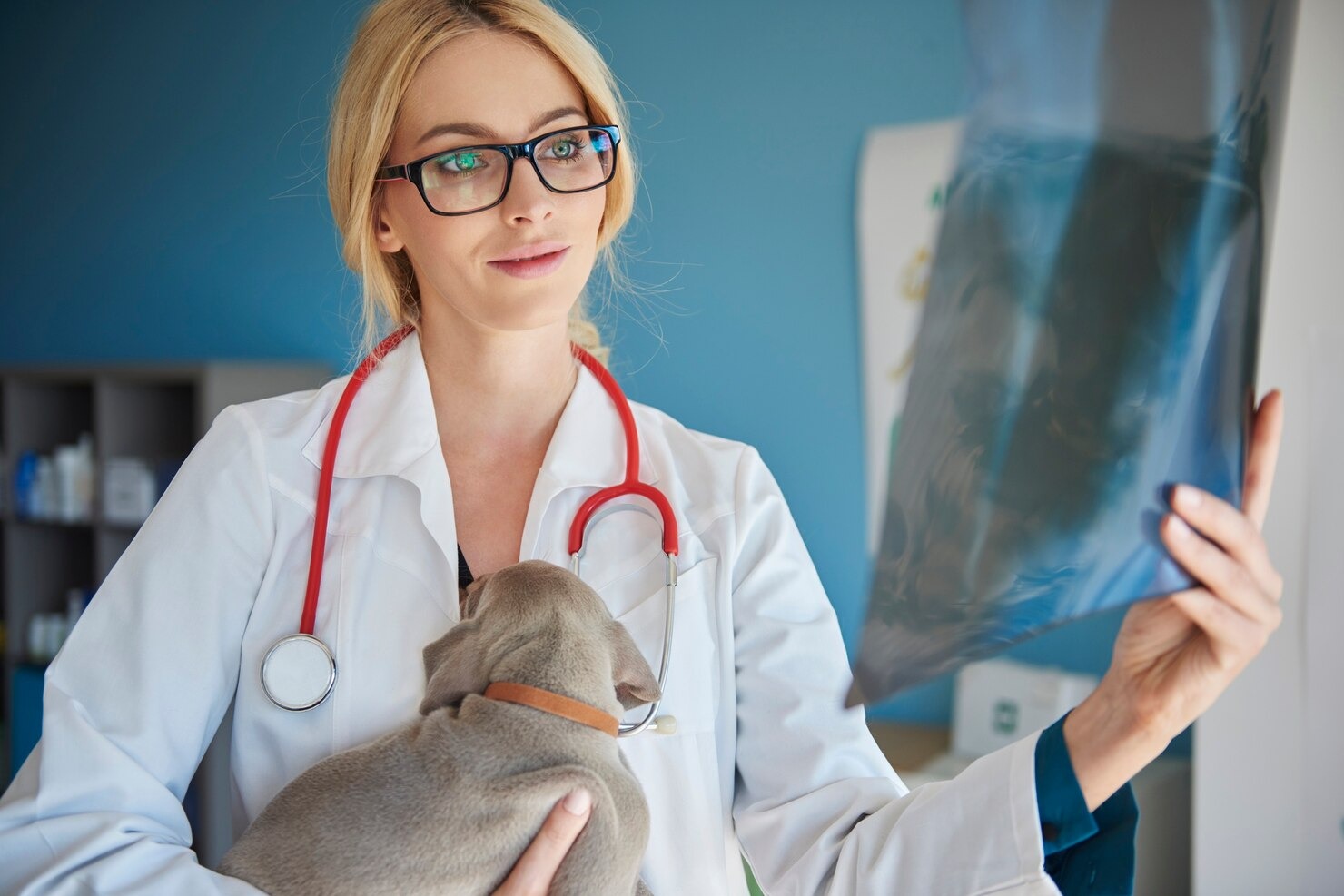
- Radiographic Imaging (X-rays): Standard digital radiographs (X-rays) are universally the first line of imaging performed. X-rays offer crucial, foundational information regarding the overall orthopedic health of the spine. While X-rays cannot physically show the soft tissue of the spinal cord or the disc material itself, they are excellent at identifying secondary signs of IVDD. Veterinarians look for narrowing of the normally uniform spaces between the vertebrae, small calcified disc material resting in the spinal canal (in situ calcification), or the collapse of the intervertebral foramen. Furthermore, X-rays are critical for ruling out other catastrophic causes of paralysis, such as severe spinal fractures, aggressive bone tumors (osteosarcoma), or vertebral infections (discospondylitis). However, they generally might not sufficiently visualize the exact nature of soft tissue compression required for surgical planning.[2]
- Advanced Cross-Sectional Imaging (MRI and CT): For a definitive diagnosis and accurate surgical planning, advanced imaging techniques are strictly required. Magnetic Resonance Imaging (MRI) is universally considered the absolute gold standard in veterinary neurology for diagnosing IVDD. An MRI provides breathtakingly detailed, three-dimensional images of the soft tissues, allowing the neurologist to directly visualize the intervertebral discs, the spinal cord, nerve roots, and any associated hemorrhage, edema (swelling), or inflammation. It precisely highlights the hydration status of every disc in the spine. If an MRI is unavailable, a Computed Tomography (CT) scan, frequently combined with a myelogram (the injection of a radiopaque contrast dye into the fluid surrounding the spinal cord), may be recommended. These imaging modalities can help pinpoint the exact anatomical location, lateralization (whether the disc ruptured to the left or right side), and severity of the disc herniation, commonly referred to as herniation of the disc, while accurately assessing the exact degree of spinal cord compression.[3]
- Cerebrospinal Fluid (CSF) Analysis: In complex or atypical cases where the MRI results are ambiguous or suggest an inflammatory disease process, a cerebrospinal fluid sample may be collected via a spinal tap while the dog is under anesthesia. The fluid is examined under a microscope by a clinical pathologist to exclude other highly destructive, inflammatory, or infectious causes of neurological symptoms, such as meningoencephalomyelitis (inflammation of the brain and spinal cord) or spinal lymphoma. Differentiating IVDD from these conditions is paramount because the treatments are entirely opposite.[4]
The diagnostic procedure for IVDD in dogs may vary depending heavily on the individual clinical presentation, the dog’s neurological grade, and the financial capabilities of the owner. However, securing an accurate, definitive diagnosis is absolutely critical to determine the most suitable, effective treatment plan and provide the best, most realistic prognosis, especially for paralyzed dogs who do not spontaneously regain normal function. In addition, thoroughly understanding the underlying condition and how IVDD occurs is crucial in managing it effectively for the remainder of the dog’s life.[5]
Treatment Options for Intervertebral Disc Degeneration in Dogs

The treatment paradigm for Intervertebral Disc Disease (IVDD) in dogs is highly nuanced and strictly dictated by the severity of the neurological clinical signs. Treatment can comprise intensive conservative medical management (non-surgical) or highly specialized, invasive surgical interventions. The ultimate decision is contingent on critical factors such as the condition’s severity (neurological Grades 1-5), the rate of symptom progression, the exact location of the slipped disc, the dog’s overall systemic health status, and the owner’s capacity to provide intensive postoperative care. Here is an exhaustive outline of both widely accepted treatment methods:[6]
Non-Surgical Treatment Options
Conservative, non-surgical management is generally the highly recommended course of action for dogs presenting with mild to moderate clinical signs—specifically Grade 1 (pain only) or Grade 2 (mild ataxia but still ambulatory). It may also be considered for owners who are financially unable to pursue advanced surgical options, though the risks of permanent paralysis in severe grades remain high without surgery. Non-surgical treatment requires immense dedication and strict compliance from the pet owner.[7]
- Strict Rest and Absolute Limitation of Movement: The absolute, non-negotiable cornerstone of conservative management is strict cage rest. This is far more intense than simply keeping the dog indoors. Strict rest and confinement are biologically imperative to curtail all unnecessary movement, allowing the torn annulus fibrosus time to scar over and heal, and to forestall any further, catastrophic extrusion of disc material into the spinal canal. Dogs require strict crate rest for a minimum of 4 to 8 continuous weeks. The crate must be comfortably padded but small enough that the dog cannot jump, pace, or stand on its hind legs. The dog must only be removed from the crate, while strictly supported by a specialized harness or sling, for highly controlled, 5-minute bathroom breaks on a short leash. Failure to adhere to this strict confinement—even for a few minutes of unsupervised wandering in the house—often results in the remaining disc material violently rupturing, instantly transforming a mildly painful dog into a paralyzed surgical emergency.[8]
- Aggressive Pharmacological Interventions (Medications): To combat the severe pain and acute inflammation associated with spinal cord compression, a multi-modal pharmacological approach is utilized. Non-steroidal anti-inflammatory drugs (NSAIDs) prescribed by your veterinarian are frequently administered to aggressively alleviate the swelling, localized inflammation, and intense pain associated with intervertebral disc issues. In certain acute, highly inflammatory scenarios, powerful prescription corticosteroids may be utilized instead of NSAIDs. It is an absolute, critical rule in veterinary pharmacology that NSAIDs and corticosteroids must never be administered simultaneously, as doing so rapidly causes catastrophic, often fatal gastrointestinal ulceration and perforation. Prescription muscle relaxants are highly effective and heavily utilized to ease the excruciating, rigid muscle spasms that universally accompany spinal pain.[9]
- Advanced Management of Discomfort: Because neuropathic pain (nerve pain) responds poorly to standard NSAIDs alone, additional analgesics are frequently employed. Nerve-pain medication prescribed by your veterinarian to quiet hyperactive nerves is a staple in IVDD pain management protocols. Additional prescription pain-blocking medication may also be added to the regimen to block specific pain receptors in the brain. Aside from oral medications, pain management might encompass highly effective alternative physical modalities such as veterinary medical acupuncture, cold laser therapy (photobiomodulation) to decrease cellular inflammation, or transcutaneous electrical nerve stimulation (TENS) applied directly over the painful spinal segments.[10]
- Controlled Rehabilitation: Once the acute inflammatory phase has passed and the dog’s condition has stabilized (usually after several weeks of strict cage rest), and strictly under the directing veterinarian’s oversight, a highly controlled rehabilitation program might be suggested to slowly bolster core strength, restore flexibility, and improve overall mobility without compromising the healing disc.[11]
Surgical Treatment Options
Surgical interventions for Intervertebral Disc Disease (IVDD) in dogs are generally contemplated and highly recommended when conservative, nonsurgical treatments fail to alleviate the condition, when pain becomes entirely refractory to maximum medical management, or when the dog presents with severe, rapidly progressing symptoms, such as significant neurological deficits, non-ambulatory paresis (Grade 3), or outright paralysis (Grades 4 and 5). Surgery aims to immediately physically decompress the spinal cord by removing the offending herniated disc material. Here are the intricate, highly specialized IVDD surgeries typically utilized by veterinary surgeons and neurologists for dogs:[12]
- Hemilaminectomy: This is by far the most prevalent and standard surgical procedure utilized for thoracolumbar IVDD (disc extrusions in the mid-to-lower back). The operation involves a deep surgical incision over the spine, where the surgeon carefully dissects the dense epaxial muscles away from the vertebrae. Utilizing a high-speed pneumatic surgical drill, the surgeon burrs away a small window of the vertebral bone (the lamina) on the lateral side of the spine. This creates a window to access the spinal canal. The surgeon then meticulously identifies, elevates, and eliminates the herniated, calcified disc material that is compressing the spinal cord, thereby instantaneously alleviating spinal cord pressure and restoring normal blood flow to the nervous tissue.[13]
- Ventral Slot Surgery: Because the anatomy of the neck differs significantly from the back, a hemilaminectomy is generally unsafe for the cervical spine. Instead, a ventral slot procedure is typically used for disc protrusions or extrusions in the cervical region (neck). The dog is positioned on its back, and the surgeon creates an incision on the underside of the neck, carefully navigating past the trachea, esophagus, and major carotid arteries to reach the bottom of the cervical vertebrae. The surgeon utilizes a specialized drill to extract a small, precise rectangular section of the bone on the underside of the adjacent vertebrae, essentially creating a ‘slot.’ This opening enables the surgeon to reach up into the spinal canal to aggressively remove the herniated disc material, achieving complete decompression of the cervical spinal cord.[14]
- Dorsal Laminectomy: This complex procedure mirrors the intent of a hemilaminectomy but is generally reserved and used for disc protrusions occurring in the lumbosacral region (the very lower back, where the spine meets the pelvis), a condition often known as Cauda Equina Syndrome. Rather than approaching from the side, the surgeon drills and extracts a portion of the bone on the vertebrae’s upper, dorsal roof to access and safely remove the herniated disc material compressing the sprawling nerve roots at the end of the spinal cord.[15]
- Prophylactic Fenestration: This preventive procedure is almost always conducted in conjunction with the primary decompression surgeries (like a hemilaminectomy or ventral slot). Knowing that chondrodystrophic breeds have multiple degenerated discs waiting to rupture, the surgeon will proactively target adjacent, currently un-ruptured discs. The process involves making small, precise incisions (windows) into the outer annulus fibrosus of multiple neighboring intervertebral discs to manually extract a significant portion of the calcified nucleus pulposus material, massively lowering the statistical risk of future catastrophic disc herniations in those specific locations.[1]
- Spinal Stabilization and Fusion: In specific, highly complex scenarios—such as trauma-induced IVDD where the sheer force fractured the vertebrae alongside rupturing the disc, or in cases of severe cervical instability (Wobbler Syndrome)—additional, highly invasive operations may be necessary to rigidly stabilize the spine if it becomes mechanically unstable following decompression surgery. This extensive procedure typically involves the intricate placement and use of heavy metal implants such as titanium pins, specialized bone screws, and bone cement to fuse the vertebrae together permanently.[2]
- Artificial Disc Prosthesis: In incredibly rare instances, and typically only offered in highly specialized, academic veterinary centers, an artificial titanium or polymer disc may be implanted into the empty disc space to actively maintain standard disc height, preserve the anatomical space for exiting nerve roots, and retain normal, dynamic spine movement after massive disc removal. While innovative, this remains largely experimental in veterinary medicine compared to standard decompression.[3]
It’s critically important for owners to acknowledge that while surgical treatments can be highly, phenomenally effective—boasting an 85% to 95% success rate for returning Grade 1-4 dogs to normal ambulation—they also carry inherent, severe risks. These include risks of general anesthesia, deep post-operative infection, severe intra-operative hemorrhage from the venous sinuses surrounding the spinal cord, and the rare but tragic potential of iatrogenic damage to the spinal cord itself. In addition, the recovery process immediately following surgery is not instantaneous; it routinely involves strict cage rest, intense, dedicated physical therapy, and ongoing, meticulously scheduled pain management for several weeks. Therefore, the suitability of any surgical treatment option for a specific dog will depend on an array of various factors, including the precise severity and location of the IVDD, the dog’s overall systemic health and age, the rapidity of the onset of paralysis, and the immediate availability of specialized surgical expertise and advanced neurosurgical equipment.[4]
Supportive Care
Regardless of whether an owner opts for conservative medical management or aggressive surgical intervention, intensive supportive and nursing care plays an absolutely critical role in the management of IVDD. Proper nursing care is often the deciding factor between a successful recovery and heartbreaking complications. This comprehensive care may include:[5]
- Intensive Physical Therapy and Rehabilitation Exercises: Once cleared by the presiding veterinarian, professional physical rehabilitation is essential to help maintain remaining muscle mass, rebuild core strength, ensure joint flexibility, and optimize overall mobility. Modalities frequently employed by certified canine rehabilitation practitioners include passive range of motion (PROM) stretching, underwater treadmill therapy (which uses water buoyancy to support the dog’s weight while encouraging normal gait patterning), targeted laser therapy, and proprioceptive retraining exercises utilizing balance boards and cavaletti poles.[6]
- Rigorous Weight Management: Immediate and sustained weight reduction is required to physically reduce the daily mechanical strain and sheer forces placed on the healing spine. Even shedding a single pound on a small dog can drastically reduce the torque applied to the intervertebral discs.[7]
- Assistive Mobility Devices: For dogs enduring a prolonged recovery, or for those who unfortunately suffer from permanent, significant weakness or irreversible paralysis, assistive devices are life-changing. Customized canine wheelchairs or carts allow paralyzed dogs to run, play, and engage with their environment freely. Specialized full-body harnesses (such as the Help ‘Em Up Harness) are vital tools that allow owners to safely and ergonomically support their dog’s heavy hindquarters during bathroom breaks without further injuring the dog’s spine or the owner’s own back.[8]
- Bladder and Bowel Management: Paralyzed dogs frequently lose the ability to empty their own bladders. Owners must be rigorously trained by veterinary staff on how to manually express their dog’s bladder by applying gentle, targeted pressure to the abdomen. This must be performed every 6 to 8 hours to prevent severe bladder over-distention, urine scalding on the skin, and life-threatening secondary urinary tract infections (UTIs).[9]
- Prevention of Decubital Ulcers (Bed Sores): Dogs lacking motor function and sensation will lie in one position indefinitely. Without intervention, the pressure over their bony prominences (hips, elbows, ankles) will cut off blood supply to the skin, causing severe, necrotic bed sores. Owners must physically turn the dog from side to side every 4 to 6 hours and provide exceptionally thick, highly supportive orthopedic bedding.[10]
The appropriate, highly tailored treatment plan for each individual dog will depend fundamentally on the specific case presentation and the presiding veterinary neurologist’s expert assessment. However, rapid, early intervention and comprehensive, dedicated aftercare are absolutely crucial to maximizing the chances of recovery, improving the long-term prognosis, and preserving an excellent quality of life for dogs suffering from intervertebral disc disease. Please remember to consult your veterinarian before making any changes to your pet’s care.[11]
How to Prevent Intervertebral Disc Degeneration in Dogs
Although completely, unequivocally preventing Intervertebral Disc Disease (IVDD) in dogs—especially in chondrodystrophic breeds burdened with an inescapable, hardwired genetic predisposition—may not be medically feasible, proactive pet owners can seamlessly implement numerous lifestyle strategies to significantly lessen the overall risk and fiercely preserve their dog’s long-term spinal health. These aggressive preventive measures can aid immensely in mitigating the statistical likelihood of an IVDD crisis, actively decelerate the microscopic progression of the degenerative disease, and dramatically improve the dog’s daily comfort:[12]
- Strict Nutritional Weight Regulation: Keeping your dog at an incredibly lean, healthy body condition score is arguably the most vital, impactful step an owner can take to reduce mechanical strain on the spine and lower the physical risk of IVDD. Canine obesity is a totally preventable exacerbating factor. Implement a balanced diet and consistently monitor your dog’s weight. Consult your primary care veterinarian to formulate an appropriate, calorie-restricted prescription diet and an absolutely safe exercise plan specifically tailored for your dog’s breed and age.[13]
- Consistent, Low-Impact Physical Activity: Involve your dog in regular, highly consistent, moderate physical exercise to help fortify their vital core epaxial muscles and retain maximum spinal flexibility. Strong core muscles act as an internal corset, supporting the spine so the discs don’t have to bear the brunt of gravity alone. Low-impact, continuous activities such as hydrotherapy (swimming) and brisk leash walking are highly recommended for dogs to dynamically support spinal health. However, actively avoiding explosive, high-impact, or excessive jumping or sharp twisting activities—such as frantic frisbee catching, roughhousing with larger dogs, or navigating agility weave poles—is absolutely crucial, as these actions can instantly apply catastrophic shear strain to the spine.[14]
- Correct, Ergonomic Lifting Methods: The way an owner physically handles a predisposed breed matters immensely. When picking up your dog, always use two hands: strictly support their broad chest behind the front legs with one hand, and firmly scoop and support their hindquarters and pelvis with the other. This keeps the spine perfectly horizontal and prevents unnecessary, downward gravitational stress on their delicate lumbar spine. This technique is especially, critically important for smaller breeds and large breeds of dogs with disproportionately long backs, such as Dachshunds, Corgis, and Basset Hounds, who are statistically highly likely to experience early degenerative problems with the same disc. Never pick a dog up by grabbing them under the armpits and letting their back half dangle vertically.[15]
- Evading Unsafe Environmental Surfaces: A home environment can be a minefield for a dog’s spine. Prevent your dog from encountering highly slick, slippery surfaces (like polished hardwood, laminate, or tile floors) or uneven outdoor terrain that could result in sudden, uncontrolled slips, falls, or severe splay-legged injuries. Ensure excellent paw traction on all indoor floors with the strategic use of non-slip rubberized rugs or textured yoga mats. Invest in sturdy, low-incline pet ramps or padded steps, and rigorously train your dog to use them to access couches and beds, thereby entirely eliminating the concussive trauma of jumping down from furniture.[1]
- Orthopedic Assistance and Supportive Bedding: Arrange a highly supportive, deeply cushioned, and warm resting surface for your dog. Investing in a high-quality, dense memory foam orthopedic bed is essential to help relieve direct pressure on their spine, hips, and joints while they sleep. Furthermore, for dogs with any history of neck pain, entirely replace standard neck collars with a high-quality, well-fitted chest harness to completely remove leash tension from the highly vulnerable cervical vertebrae and trachea during daily walks.[2]
- Routine, Proactive Veterinary Examinations: Regular, bi-annual veterinary check-ups can heavily assist in closely monitoring your dog’s overall orthopedic health, assessing subtle changes in their neurological reflexes, and detecting any initial, incredibly mild signs of IVDD before they escalate into a catastrophic rupture. Early detection and rapid medical intervention can vastly enhance the prognosis and lifelong management of the condition, severely reducing the statistical risk of recurring, paralyzing IVDD flare-ups.[3]
By strictly, uncompromisingly adhering to these preventative, highly protective measures, you can assist immensely in maintaining your dog’s optimal spinal health and actively lessen its overall risk of developing severe Intervertebral Disc Disease. However, it is fundamentally critical to acknowledge that certain inescapable factors, such as complex genetics and inherent breed conformation predisposition, may still strongly contribute to the likelihood of disc disease manifesting in specific dogs. Despite executing these preventative measures flawlessly, IVDD remains most common, and sometimes unavoidable, in certain genetically predisposed canine breeds.[4]
Frequently Asked Questions
What is the absolute longest a dog can go without deep pain perception before surgery becomes ineffective?
In the realm of veterinary neurology, the loss of “deep pain perception” (the conscious ability to feel a severe pinch to the toes) is considered a catastrophic surgical emergency. While every dog’s biology is unique, the generally accepted, critical medical window for surgical intervention is between 12 to 24 hours from the exact moment deep pain is lost. If a dog undergoes decompressive surgery (like a hemilaminectomy) within this narrow window, the prognosis for regaining the ability to walk is roughly 50%. However, if the spinal cord remains severely compressed without deep pain for longer than 24 to 48 hours, the spinal cord tissue begins to undergo irreversible necrosis (tissue death) due to prolonged ischemia. Beyond this timeframe, the surgical success rate plummets dramatically, often dropping below 5%, and the paralysis is highly likely to be permanent, regardless of surgical intervention.
Are harnesses truly better than standard collars for dogs diagnosed with or prone to IVDD?
Yes, absolutely. For any dog diagnosed with Intervertebral Disc Disease, especially those with a history of cervical (neck) IVDD, a well-fitted, Y-shaped chest harness is vastly superior to a standard neck collar, and its use is strictly recommended by veterinary professionals. When a dog pulls on a standard neck collar, or when an owner abruptly jerks the leash, massive amounts of concentrated mechanical force are transmitted directly into the delicate cervical vertebrae and the fragile intervertebral discs of the neck. This acute sheer force can easily precipitate a sudden, agonizing disc herniation or exacerbate an existing, slowly bulging disc. A proper chest harness safely distributes this mechanical leash pressure widely across the robust, bony sternum, the heavy muscles of the chest, and the shoulders, entirely bypassing the vulnerable spinal column of the neck.
Can a dog fully recover from Stage 5 IVDD without undergoing spinal surgery?
It is exceptionally, exceedingly rare for a dog to fully recover from Grade 5 IVDD (complete paralysis accompanied by the total loss of deep pain perception) using strictly conservative, non-surgical medical management. Grade 5 indicates that the spinal cord compression is so massive and severe that even the deepest, most protected nerve fibers within the core of the spinal cord have ceased functioning. Without immediate surgical decompression to physically remove the extruded disc material and restore critical blood flow, the severe pressure and resulting localized inflammation will almost inevitably cause permanent, irreversible death of the spinal cord tissue (myelomalacia). While strict crate rest and high-dose medications are highly effective for Grade 1 and Grade 2 IVDD, relying solely on conservative management for a Grade 5 presentation generally results in a permanent, lifelong necessity for a canine wheelchair and manual bladder expression.
Schedule a Veterinary Appointment
If you notice any signs of spinal discomfort, mobility issues, or suspect your dog may be suffering from Intervertebral Disc Disease, time is of the essence. Please schedule an appointment with a veterinarian immediately to ensure your pet receives the proper diagnosis and specialized care they need for a successful recovery.
References
- VCA Hospitals. Intervertebral Disc Disease in Dogs. VCA Animal Hospitals, 2023.
- Merck Veterinary Manual. Degenerative Diseases of the Spinal Column in Dogs. Merck & Co., 2022.
- Dickinson PJ, Bannasch DL. Current Understanding of the Genetics of Intervertebral Disc Degeneration. Frontiers in Veterinary Science, 2020.
- Olby NJ, et al. Prognostic Factors in Canine Acute Intervertebral Disc Disease. Journal of Veterinary Internal Medicine, 2020.
- American College of Veterinary Surgeons (ACVS). Canine Intervertebral Disc Disease. ACVS, 2021.
- Freeman P, et al. Magnetic resonance imaging of the canine spine. Veterinary Record, 2012.
- Moore SA, et al. Clinical trial protocols for intervertebral disc disease. Journal of Veterinary Internal Medicine, 2017.
- Levine JM, et al. Rehabilitation therapy for dogs with intervertebral disc herniation. Journal of the American Veterinary Medical Association, 2007.
- Jeffery ND, et al. Surgery for canine thoracolumbar disc extrusion. Veterinary Surgery, 2013.
- AVMA. Weight Management and Canine Health. American Veterinary Medical Association, 2022.
- Coates JR. Intervertebral Disc Disease. Veterinary Clinics of North America: Small Animal Practice, 2000.
- Webb AA, et al. Neurological examination and localization. Canadian Veterinary Journal, 2009.
- Cornell University College of Veterinary Medicine. Canine Spinal Cord Injuries. Cornell University, 2021.
- ASPCA. Managing Paralyzed Pets and Spinal Health. ASPCA Pet Health Insurance, 2023.
- Brown EA, et al. FGF4 retrogene on CFA12 is responsible for chondrodystrophy and intervertebral disc disease in dogs. PNAS, 2017.


