


 March 4, 2023
 Phil Good, DVM
What Are Traumatic Injuries In Dogs?
This content was prepared with AI assistance and reviewed by a licensed professional for accuracy.
As a veterinary professional, I have witnessed firsthand the devastating impact that unexpected accidents can have on our canine companions. Traumatic injuries in dogs represent some of the most critical and time-sensitive emergencies we treat in veterinary medicine. Whether the result of an unforeseen vehicular accident, a confrontation with wildlife, or a sudden fall during vigorous play, dog trauma requires swift identification, immediate triage, and comprehensive medical intervention. Traumatic injuries in dogs can range from superficial skin abrasions to life-threatening internal hemorrhaging and complex fractures. Understanding the pathophysiology of trauma, recognizing the subtle early signs of shock, and knowing exactly how veterinary teams approach these crises can empower pet owners to act decisively when every second counts. In this comprehensive guide, we will explore the diverse spectrum of trauma caused by injuries, detailing the specific clinical manifestations, diagnostic protocols, and advanced therapeutic strategies used to stabilize and heal patients suffering from severe trauma in dogs.[1]
The field of veterinary traumatology relies on the concept of the “golden hour,” a critical window of time immediately following an injury during which prompt medical and surgical interventions are most likely to prevent irreversible damage and death. When a dog experiences a high-impact event, the body immediately mounts a profound systemic response. The sympathetic nervous system releases catecholamines like epinephrine and norepinephrine, leading to an increased heart rate and the constriction of blood vessels in an attempt to maintain blood pressure and redirect oxygenated blood to vital organs. However, if the trauma is severe enough to cause significant blood loss or tissue destruction, this compensatory mechanism eventually fails, leading to hypovolemic shock, cellular hypoxia, and potentially multiple organ dysfunction syndrome (MODS). Consequently, addressing a puncture wound, managing complex bone breaks, or stabilizing a patient after a blunt force impact requires a highly coordinated, multidisciplinary approach that combines critical care, surgery, and intensive nursing.[2]
Types of Traumatic Injuries in Dogs
Trauma is a broad medical classification that encompasses any physical injury or wound caused by external force or violence. In the context of veterinary medicine, we categorize these injuries based on the type of tissue damaged, the mechanism of the injury, and the severity of the structural disruption. Dogs, by nature of their active, exploratory, and sometimes impulsive behaviors, are susceptible to a wide array of traumatic events. The physical forces involved in trauma—such as compression, tension, shearing, and bending—dictate the specific type of tissue failure that occurs. For instance, a direct, high-velocity impact may cause bones to shatter under compressive forces, while a sudden twisting motion during a game of fetch may apply excessive tension and rotational forces to a joint, leading to ligamentous tearing. Understanding these distinct categories of injury allows veterinarians to anticipate associated complications, formulate targeted diagnostic plans, and implement the most effective treatment modalities to restore the patient’s form and function.[3]
Fractures
Fractures, commonly known as broken bones, represent a significant portion of orthopedic trauma in canine patients. A fracture occurs when the physical stress placed upon a bone exceeds its mechanical limits, leading to a disruption in the continuity of the osseous tissue. The canine skeleton is a dynamic, living structure composed of a rigid mineral matrix interwoven with flexible collagen fibers, providing both strength and elasticity. However, high-impact events like motor vehicle collisions or falls from significant heights can easily overcome these mechanical defenses. When we evaluate fractures in dogs, we classify them based on several distinct parameters: the specific bone involved, the anatomical location of the break (such as the diaphysis, metaphysis, or epiphysis), the configuration of the fracture line, and whether the overlying skin has been breached.[4]
The classification of the fracture dictates the surgical or conservative approach required for optimal healing. A simple or closed fracture involves a single break where the bone fragments remain safely contained within the skin barrier, minimizing the immediate risk of environmental contamination. In contrast, a compound or open fracture is an absolute orthopedic emergency; the sharp edges of the fractured bone penetrate through the muscle, subcutaneous tissue, and skin, exposing the sterile bone marrow cavity and surrounding soft tissues to environmental debris and opportunistic bacteria. Open fractures carry a profound risk of osteomyelitis—a severe, deeply seated bone infection that can delay healing, necessitate multiple surgeries, or even lead to limb amputation if aggressive intravenous antibiotic therapy and meticulous surgical debridement are not immediately initiated.[5]
The geometric configuration of the fracture line also plays a crucial role in surgical planning. Transverse fractures occur at a direct right angle to the long axis of the bone, typically resulting from direct, localized blunt force. Oblique and spiral fractures, which curve or wrap around the bone shaft, are usually the consequence of torsional or twisting forces applied while the limb is firmly planted. Comminuted fractures are highly complex injuries where the bone shatters into three or more distinct fragments; these high-energy injuries often require advanced internal fixation techniques, utilizing specialized stainless steel or titanium bone plates, interlocking nails, and precisely placed cortical screws to bridge the defect and bear the animal’s weight while the bone heals. In growing puppies, whose bones are softer and highly porous, we frequently encounter greenstick fractures—where the bone bends and only partially cracks—as well as Salter-Harris fractures, which involve the fragile cartilaginous growth plates at the ends of long bones. Injuries to the growth plates require immediate, precise surgical alignment to prevent premature closure, which can lead to severe angular limb deformities and secondary osteoarthritis as the dog matures.[6]
Dislocations
Dislocations, clinically referred to as luxations, occur when extreme force completely disrupts the normal anatomical alignment of the articular surfaces within a joint, tearing the surrounding stabilizing structures, including the joint capsule and collateral ligaments. A subluxation refers to a partial dislocation where some contact between the articular surfaces remains. Joints are highly complex biomechanical hinges designed to provide smooth, frictionless movement while maintaining strict stability under heavy loads. When a joint is dislocated, the immediate consequences are intense, sharp pain, acute non-weight-bearing lameness, severe soft tissue swelling, and a distinct loss of normal range of motion. The longer a joint remains dislocated, the more difficult it becomes to reduce (put back into place) due to severe muscle contracture and the organization of inflammatory fibrin within the empty joint space.[7]
One of the most frequently encountered traumatic luxations in veterinary emergency rooms is coxofemoral (hip) luxation. This typically occurs when a dog is struck by a vehicle from the side, forcing the head of the femur to violently pop out of the acetabulum (the cup-like socket of the pelvis). In the vast majority of these cases, the femoral head is displaced in a craniodorsal direction—upward and forward. Treating a hip dislocation requires the dog to be placed under heavy general anesthesia to achieve complete muscle relaxation. The veterinarian then performs a closed reduction, applying specific manual traction and rotational forces to guide the femoral head back into the socket. Once reduced, the limb is typically immobilized using a specialized bandage known as an Ehmer sling to hold the hip in internal rotation and abduction, allowing the torn joint capsule to heal. If closed reduction fails or the joint remains highly unstable, surgical interventions, such as a toggle-pin stabilization or a femoral head and neck ostectomy (FHO), become necessary.[8]
Other joints are also highly susceptible to traumatic luxation. Traumatic elbow luxations, while less common due to the inherently stable, highly congruent nature of the joint, can occur following severe twisting injuries or falls where the limb is trapped. Shoulder dislocations (glenohumeral luxations) frequently damage the supportive medial or lateral glenohumeral ligaments and often require surgical stabilization using heavy non-absorbable sutures or synthetic mesh to rebuild the joint capsule. Additionally, while often a chronic developmental issue, sudden trauma can exacerbate or acutely present as a Patellar luxation: a condition where the kneecap slips out of the femoral groove, causing a distinctive skipping lameness that is highly prevalent in small and toy breed dogs. Regardless of the joint involved, rapid veterinary assessment is crucial to minimize permanent damage to the delicate articular cartilage, which can lead to early-onset, debilitating osteoarthritis.[9]
Lacerations and Abrasions
The skin is the body’s largest organ and its primary defense against environmental pathogens, chemical irritants, and physical trauma. When this vital barrier is breached by mechanical forces, the resulting wounds are broadly categorized as either lacerations or abrasions, depending on the depth of tissue damage and the specific nature of the causative force. Abrasions are superficial friction injuries—commonly referred to as “road rash”—that occur when a dog is dragged across a rough, unyielding surface, such as asphalt or gravel. In an abrasion, only the outermost layers of the skin (the epidermis and superficial dermis) are sheared away. While abrasions typically do not cause life-threatening blood loss, they strip away the protective dermal layers, exposing highly sensitive nerve endings. This results in significant, burning pain and creates a large surface area highly susceptible to environmental bacterial colonization and subsequent superficial skin infections.[10]
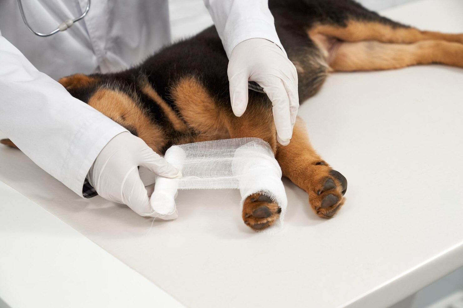
Lacerations, conversely, involve a complete tearing or slicing through the full thickness of the skin and often extend deep into the underlying subcutaneous fat, fascia, muscle bellies, or even down to the bone. Lacerations are typically caused by sharp implements, such as broken glass, jagged metal fencing, or the teeth of another animal. A clean, linear laceration with smooth, well-defined edges (like those caused by a scalpel or a piece of glass) is generally straightforward to repair, provided it is addressed promptly before massive bacterial replication occurs. The veterinarian will typically sedate or anesthetize the dog, thoroughly lavage (flush) the wound with copious amounts of sterile saline to remove debris, carefully trim away any devitalized (dead) tissue, and meticulously suture the wound edges together in multiple layers to eliminate dead space where fluid could accumulate.[11]
The most severe, specialized form of laceration is a shearing or degloving injury. These horrific injuries almost exclusively occur when a dog’s limb is trapped beneath a moving vehicle tire. The massive frictional force grips the skin and literally strips it away from the underlying deep fascial planes, stripping the limb bare of skin, subcutaneous tissue, and sometimes blood vessels and nerves, leaving the muscles and bones completely exposed. Degloving injuries require months of intensive, specialized open wound management. Treatment involves frequent, painful bandage changes, the application of specialized hydrocolloid or advanced topical antimicrobial dressings, and eventually, complex reconstructive surgeries, including skin grafts or rotational skin flaps, to finally close the massive defect.[12]
Contusions
A contusion is the medical term for a bruise, representing localized damage to the microscopic blood vessels (capillaries and small venules) that run through the subcutaneous tissue, fat, and muscle, without a concurrent breach in the overlying skin. Contusions are the direct result of blunt force trauma—such as a kick, a strike from a heavy falling object, or the generalized impact of a vehicle collision. When these tiny vessels rupture due to the crushing force, red blood cells leak out of the circulatory system and pool into the surrounding interstitial tissue spaces. Initially, a contusion may present as a swollen, exquisitely tender area that feels firm and warm to the touch due to the acute inflammatory cascade. As the leaked hemoglobin breaks down into biliverdin and bilirubin over the following days and weeks, the skin overlying the contusion will transition through a predictable spectrum of colors, changing from dark purple or black to shades of green, yellow, and finally a faded brown.[13]
While minor contusions are largely self-limiting and resolve with rest, severe blunt force trauma can lead to massive deep tissue bruising and the formation of a hematoma—a large, localized, clotted collection of blood located outside the blood vessels. Large hematomas can cause significant problems; they can exert mechanical pressure on adjacent nerves leading to intense pain or temporary paralysis, or they can compromise the blood supply to overlying skin, leading to tissue necrosis. Furthermore, massive muscle contusions can result in the widespread destruction of muscle cells (rhabdomyolysis), releasing large quantities of the protein myoglobin into the bloodstream. Myoglobin is directly toxic to the delicate filtering tubules of the kidneys, and in severe cases, extensive muscle trauma can precipitate acute renal failure. Therefore, any dog that has sustained significant blunt force trauma requires careful monitoring of their renal function and urine output, alongside supportive intravenous fluid therapy to flush these toxic byproducts from their system.[14]
Sprains and Strains
The intricate movements of the canine skeleton are made possible by a complex network of soft tissues, primarily ligaments, tendons, and muscles. Sprains and strains are common injuries in dogs that involve damage to the soft tissues, but they represent injuries to fundamentally different anatomical structures. A precise understanding of the difference between these two conditions is essential for proper diagnosis and the implementation of appropriate rehabilitation protocols. While both injuries result in acute inflammation, localized swelling, pain upon palpation, and varying degrees of lameness, their healing trajectories and potential for long-term joint instability differ significantly.[15]
A sprain refers strictly to an injury involving a ligament. Ligaments are dense, highly organized bands of fibrous connective tissue that connect one bone to another bone, serving as the primary passive stabilizers of a joint. Sprains occur when a joint is subjected to sudden, abnormal twisting or shear forces that stretch the ligament beyond its physiological yield point. Veterinary medicine categorizes sprains into three grades of severity. A Grade I sprain involves microscopic tearing of the ligament fibers with minimal swelling and no joint instability. A Grade II sprain represents a partial tear of the ligament; the joint may exhibit slight instability and moderate pain. A Grade III sprain is a catastrophic, complete rupture of the ligament, resulting in severe joint instability, profound pain, and total loss of function. The most notorious Grade III sprain in veterinary orthopedics is the rupture of the cranial cruciate ligament (CCL) in the stifle (knee), a debilitating injury that usually necessitates complex surgical intervention, such as a Tibial Plateau Leveling Osteotomy (TPLO), to restore dynamic joint stability.[16]
Conversely, a strain represents an injury to a muscle or a tendon. Tendons are the tough, resilient cords of fibrous tissue that anchor the fleshy muscle belly to the bone, transmitting the mechanical force generated by muscle contraction into skeletal movement. Strains typically result from massive, acute overexertion, explosive acceleration, or repetitive eccentric overloading of the muscle-tendon unit. Working dogs, agility competitors, and highly active sporting breeds are particularly prone to severe strains, often involving the Achilles tendon mechanism, the biceps brachii tendon, or the large muscle groups of the hindlimbs. Severe strains can result in tearing at the musculotendinous junction (where the muscle meets the tendon) or even an avulsion fracture, where the sheer force of the contracting tendon violently rips a small fragment of bone away from its attachment site. Recovery from severe strains requires extended periods of strict confinement, followed by carefully structured, progressive physical rehabilitation to rebuild muscle strength and restore tendon elasticity without causing re-injury.[17]
Bite Wounds
Bite wounds are among the most deceiving and frequently underestimated traumatic injuries seen in veterinary practice. When a dog is bitten by another dog, a coyote, or any other predator, the initial visual assessment often reveals only a few seemingly minor, circular puncture marks on the skin’s surface. However, veterinary professionals refer to bite wounds as exhibiting the “iceberg effect.” The visible punctures represent only a tiny fraction of the massive, catastrophic tissue damage lurking hidden beneath the skin. When an attacking animal bites down, its canine teeth act like thick, unsterile nails, driving deeply into the victim’s flesh. As the attacker violently shakes its head—a primal instinct designed to disable prey—the teeth shear and rip the underlying subcutaneous tissues, tearing blood vessels, shredding muscle fascia, and creating massive pockets of dead space beneath the intact skin.[18]
The mechanical crushing force exerted by the jaws of a large breed dog can exceed several hundred pounds of pressure per square inch. This extreme pressure causes profound blunt force trauma, crushing delicate cellular structures and severely compromising the local blood supply, leading to extensive tissue necrosis in the days following the attack. Furthermore, the mouth of a dog or cat is teeming with millions of pathogenic bacteria, including aggressive anaerobic species and highly virulent organisms like Pasteurella multocida, Capnocytophaga canimorsus, and various species of Staphylococcus and Streptococcus. The biting teeth act as hypodermic needles, injecting these virulent bacteria deep into the dark, low-oxygen environment of the crushed tissue—the absolute perfect breeding ground for a severe, rapidly spreading infection.[19]
Because of this massive underlying damage and heavy bacterial contamination, a “simple” bite wound can rapidly evolve into a life-threatening systemic infection or sepsis if not managed aggressively. Bite wounds located on the thorax (chest) or abdomen are particularly critical, as the teeth may have penetrated the body wall, introducing bacteria directly into the pleural or peritoneal cavities, leading to fatal pyothorax or septic peritonitis. Proper veterinary management of significant bite wounds almost always requires general anesthesia. The veterinarian must surgically open the wound, meticulously explore the full extent of the dead space, aggressively flush out bacteria and debris with liters of sterile fluid, surgically excise all dead and dying tissue (debridement), and often place active suction drains (such as a Jackson-Pratt drain) to prevent fluid accumulation and abscess formation while the area heals. Broad-spectrum systemic antibiotic therapy, guided whenever possible by aerobic and anaerobic bacterial cultures, is an absolute necessity.[20]
Internal Injuries
While broken bones and severe lacerations are highly visible and immediately alarming to pet owners, the most lethal consequence of trauma is often the damage that cannot be seen from the outside. Internal injuries—trauma to the delicate, blood-rich organs locked within the rigid cavities of the chest and abdomen—are the primary cause of rapid mortality following blunt force trauma, such as vehicular accidents or high-altitude falls. When a dog’s body is subjected to massive, sudden deceleration forces, the internal organs continue moving forward, crashing violently against the inside of the ribcage or abdominal wall. This sheer kinetic energy tears vital blood vessels, ruptures hollow organs, and crushes highly vascular solid tissues, creating life-threatening physiological emergencies that require immediate, aggressive stabilization.[21]
Trauma to the abdomen frequently results in a condition known as hemoabdomen—massive internal bleeding that pools freely within the peritoneal cavity. The spleen and the liver are the most commonly injured abdominal organs due to their large size, fragile tissue architecture, and massive blood supply. A ruptured spleen can bleed so profusely that the dog can entirely exsanguinate (bleed to death) internally within a matter of hours. Clinical signs of acute internal bleeding include profound weakness, completely white or pale mucous membranes (gums), a dangerously rapid heart rate (tachycardia) as the heart desperately tries to pump the dwindling blood volume, and a weak, thready pulse. If diagnostic imaging confirms a severely bleeding ruptured spleen or liver lobe, emergency exploratory laparotomy (abdominal surgery) is required to ligate the bleeding vessels and remove the irreparably damaged organ.[22]
In addition to hemorrhage, trauma can cause the rupture of hollow organs. A full urinary bladder is highly susceptible to bursting upon blunt impact. A ruptured bladder allows caustic, metabolic waste-laden urine to leak freely into the abdomen (uroabdomen). This causes severe chemical peritonitis and allows dangerous levels of potassium and urea to be reabsorbed into the bloodstream. High potassium levels (hyperkalemia) can cause fatal cardiac arrhythmias. Similarly, severe trauma or a urinary blockage can compromise the urinary tract’s integrity. If the gastrointestinal tract is perforated by trauma, highly infectious intestinal contents leak into the abdomen, causing catastrophic septic peritonitis that carries a very high mortality rate, leading to symptoms such as acute vomiting, bloody diarrhea, and profound shock. Furthermore, it’s vital to note that not all internal damage is caused by physical impact; ingestion of toxic substances can cause internal damage that rapidly destroys liver or kidney function, mirroring the devastating systemic collapse seen in physical trauma.[23]
Thoracic (chest) trauma is equally perilous. Blunt force to the chest frequently causes pulmonary contusions (bruising of the lungs), where blood and inflammatory fluid fill the tiny air sacs (alveoli), severely impairing the dog’s ability to absorb oxygen. Trauma can also cause a pneumothorax, a condition where a ruptured lung lobe allows free air to leak into the chest cavity. Because dogs, like humans, rely on a negative-pressure vacuum within the chest to expand their lungs, the accumulation of free air destroys this vacuum, causing the lungs to collapse and leading to severe, life-threatening respiratory distress. Treatment often requires the emergency placement of a thoracostomy tube (chest tube) to continuously suction out the free air and re-establish the vital negative pressure required for breathing.[24]
Head Injuries
Traumatic Brain Injury (TBI) and severe head trauma represent some of the most complex and neurologically challenging emergencies handled in critical care veterinary medicine. The brain is an incredibly delicate organ, housed within the rigid, unyielding bony vault of the skull. While the skull provides excellent protection against minor bumps, it becomes a distinct liability when severe trauma causes the brain tissue to swell. Veterinary neurologists divide head trauma into two distinct phases: primary injury and secondary injury. The primary injury occurs at the exact millisecond of impact. This includes the direct, irreversible physical disruption of brain tissue, such as skull fractures, cerebral lacerations, contusions (bruises on the brain surface), and diffuse axonal injury, where the violent rotational forces of the impact physically shear and tear the microscopic nerve fibers (axons) connecting different regions of the brain. The primary injury cannot be treated or reversed; the damage is done the moment the accident occurs.[25]
The fundamental goal of veterinary emergency treatment for head trauma is to aggressively prevent and manage the secondary injury. Secondary injury is a complex, cascading biochemical and physiological process that unfolds in the hours and days following the initial impact. As the injured brain tissue becomes inflamed, it begins to swell (cerebral edema). Because the skull cannot expand to accommodate this swelling, the pressure inside the head (intracranial pressure, or ICP) rises dangerously. As ICP increases, it begins to crush the brain’s blood vessels, choking off the vital supply of oxygen and glucose to the surviving brain cells. This ischemia leads to further cell death, releasing toxic excitatory neurotransmitters like glutamate, which cause calcium to flood into cells, triggering even more swelling and cellular destruction in a deadly, self-perpetuating cycle. If the intracranial pressure continues to rise unchecked, the brain will be physically pushed out of the only exit available—the hole at the base of the skull where the spinal cord exits (the foramen magnum). This catastrophic event, known as brain herniation, causes immediate, fatal crushing of the respiratory and cardiovascular centers in the brainstem.[26]
Clinical signs of severe head trauma and elevated intracranial pressure are progressive and highly alarming. An affected dog may exhibit profound alterations in consciousness, ranging from dullness and disorientation to a deep stupor or unarousable coma. Neurological assessments frequently reveal abnormal pupil sizes (anisocoria, where one pupil is large and the other is small), an inability of the pupils to constrict in response to bright light, and abnormal, rigid body postures, such as decerebrate rigidity, where all four limbs are held stiffly extended while the head is arched backward. Additionally, trauma to the head frequently involves collateral damage to the eyes and facial structures, including painful corneal ulcerations, forward displacement of the eyeball (proptosis), or fractures of the jaw that complicate airway management. Treating severe head trauma requires advanced intensive care, including elevating the dog’s head to promote venous drainage, administering specialized hyperosmolar intravenous fluids to medically draw fluid out of the swollen brain, strictly maintaining normal oxygen and carbon dioxide levels, and providing robust anti-seizure medication.[27]
Causes of Traumatic Injuries in Dogs

Understanding the root causes of traumatic injuries is essential not only for veterinary professionals trying to anticipate the hidden internal damage a patient might have sustained but also for dog owners seeking to implement effective preventative measures. The causes of canine trauma are as varied as the environments in which dogs live and play, encompassing a wide spectrum of physical, kinetic, and environmental hazards. In an urban or suburban setting, vehicular trauma remains the undisputed leading cause of catastrophic, multi-systemic injuries. Dogs can be injured by being hit by a car, motorcycle, or bicycle, and the massive kinetic energy transferred from a moving two-ton vehicle to a dog’s relatively small body causes devastating blunt and shearing forces. These patients, often referred to clinically as “HBC” (Hit By Car), frequently present with a complex constellation of injuries, simultaneously suffering from shattered pelvises, ruptured diaphragms, pulmonary contusions, and severe traumatic brain injury.[28]
Another profound source of trauma, particularly in densely populated urban environments with high-rise apartment buildings, is a phenomenon documented as “high-rise syndrome.” This occurs when a dog falls or jumps from a significant height, such as a third-story balcony or an open window. Interestingly, the specific pattern of injuries sustained in a fall is highly dependent on the height. Shorter falls often result in severe fractures of the forelimbs as the dog attempts to brace for impact, while falls from greater heights allow the dog to reach terminal velocity, resulting in horrific facial and jaw fractures, spinal cord trauma, and massive internal organ rupture as the body absorbs the terminal impact. Even relatively minor falls—such as a small breed dog leaping from the arms of its owner or jumping off a high bed—can result in devastating fractures of the delicate radius and ulna bones in the front legs.[29]
Interactions with other animals constitute another major category of veterinary trauma. Dog-on-dog aggression, particularly fights involving significant size disparities (a large dog attacking a small dog), frequently results in severe crushing bite wounds, deep lacerations, and life-threatening neck and throat injuries that can crush the trachea and disrupt the jugular veins. In rural or wooded areas, encounters with wildlife pose significant risks. Dogs may be gored by deer, attacked by coyotes, or suffer specialized penetrating trauma from porcupine quills, which possess microscopic backward-facing barbs that continuously migrate deeper into the dog’s tissues, requiring painstaking surgical removal. Environmental hazards, such as impalement on sharp sticks during a game of fetch, running through plate glass doors, or suffering deep lacerations from hidden barbed wire, also contribute heavily to the trauma caseload. Finally, thermal trauma (burns from house fires, boiling water spills, or chewing electrical cords) and malicious human-induced trauma (blunt force abuse, gunshot wounds, or arrow injuries) require specialized forensic and critical care management to stabilize the patient and preserve the integrity of severely compromised tissues.[30]
Symptoms of Canine Traumatic Injuries
The clinical presentation of a dog that has recently suffered a traumatic injury can be wildly variable, ranging from glaringly obvious orthopedic deformities to highly subtle, insidious signs of internal, life-threatening shock. Identifying these symptoms rapidly is paramount, as early recognition allows for immediate intervention during the critical “golden hour.” When a dog has sustained orthopedic trauma—such as a fracture, dislocation, or severe ligamentous tear—the symptoms are primarily localized to the musculoskeletal system. The dog will typically exhibit acute, non-weight-bearing lameness, actively holding the affected limb off the ground. There may be visible swelling, localized heat indicating acute inflammation, and a distinct unwillingness to let the limb be touched or manipulated. In cases of complete bone fracture, the limb may hang at an unnatural, distinctly abnormal angle, and you may hear or feel “crepitus”—a sickening, grating sensation caused by the sharp ends of the broken bone grinding against one another.[31]
However, it is the systemic signs of trauma—the symptoms indicating that the cardiovascular, respiratory, or neurological systems are failing—that are the most critical for pet owners to recognize. When a dog experiences severe internal bleeding or massive fluid shifts due to tissue damage, they enter a state of hypovolemic shock. The clinical hallmarks of shock include profoundly pale, white, or ashen mucous membranes (gums) instead of the normal healthy pink. Capillary refill time—the time it takes for color to return to the gums after applying gentle finger pressure—will be significantly prolonged (greater than 2 seconds), indicating poor peripheral blood perfusion. The dog’s heart rate will be excessively rapid (tachycardia) as the heart attempts to circulate the diminishing blood volume, yet the pulses felt on the inside of the hind leg (femoral pulses) will be weak, thready, and difficult to detect. The dog’s extremities (paws and ears) may feel uncharacteristically cold to the touch due to the body actively shunting blood away from the skin to preserve flow to the brain and heart. Lethargy, profound weakness, and an inability or unwillingness to stand are grave signs that the dog’s cardiovascular system is collapsing.[32]
Respiratory distress is another terrifying and common symptom of severe trauma, indicating conditions such as pulmonary contusions, pneumothorax, or a ruptured diaphragm. A dog in respiratory distress will exhibit an increased respiratory rate (tachypnea) and increased respiratory effort. They may stand with their elbows abducted (bowed outward) and their neck extended in a desperate attempt to maximize airflow into their lungs. Breathing may appear paradoxical, where the abdomen moves violently opposite to the chest wall. If oxygen levels in the blood drop to critically low levels, the dog’s gums and tongue may turn a deep, dusky blue or purple—a condition known as cyanosis, which is an absolute, immediate life-or-death emergency. Neurological symptoms following head or spinal trauma include profound disorientation, stumbling or uncoordinated walking (ataxia), complete paralysis of one or more limbs, abnormal pupil sizes, generalized seizures, or a progressive decline into a comatose state. Additionally, the presence of blood dripping from the nose (epistaxis), mouth, or rectum, or coughing up frothy blood (hemoptysis), strongly suggests severe internal organ disruption.[33]
Veterinary Diagnosis of Traumatic Injuries in Dogs

The diagnostic approach to a traumatized dog is inherently different from a routine veterinary visit. In trauma medicine, diagnostics and life-saving treatments occur simultaneously. The primary goal of the initial assessment is not to definitively diagnose every single broken bone or minor laceration, but rather to rapidly identify and immediately treat rapidly fatal conditions—specifically, obstructions of the airway, catastrophic bleeding, and severely impaired breathing. This structured, algorithmic approach to diagnostics ensures that the veterinary team prioritizes the physiological systems that sustain life before focusing on secondary injuries. Only after the patient has been stabilized through aggressive emergency interventions will the veterinarian proceed with a more detailed, comprehensive diagnostic workup to catalog the full extent of the damage.[34]
History
While gathering a thorough medical history is a staple of veterinary medicine, taking a history in a trauma scenario requires a rapid, highly focused approach, often utilizing the “AMPLE” mnemonic: Allergies, Medications currently taking, Past medical history, Last meal (crucial information if emergency anesthesia is required), and the Events leading up to the trauma. The veterinarian or triage technician will rapidly question the owner to establish the mechanism of injury. Knowing precisely what happened provides vital clues regarding the physical forces the dog absorbed. For example, knowing a dog was hit by a car traveling at 45 mph dictates a completely different level of internal suspicion than knowing a dog fell off a living room couch. Time is of the essence; establishing exactly how long ago the trauma occurred helps the medical team determine the timeline of shock progression and the viability of highly contaminated wounds.[35]
The history also involves determining the dog’s baseline health status. The team must know if the dog has preexisting cardiovascular disease, chronic renal failure, or a history of seizures, as these comorbidities drastically alter how the dog will respond to trauma, fluid therapy, and heavy pain medications. A thorough review of the dog’s vaccination status and any preventive medications is particularly vital if the trauma involved an animal bite, as rabies status must be strictly verified. Furthermore, the veterinarian will consider breed predispositions, as some dog breeds are predisposed to specific diseases that complicate trauma management; for example, brachycephalic breeds (like Pugs and Bulldogs) are at a massive risk for acute airway obstruction when stressed, while Doberman Pinschers may have underlying von Willebrand’s disease, a genetic bleeding disorder that can turn a minor laceration into a fatal hemorrhagic event.[36]
Physical Examination
The physical examination of a trauma patient is divided into two distinct phases: the primary survey and the secondary survey. The primary survey is an ultra-rapid triage assessment focused solely on the “ABCs” of critical care: Airway, Breathing, and Circulation (with D for Neurological Dysfunction). The veterinarian immediately checks if the airway is clear of blood, vomit, or broken teeth. They assess the respiratory rate, effort, and auscultate the lungs with a stethoscope to listen for the dull, muffled sounds indicative of a pneumothorax or the harsh crackles of pulmonary contusions. Cardiovascular status is evaluated instantly by assessing heart rate, pulse quality, mucous membrane color, and capillary refill time. If the dog is in active respiratory or cardiovascular collapse, the physical exam pauses immediately to provide life-saving interventions, such as intubation, oxygen therapy, or massive intravenous fluid resuscitation.[37]
Once the ABCs are secured and the dog’s vital signs are stabilized, the veterinarian performs the secondary survey. This is a meticulous, deliberate, nose-to-tail examination utilizing the “A CRASH PLAN” mnemonic (Airway, Cardiovascular, Respiratory, Abdomen, Spine, Head, Pelvis, Limbs, Arteries, Nerves). The veterinarian will thoroughly examine the skull for fractures, assess the eyes for proper pupillary light reflexes and evidence of internal ocular bleeding (hyphema), and inspect the oral cavity. They will systematically palpate the spine, feeling for step-defects or areas of profound hyperesthesia (pain) that indicate a vertebral fracture. The abdomen is carefully evaluated for distension, bruising, or the presence of a fluid wave, which suggests internal bleeding. The veterinarian will also check for signs of urinary tract integrity by examining the perineum and assessing if the dog can urinate normally. This comprehensive physical exam ensures that no subtle, secondary injuries are missed in the chaotic aftermath of a severe traumatic event.[38]
Palpation and Manipulation
Palpation and manipulation form the cornerstone of the orthopedic and soft tissue assessment during the secondary survey. Palpation involves the veterinarian using their hands to apply calculated pressure to the body’s structures, feeling for abnormal swelling, heat, defects in the muscle fascia, and specific localized pain responses. When evaluating the abdomen, deep, gentle palpation allows the veterinarian to assess the size and integrity of the spleen, liver, and kidneys, and to detect the rigid, board-like abdominal muscle guarding that classically indicates severe internal peritonitis or a ruptured organ. Carefully sliding the fingers along the ribs can detect painful rib fractures or a “flail chest,” a life-threatening condition where a segment of the ribcage is broken in multiple places and moves independently of the rest of the chest wall during breathing.[39]
Manipulation refers to the deliberate, guided movement of the dog’s limbs and joints through their normal physiological range of motion. The veterinarian will carefully flex and extend the shoulders, elbows, carpi, hips, stifles, and tarsi. During this manipulation, the practitioner is feeling for resistance, joint effusion (swelling within the joint capsule), and “crepitus,” the distinct grinding sensation that confirms the presence of bone fragments or the complete loss of smooth articular cartilage. Specific orthopedic manipulation tests are utilized to diagnose ligament ruptures. For example, the “cranial drawer test” and the “tibial compression test” are specific physical manipulations used to definitively diagnose a ruptured cranial cruciate ligament in the stifle. Palpation and manipulation must be performed with extreme care and adequate analgesia, as aggressively moving a severely broken bone or unstable spinal fracture can inadvertently sever major nerves or lacerate vital blood vessels, drastically worsening the patient’s condition.[40]
Laboratory Tests
Laboratory diagnostics in the immediate aftermath of trauma are designed to provide a rapid, objective snapshot of the patient’s internal physiological stability, guiding fluid therapy and transfusion requirements. The most common immediate tests are collectively known as the “Big Four” or point-of-care database: Packed Cell Volume (PCV), Total Solids (TS) or Total Protein (TP), Blood Glucose (BG), and Blood Urea Nitrogen (BUN). The PCV measures the percentage of red blood cells in the circulation; a rapidly dropping PCV strongly suggests active, severe internal hemorrhage. Total Solids measure the protein content in the blood; low proteins combined with a low PCV confirm acute whole-blood loss, guiding the decision to administer life-saving blood plasma or packed red blood cell transfusions. Blood glucose can fluctuate wildly during trauma; massive stress and pain cause it to spike, while severe systemic infection (sepsis) from a ruptured bowel can cause it to plummet to dangerously low levels.[41]
In cases of severe trauma and hypovolemic shock, blood lactate measurement is an absolutely vital diagnostic tool. Lactate is a metabolic byproduct produced when the body’s cells are starved of oxygen and are forced to generate energy anaerobically. A high blood lactate level objectively confirms that the dog’s tissues are poorly perfused and dying of hypoxia. Serial lactate measurements are used to monitor the effectiveness of intravenous fluid therapy; if the lactate levels rapidly decrease (lactate clearance), it proves that the cardiovascular system is stabilizing and blood flow is returning to the organs. Comprehensive blood chemistry panels are run to evaluate liver enzymes (ALT, AST), kidney function (creatinine, BUN), and critical electrolyte balances (sodium, potassium, chloride). Coagulation panels (Prothrombin Time [PT] and Activated Partial Thromboplastin Time [aPTT]) are essential, as massive trauma frequently triggers Disseminated Intravascular Coagulation (DIC), a catastrophic failure of the body’s clotting system where the dog simultaneously bleeds uncontrollably while forming microscopic clots throughout their organs.[42]
Additional Diagnostics
Modern veterinary imaging has revolutionized the speed and accuracy with which we diagnose traumatic internal injuries. One of the most critical imaging modalities used immediately upon a trauma patient’s arrival is the FAST ultrasound scan. FAST stands for Focused Assessment with Sonography for Trauma. This is a rapid, non-invasive ultrasound technique performed cage-side while the dog is receiving oxygen and fluids. An AFAST (Abdominal FAST) scan specifically looks for the presence of free fluid (blood or urine) pooling in four specific quadrants of the abdomen. A TFAST (Thoracic FAST) scan evaluates the chest cavity for the presence of free air (pneumothorax), free fluid (hemothorax), and signs of severe heart trauma (pericardial effusion). FAST scans take only minutes to perform, do not require the dog to be restrained in stressful positions, and provide immediate, life-saving information regarding the need for emergency surgery.[43]
Once the patient is cardiovascularly stable, comprehensive diagnostic imaging is pursued. Radiographs (X-rays) are the gold standard for diagnosing skeletal fractures, joint luxations, and evaluating the chest for broken ribs, diaphragmatic hernias, and the severity of pulmonary contusions. Orthogonal views (taking X-rays from two different, perpendicular angles) are mandatory to fully visualize the three-dimensional displacement of a broken bone. In complex trauma cases, particularly those involving severe head trauma, intricate pelvic shattering, or suspected spinal fractures, advanced imaging such as Computed Tomography (CT) or Magnetic Resonance Imaging (MRI) is utilized. A CT scan provides highly detailed, three-dimensional cross-sectional slices of the body, allowing surgeons to precisely map out the configuration of shattered bones and detect microscopic brain hemorrhages that traditional X-rays simply cannot see. In cases involving joint or spinal trauma, diagnostic procedures like joint fluid analysis (arthrocentesis) or cerebrospinal fluid taps may be performed to assess for infection or intense inflammatory markers.[44]
Emergency Treatments for Traumatic Injuries in Dogs

The medical and surgical management of a severely traumatized dog is an intense, dynamic process that requires a highly skilled, multidisciplinary veterinary team. The overarching goal of emergency treatment is to halt the progression of shock, restore adequate tissue oxygenation, control severe pain, and surgically repair damaged structures. The immediate priority is massive volume resuscitation. When a dog bleeds or experiences massive fluid shifts due to crush injuries, their circulating blood volume plummets, leading to cardiovascular collapse. Veterinarians aggressively combat this by administering rapid, calculated boluses of balanced intravenous crystalloid fluids through large-bore IV catheters. In cases of profound shock or severe head trauma, specialized intravenous fluids or synthetic colloids may be utilized to rapidly pull fluid from the interstitial spaces back into the vascular system, expanding the blood volume and instantly improving cardiac output. If blood loss is catastrophic, life-saving whole blood or packed red blood cell transfusions, typed and cross-matched for compatibility, are administered to restore the oxygen-carrying capacity of the blood.[45]
Pain management in trauma patients is not just a matter of compassion; it is a critical physiological necessity. Uncontrolled, agonizing pain triggers a massive sympathetic nervous system response, driving up the heart rate, increasing oxygen demand, suppressing the immune system, and contributing directly to the downward spiral of shock. Veterinary emergency medicine utilizes a multimodal approach to analgesia, attacking pain pathways at various levels of the nervous system simultaneously. Potent prescription pain medication administered by your veterinarian forms the backbone of trauma analgesia, providing profound, rapid-onset pain relief. These are often combined with continuous intravenous infusions of specialized pain-modulating medications to prevent the nervous system from becoming hyper-sensitized to pain. Once the patient’s blood pressure is thoroughly stabilized and renal function is confirmed, prescription veterinary anti-inflammatory medications may be carefully introduced to reduce profound tissue inflammation.[46]
Wound management and surgical intervention represent the definitive treatment phase. Severe, contaminated wounds and deep lacerations undergo meticulous surgical debridement under general anesthesia. The veterinary surgeon carefully excises all necrotic, devitalized tissue—which serves as a breeding ground for bacteria—until clean, healthy, bleeding margins are achieved. For orthopedic trauma, the timing of surgical repair is critical. While open fractures require immediate surgical flushing and stabilization to prevent osteomyelitis, the definitive internal fixation (plating and screwing) of closed fractures is often delayed for 24 to 48 hours. This delay allows the critical care team to medically stabilize the patient’s cardiovascular system, treat life-threatening pulmonary contusions, and ensure the dog is strong enough to survive a lengthy, complex orthopedic surgery under prolonged general anesthesia. In cases involving a ruptured spleen, a torn diaphragm, or a lacerated liver lobe, however, emergency exploratory laparotomy is performed immediately, as the ongoing internal hemorrhage is rapidly fatal.[47]
How to Prevent Traumatic Injuries in Dogs
While the highly specialized medical interventions available in modern veterinary emergency rooms are miraculous, the most effective strategy for managing trauma is absolute prevention. Preventing traumatic injuries in dogs requires dog owners to maintain a state of active vigilance, implement robust safety protocols in the dog’s daily environment, and profoundly understand the instinctual behaviors that often drive dogs into dangerous situations. The implementation of these safety measures can dramatically reduce the incidence of catastrophic, life-altering injuries, preserving the health and longevity of your canine companion.[48]
The single most effective preventative measure a dog owner can take is the strict, unwavering use of a leash when outside of a securely fenced area. The vast majority of vehicular trauma cases involve dogs that were allowed to roam freely or were being walked off-leash in urban or suburban environments. Even the most highly trained, obedient dog can be suddenly startled by a loud noise, or have their prey drive intensely triggered by a fleeing squirrel, causing them to bolt blindly into heavy traffic. A sturdy leash, attached to a well-fitted, escape-proof harness rather than a simple neck collar (which can cause tracheal trauma if the dog pulls violently), provides the owner with absolute physical control over their pet’s movements. Furthermore, ensuring that residential yards are secured with high-quality, deeply rooted fencing—and regularly inspecting the perimeter for dug holes or broken slats—is vital to prevent escape and subsequent encounters with vehicles, aggressive stray animals, or local wildlife.[49]
Safe transportation practices are heavily overlooked but critically important. Allowing a dog to ride loose in the bed of a pickup truck or unrestrained in the cabin of a car is incredibly dangerous. In the event of a sudden stop or a collision, an unrestrained dog becomes a high-velocity projectile, leading to devastating blunt force trauma, severe spinal injuries, or ejection from the vehicle. Dogs should always be secured in the back seat using an crash-tested canine seatbelt harness, or ideally, confined within a robust, crash-tested travel crate that is firmly anchored to the vehicle’s frame. Additionally, mitigating environmental hazards inside the home is essential. Keep caustic chemicals, toxic human medications, and sharp objects securely locked away. During extreme weather, protect your dog from profound thermal trauma by limiting exposure to freezing temperatures (preventing frostbite to the extremities) and never leaving a dog inside a parked car during warm weather, which rapidly leads to fatal hyperthermia and multi-organ coagulation. Finally, regular veterinary check-ups are crucial, as health issues that could heighten your dog’s susceptibility to injury—such as failing vision, early-onset arthritis, or vestibular disease—can be identified and managed early, preventing devastating falls or accidents. Always consult your veterinarian before making any changes to your pet’s care or implementing new training regimens.[50]
Frequently Asked Questions
How do you know if a dog has internal bleeding?
Detecting internal bleeding in dogs requires careful observation, as the hemorrhage occurs invisibly within the chest or abdomen. The most common signs are related to hypovolemic shock resulting from massive blood volume loss. You may notice your dog’s gums becoming profoundly pale, white, or ashen. They will exhibit profound lethargy, extreme weakness, and may collapse or be entirely unable to stand. Their heart rate will become very rapid, and their breathing may become fast and shallow. In cases of abdominal bleeding, the belly may appear distended, swollen, and feel tense or painful to the touch. In extreme cases, you may see blood pooling in the whites of their eyes or dark, tarry stools (melena). Internal bleeding is an absolute, critical emergency requiring immediate veterinary surgical and medical intervention to prevent fatal cardiovascular collapse.
How long do soft tissue injuries take to heal in dogs?
The healing timeline for soft tissue injuries (such as muscle strains, ligament sprains, and deep lacerations) varies drastically based on the severity of the tissue disruption and the dog’s compliance with rest protocols. Minor Grade I strains or superficial skin lacerations may heal relatively quickly, within 10 to 14 days, provided they are kept clean and the dog is rested. However, severe injuries, such as a partial ligament tear or a deeply bruised muscle group, can take 6 to 8 weeks to adequately heal. A complete ligament rupture, such as a torn cranial cruciate ligament, will not heal on its own and requires complex surgical stabilization, followed by 12 to 16 weeks of highly restricted activity and intensive, progressive physical rehabilitation to restore full, pain-free mobility.
Can a dog be traumatized after being attacked?
Yes, dogs can experience profound, long-lasting emotional and psychological trauma following a violent attack or a severely frightening event, much like Post-Traumatic Stress Disorder (PTSD) in humans. The psychological impact of being attacked by another dog or abused can manifest in profound behavioral shifts. A previously confident dog may become highly fearful, constantly anxious, and hyper-vigilant. They may develop intense phobias of other dogs, loud noises, or specific locations associated with the attack. This fear can rapidly transition into fear-based aggression, where the dog growls, lunges, or snaps defensively to protect itself from perceived threats. Treating psychological trauma requires extreme patience, a highly controlled environment, and often the expertise of a board-certified veterinary behaviorist to implement systematic desensitization, positive counter-conditioning, and potentially the use of anti-anxiety medications.
Don’t Wait in an Emergency
If your dog has experienced a traumatic injury, every second counts. Prompt medical attention can make the difference between a rapid recovery and severe complications.
References
- American Veterinary Medical Association. Emergency Care for Your Pet. AVMA, 2023.
- Silverstein, D. Shock in Animals. Merck Veterinary Manual, 2022.
- Simpson, S. A., et al. “Retrospective evaluation of the causes and severity of trauma in dogs.” Journal of Veterinary Emergency and Critical Care, 2011.
- VCA Hospitals. Fractures in Dogs. VCA Animal Hospitals, 2023.
- Harasen, G. Overview of Fracture Management in Animals. Merck Veterinary Manual, 2022.
- DeCamp, C. E., et al. Brinker, Piermattei and Flo’s Handbook of Small Animal Orthopedics and Fracture Repair. Elsevier, 2015.
- Veterinary Information Network. Management of Traumatic Luxations. VIN, 2008.
- VCA Hospitals. Hip Dislocation in Dogs. VCA Animal Hospitals, 2023.
- Di Dona, F., et al. “Patellar luxation in dogs.” Veterinary Medicine: Research and Reports, 2018.
- Pavletic, M. Wound Management in Animals. Merck Veterinary Manual, 2022.
- VCA Hospitals. Care of Open Wounds in Dogs. VCA Animal Hospitals, 2023.
- Bohling, M. W. “Skin grafts and reconstructive surgery in the dog.” Veterinary Clinics of North America: Small Animal Practice, 2010.
- Veterinary Information Network. Blunt Force Trauma Diagnostics. VIN, 2009.
- Tivers, M., et al. “Management of blunt trauma in dogs.” In Practice, 2014.
- Johnston, S. Overview of Lameness in Dogs. Merck Veterinary Manual, 2022.
- VCA Hospitals. Cruciate Ligament Rupture in Dogs. VCA Animal Hospitals, 2023.
- Zink, M. C., et al. “Rehabilitation and physical therapy for the sporting dog.” Veterinary Clinics: Small Animal Practice, 2013.
- Veterinary Information Network. Management of Bite Wounds. VIN, 2011.
- Meyers, B., et al. “Bacteriology of dog and cat bite wounds.” Journal of Veterinary Emergency and Critical Care, 2008.
- VCA Hospitals. Bite Wounds in Dogs. VCA Animal Hospitals, 2023.
- Macintire, D. Initial Management of the Trauma Patient. Merck Veterinary Manual, 2022.
- Mongil, C. M., et al. “Traumatic hemoabdomen in 54 dogs.” Veterinary Surgery, 2014.
- VCA Hospitals. Peritonitis in Dogs. VCA Animal Hospitals, 2023.
- Sigrist, N. E., et al. “Thoracic trauma in dogs.” Journal of Veterinary Emergency and Critical Care, 2012.
- Platt, S. Traumatic Brain Injury in Animals. Merck Veterinary Manual, 2022.
- Sande, A., et al. “Traumatic brain injury in the dog.” Veterinary Clinics of North America: Small Animal Practice, 2010.
- VCA Hospitals. Head Trauma in Dogs. VCA Animal Hospitals, 2023.
- American Veterinary Medical Association. Road Accidents and Pets. AVMA, 2022.
- Gordon, L. E., et al. “High-rise syndrome in dogs.” Journal of the American Veterinary Medical Association, 2004.
- Veterinary Information Network. Penetrating and Ballistic Trauma. VIN, 2009.
- VCA Hospitals. Recognizing Pain in Dogs. VCA Animal Hospitals, 2023.
- Mazzaferro, E. Evaluation of the Trauma Patient. Merck Veterinary Manual, 2022.
- Hall, K. E., et al. “Emergency management of respiratory distress.” Veterinary Clinics of North America: Small Animal Practice, 2016.
- Veterinary Information Network. Triage of the Trauma Patient. VIN, 2011.
- Rozanski, E., et al. “Triage and initial assessment of the emergency patient.” Journal of Veterinary Emergency and Critical Care, 2009.
- VCA Hospitals. Von Willebrand’s Disease in Dogs. VCA Animal Hospitals, 2023.
- Fletcher, D. Cardiopulmonary Resuscitation in Animals. Merck Veterinary Manual, 2022.
- Drobatz, K. J. “The physical examination in the emergency patient.” Journal of Veterinary Emergency and Critical Care, 2012.
- VCA Hospitals. Rib Fractures and Flail Chest in Dogs. VCA Animal Hospitals, 2023.
- Piermattei, D. L. Atlas of Surgical Approaches to the Bones and Joints of the Dog and Cat. Saunders, 2004.
- Giger, U. Point-of-Care Testing in Veterinary Medicine. Merck Veterinary Manual, 2022.
- Pang, D. S., et al. “Lactate in veterinary critical care: pathophysiology and management.” Journal of the American Animal Hospital Association, 2011.
- Boysko, M., et al. “Focused Assessment with Sonography for Trauma (FAST) in animals.” Veterinary Clinics of North America: Small Animal Practice, 2009.
- Veterinary Information Network. Advanced Imaging in Trauma. VIN, 2011.
- Mazzaferro, E. M. “Fluid therapy for the trauma patient.” Journal of Veterinary Emergency and Critical Care, 2013.
- Epstein, M. Pain Management in Animals. Merck Veterinary Manual, 2022.
- Beal, M. W. “Approach to the acute abdomen.” Veterinary Clinics of North America: Small Animal Practice, 2013.
- American Veterinary Medical Association. Household Hazards and Preventative Care. AVMA, 2023.
- VCA Hospitals. Preventing Accidents in Dogs. VCA Animal Hospitals, 2023.
- Miller, M. “Safety and restraint in canine transport.” Journal of Veterinary Behavior, 2015.


