


 March 4, 2023
 Phil Good, DVM
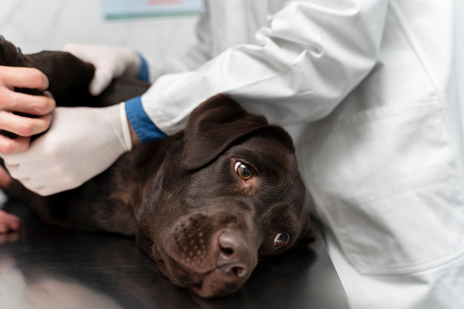
What is Soft Tissue Trauma in Dogs?
This content was prepared with AI assistance and reviewed by a licensed professional for accuracy.
When it comes to the active, energetic lives of our canine companions, understanding the nuances of soft tissue trauma in dogs is essential for any responsible pet owner. This encompassing term covers a vast array of injuries involving the non-bony structures of the body, including muscles, tendons, ligaments, skin, and underlying fascia. Common examples include sprains and strains, as well as surface-level wounds like Lacerations. It is important to distinguish soft tissue injuries from Fractures, which specifically involve the breaking of bone tissue. While a fracture requires a very distinct orthopedic intervention, soft tissue trauma presents its own complex set of diagnostic and therapeutic challenges that can significantly impact a dog’s mobility and quality of life.[1] Understanding how these injuries occur, how to recognize the clinical signs, and when to seek professional veterinary care is the cornerstone of proactive pet health management. Whether you have a highly trained working dog, an agility competitor, or a beloved household pet who simply enjoys a vigorous game of fetch, soft tissue injuries are a common reality of canine life.
The intricate musculoskeletal system of a dog relies heavily on the healthy function of its soft tissues. Muscles provide the force necessary for movement; tendons serve as the resilient cables anchoring those muscles to the bones; and ligaments act as the crucial stabilizing bands that hold the bones together at the joints. When any of these soft tissue structures are subjected to forces that exceed their natural tensile strength, cellular damage occurs.[2] This damage triggers a rapid physiological response, initiating an inflammatory cascade designed to protect the area and begin the healing process. However, unchecked inflammation can lead to further tissue degradation and the formation of restrictive scar tissue, which can permanently alter a dog’s biomechanics. Therefore, proper management of soft tissue trauma in dogs requires far more than just “waiting it out.” It demands a targeted approach to control inflammation, manage pain, and restore normal tissue function through a combination of medical intervention, physical rehabilitation, and strategic rest.[3]
Introduction
Imagine the scenario: you are enjoying a sunny afternoon in the park, throwing a frisbee for your athletic Australian Shepherd. The dog leaps into the air, twists to catch the disc, and lands awkwardly on a single hind leg, letting out a sudden, sharp yelp. Instantly, the dog refuses to bear weight on the limb, holding it tightly against its body. This sudden shift from joyful play to acute distress is the classic presentation of a soft tissue injury. The sudden mechanical overload on the dog’s joint likely caused the structural failure of a ligament or tendon. As a veterinarian, I frequently see pet owners who are understandably alarmed by these sudden lameness episodes. Because the injury is beneath the skin and does not involve an obvious bone deformity, it can be difficult for an owner to assess the severity of the situation.[4] This is why a comprehensive understanding of soft tissue trauma is so valuable; it empowers you to make informed decisions about your pet’s immediate care and ongoing recovery.
Soft tissue trauma in dogs ranges significantly in severity, from minor contusions that resolve over a weekend to catastrophic ligament ruptures that require advanced surgical reconstruction. Working breeds, large breed dogs, and older pets with degenerative joint changes are particularly susceptible to these types of injuries, though no dog is entirely immune.[5] The long-term implications of poorly managed soft tissue trauma can be severe. Without appropriate veterinary intervention, minor injuries can develop into chronic conditions. Compensatory gait changes—where a dog shifts its weight to avoid pain in an injured limb—can place abnormal stress on other joints, leading to secondary injuries and the premature onset of osteoarthritis. A dog’s overall comfort, willingness to engage in play, and fundamental quality of life are deeply intertwined with the health of its musculoskeletal system. Consequently, timely diagnosis and a rigorous, structured treatment plan are non-negotiable elements of veterinary care when dealing with any suspected soft tissue damage.[6]
Common Types of Soft Tissue Trauma in Dogs
1. Bruises and Contusions
Bruises and contusions represent the most basic form of soft tissue trauma in dogs. These injuries occur when a blunt force impacts the body, causing the microscopic capillaries and smaller blood vessels situated just beneath the skin and within the muscle layers to rupture.[7] Because the overlying skin remains intact, the escaping red blood cells pool in the surrounding interstitial spaces, creating a localized collection of blood. Depending on the size of the hemorrhage, veterinarians refer to these as petechiae (pinpoint spots of bleeding) or ecchymosis (larger, diffuse areas of bruising). You might notice the skin in the affected area turning red, purple, or dark blue, though this discoloration can be notoriously difficult to spot on dogs with thick or darkly pigmented coats.
As the dog’s body begins the natural healing process, specialized white blood cells called macrophages arrive at the site of the contusion to clean up the cellular debris. They break down the pooled hemoglobin into biliverdin and bilirubin, which causes the bruise to change colors over time—often shifting to a greenish or yellowish hue before fading completely.[8] While minor bruises generally cause only mild discomfort and transient tenderness, deep muscle contusions can be profoundly painful. A severe contusion in a major muscle group, such as the quadriceps or hamstrings, can significantly impair a dog’s mobility, resulting in a noticeable limp and a reluctance to exercise. In extreme cases of blunt force trauma, what appears to be a simple bruise on the surface may actually indicate deeper, more dangerous internal bleeding, which is why veterinary evaluation is always recommended after any significant traumatic impact.
2. Sprains
Sprains are a highly specific classification of soft tissue injury that solely affects ligaments, the tough, fibrous bands of connective tissue responsible for attaching bone to bone and stabilizing joints. Sprains occur when a joint is forced beyond its normal, physiological range of motion. This excessive stretching force causes the collagen fibers within the ligament to fray or tear. In canine sports medicine, sprains are most commonly seen in the carpus (the “wrist” joint of the front leg) and the tarsus (the “ankle” joint of the back leg), as well as the stifle (the knee joint).[9] The sudden, multidirectional forces exerted during activities like agility weaving, jumping, or sudden braking can easily overwhelm the structural integrity of these critical stabilizing ligaments.
Veterinarians grade sprains on a scale from I to III to categorize their severity. A Grade I sprain involves microscopic tearing of the ligament fibers with no loss of joint stability; the joint remains tight, but the dog experiences localized pain and minor swelling. A Grade II sprain represents a partial tear of the ligament, leading to increased pain, moderate swelling, and a detectable looseness or instability within the joint. Finally, a Grade III sprain is a catastrophic, complete rupture of the ligament, resulting in severe pain, massive joint effusion (fluid accumulation), and total joint failure.[10] A dog with a Grade III sprain will typically exhibit a “toe-touching” lameness or carry the limb entirely. Due to the poor blood supply inherent to ligamentous tissue, sprains heal notoriously slowly, and severe grades often necessitate surgical stabilization to restore function and prevent debilitating, long-term osteoarthritis.
3. Strains
While sprains affect ligaments, strains are injuries that specifically involve muscles and the tendons that attach them to bones. A strain happens when a musculo-tendinous unit is stretched forcefully or forced to contract against too much resistance, leading to a disruption of the muscle fibers or the tendon itself. This type of soft tissue trauma in dogs is frequently observed as an “overuse” injury or as a result of an inadequate warm-up prior to explosive physical activity. Common sites for strains in dogs include the iliopsoas muscle (a deep groin muscle involved in flexing the hip), the biceps tendon in the shoulder, and the superficial digital flexor tendon in the lower limbs.[11] High-speed, explosive athletes like Greyhounds and Whippets are notoriously prone to severe muscle strains.
Similar to sprains, strains are diagnostically graded from I to III. Grade I strains involve minor tearing of a few muscle fibers, presenting as mild soreness and stiffness, particularly after resting following exercise. Grade II strains entail a more significant tear within the muscle belly or tendon, accompanied by localized hemorrhage, noticeable swelling, and a distinct functional deficit, such as a shortened stride or consistent limping. Grade III strains represent a complete tear or avulsion (where the tendon pulls a piece of bone away with it).[12] These severe injuries cause sudden, excruciating pain, a palpable gap within the muscle tissue, and an immediate inability to use the affected limb. Strains require a carefully managed rehabilitation process; rushing a dog back into activity before the muscle fibers have adequately healed and realigned frequently leads to chronic, recurrent injuries and dense scar tissue formation.
4. Lacerations
Lacerations are traumatic tearing or cutting injuries to the skin and underlying tissues, typically caused by contact with sharp environmental objects such as broken glass, jagged metal fencing, or hidden debris in underbrush. Unlike clean, surgical incisions, lacerations often have irregular, jagged edges and can be contaminated with dirt, debris, and environmental bacteria. The severity of a laceration depends heavily on its location, depth, and the specific structures involved. A superficial laceration may only breach the epidermis and dermis, causing minor capillary bleeding. However, deep lacerations can slice through subcutaneous fat, sever major blood vessels, transect peripheral nerves, and even slice into underlying muscle bellies and tendons.[13]
The immediate clinical priority when managing a laceration is achieving hemostasis (stopping the bleeding) and preventing severe infection. If a laceration occurs, pet owners should apply direct, firm pressure with a clean cloth or sterile gauze and seek emergency veterinary care. Veterinarians assess lacerations to determine the best method of closure. Wounds evaluated within the “golden period” (typically the first 6-8 hours post-injury) that are relatively clean can often be sutured closed primarily after thorough flushing and debridement. Conversely, older, highly contaminated, or infected lacerations may need to be left open to heal by secondary intention, requiring meticulous daily bandage changes and advanced wound management techniques to manage exudate and promote healthy granulation tissue formation.[14]
5. Abrasions
Abrasions, colloquially known as “road rash” or scrapes, are superficial, friction-based injuries that sheer away the epidermis (the outermost layer of skin) and potentially the upper layers of the dermis. These soft tissue injuries are incredibly common in active dogs and typically result from sliding on rough, unyielding surfaces like asphalt, concrete, gravel, or hard-packed dirt. While an abrasion may not look as dramatic or immediately life-threatening as a deep, bleeding laceration, it should not be underestimated. The skin is densely populated with microscopic nerve endings, making abrasions exquisitely painful and highly sensitive to touch and environmental irritants.[15]
The primary concern with abrasions, aside from managing the dog’s localized pain, is the high risk of secondary bacterial infection. The protective barrier of the skin has been stripped away, leaving a large, weeping surface area exposed to environmental pathogens. Furthermore, the friction that causes the abrasion often forcefully embeds microscopic particulate matter—like dirt, sand, and asphalt granules—deep into the raw tissue. Proper veterinary treatment for a severe abrasion involves sedation and meticulous, high-volume lavage (flushing) to physically remove these foreign contaminants. The wound is then typically treated with a generic topical antimicrobial ointment and protected with a non-adherent, semi-occlusive bandage to create a moist healing environment, which has been clinically proven to accelerate the rate of epithelialization (the growth of new skin cells).[16]
6. Puncture Wounds
Puncture wounds occur when a sharp, narrow object forcibly penetrates the protective layers of the skin, driving deep into the underlying soft tissues. In veterinary medicine, the most common causes of puncture wounds are bites from other dogs or wild animals, though stepping on nails, thorns, or sharp sticks are also frequent culprits. Puncture wounds are notoriously deceptive; they present with a very small surface entry hole that rapidly seals over, trapping bacteria, debris, and hair deep within the underlying muscle and fascial planes. This creates a dangerous “iceberg effect,” where the minimal visible damage on the surface belies a massive pocket of crushed, contaminated, and necrotic tissue beneath.[17]
Because the entry wound seals so quickly, it creates the perfect, oxygen-deprived environment for anaerobic bacteria to rapidly multiply. This frequently leads to the formation of painful, fluctuant abscesses or rapidly spreading cellulitis. Animal bite punctures are particularly dangerous, as the canine or feline mouth is teeming with virulent bacteria like Pasteurella multocida and Staphylococcus species. Additionally, the sheer force of a bite involves a “crush and tear” mechanism, meaning the underlying tissues are violently separated from their blood supply. Treating puncture wounds requires veterinary expertise; they must be surgically explored, aggressively flushed with sterile saline, and frequently require the placement of a Penrose drain to allow infected fluid to escape while the deeper tissues heal. Broad-spectrum systemic antibiotics are almost always a clinical necessity.[18]
7. Hematomas
A hematoma is a localized, dense pooling of blood that accumulates outside of the blood vessels, forming a distinct, blood-filled pocket or swelling within the tissue spaces. Unlike a simple bruise where blood is diffusely spread through the tissue, a hematoma forms a palpable, often highly pressurized mass. They are the direct result of significant trauma that severs a larger vein or artery within an enclosed anatomical space. While hematomas can develop anywhere in the body following blunt force trauma or a surgical procedure, one of the most frequently encountered forms in veterinary practice is the aural hematoma, which affects the pinna (the flap of the ear).
An aural hematoma typically develops secondary to an underlying condition, such as a severe ear infection (otitis externa) or an allergic skin disease that causes intense pruritus (itching). The dog violently shakes its head or aggressively scratches at its ear, causing the delicate blood vessels situated between the skin and the auricular cartilage to rupture. The ear flap rapidly fills with blood, becoming swollen, heavy, and extremely painful. If left untreated, the body will eventually reabsorb the blood, but the profound inflammatory process will cause the cartilage to permanently crinkle and deform, resulting in a scarred, “cauliflower ear.” Veterinary treatment usually involves surgically draining the hematoma, flushing out the fibrin clots, and placing specialized sutures to tack the skin back down to the cartilage, eliminating the dead space so the pocket cannot refill.[19]
8. Crush Injuries
Crush injuries represent some of the most catastrophic and complex forms of soft tissue trauma in dogs. These injuries occur when a part of the dog’s body is subjected to a massive, prolonged compressive force. In clinical practice, these injuries can result from accidents involving heavy objects falling on the dog, being hit by a car, or getting caught in a tight space such as a closing electric gate or farm machinery. The sheer mechanical force of a crush injury obliterates multiple tissue layers simultaneously—devastating the skin, crushing the underlying muscle beds, obliterating nerves, and shattering the vascular supply. Even if the bones remain intact, the soft tissue devastation can be life-threatening.[20]
The danger of a crush injury extends far beyond the immediate structural damage. When heavy pressure is applied to a large muscle mass, it cuts off the blood supply, leading to acute tissue ischemia (lack of oxygen). As the muscle cells die, they release massive amounts of potassium, lactic acid, and a specialized protein called myoglobin into the local environment. Once the crushing force is removed and blood flow returns—a process known as reperfusion—these toxic byproducts are flushed directly into the systemic circulation. This “crush syndrome” can lead to deadly cardiac arrhythmias due to the potassium spike, and the large myoglobin molecules can physically clog the delicate filtration mechanisms of the kidneys, precipitating acute and often fatal renal failure. Therefore, any dog subjected to a crush injury requires immediate, aggressive intravenous fluid therapy and intensive care monitoring, even if they appear stable initially.[1]
9. Tendon and Ligament Injuries
Beyond the typical acute sprains and strains discussed earlier, dogs can suffer from specific, highly debilitating injuries to major tendon and ligament structures. Tendons, which anchor muscles to bones, and ligaments, which connect bones together, are incredibly dense, fibrous tissues with notoriously poor blood supplies. This physiological reality means that when they are injured, their healing capacity is inherently limited. Common clinical presentations include injuries to the Achilles mechanism (the common calcaneal tendon), which is crucial for extending the hock joint, and injuries to the flexor tendons of the toes, often seen in agility dogs or hunting breeds working in rough terrain.[2]
These soft tissue injuries can be acute, resulting from a sudden traumatic event like getting a foot caught in a fence, or chronic, developing slowly over time due to repetitive micro-trauma, overuse, or breed-specific degenerative conditions. When a major tendon like the Achilles is partially torn or completely ruptured, the dog will exhibit a classic “plantigrade” stance, walking flat-footed on its hock rather than up on its toes. Repairing these major connective tissues is a formidable surgical challenge. Veterinarians must use specialized, heavy-duty suture patterns to carefully appose the frayed tendon ends. Following surgery, the limb must be rigidly immobilized in a cast or specialized orthotic brace for many weeks to protect the delicate repair while the slow process of fibroplasia and collagen remodeling occurs.[3]
10. Internal Injuries
While we often conceptualize soft tissue trauma as damage to the muscles or skin, internal injuries involving the body’s major organs are a critical, often hidden, manifestation of severe trauma. When a dog is subjected to massive blunt force—such as during a high-speed motor vehicle accident, a severe fall from a balcony, or being kicked by a large farm animal—the sudden deceleration forces transmit shockwaves deep into the body cavity. This can result in devastating injuries to the soft, highly vascular organs nestled within the abdomen and the thorax.[4]
Common internal soft tissue injuries include splenic ruptures or liver lacerations, both of which can cause massive, life-threatening internal bleeding known as a hemoabdomen. A dog with a hemoabdomen may initially show no external signs of trauma but will rapidly present with pale gums, a racing heart rate, profound weakness, and eventual collapse as they go into hypovolemic shock. Thoracic trauma can lead to pulmonary contusions (bruising of the lung tissue), which leak blood and fluid into the microscopic airways, severely compromising the dog’s ability to oxygenate its blood. Bladder ruptures and diaphragmatic hernias (where the muscular wall separating the chest and abdomen tears, allowing abdominal organs to slide into the chest cavity) are also critical internal soft tissue injuries that require immediate, life-saving surgical intervention.[5]
Causes of Soft Tissue Trauma in Dogs

Understanding the common mechanisms of injury is vital for prevention and rapid identification. Soft tissue trauma in dogs is rarely spontaneous; it is almost always precipitated by a distinct physical event or a pattern of persistent mechanical stress. The most dramatic and severe injuries stem from high-velocity impact events. Motor vehicle accidents are a leading cause of massive soft tissue destruction, often resulting in a complex combination of degloving injuries (where the skin and fat are violently sheared away from the underlying muscle), severe road rash abrasions, deep muscular contusions, and internal organ damage. Similarly, falls from significant heights—often referred to as “high-rise syndrome”—generate immense axial loading forces that can rupture joint capsules, tear the cruciate ligaments in the knee, and cause severe abdominal trauma.[6]
Beyond accidents, animal interactions are a primary vector for trauma. Dog fights and predator attacks generate severe puncture wounds and crushing bite injuries. The powerful jaws of a large dog can inflict shearing forces that tear muscles away from their fascial attachments and crush the delicate neurovascular bundles traversing the limbs. On the opposite end of the spectrum are injuries caused by athletic exertion. Sporting dogs participating in agility, flyball, or dock diving subject their musculoskeletal systems to extreme torsional (twisting) forces. Repetitive jumping, sharp turning, and rapid acceleration frequently lead to acute muscle strains, tendonitis, and ligamentous sprains. Environmental hazards also play a daily role; dogs running through dense woods frequently impale themselves on hidden branches, resulting in deep penetrating wounds, or slice their paw pads on broken glass hidden in tall grass. Even seemingly innocuous activities, like a dog leaping awkwardly off a couch or slipping on a hardwood floor, can cause an explosive, uncoordinated muscle contraction that leads to a painful groin or shoulder strain.[7]
Symptoms of Soft Tissue Trauma in Dogs

Identifying that your dog is suffering from a soft tissue injury requires keen observation, as animals intuitively hide their pain as a primal survival mechanism. The symptoms of soft tissue trauma in dogs can range broadly based on the nature and gravity of the injury, but certain cardinal signs of inflammation and biomechanical failure are universally present. The most obvious and immediate indicator is a sudden alteration in gait. Lameness can range from a subtle head bob when the dog places weight on an injured front limb, to a non-weight-bearing, three-legged hop indicative of a severe sprain or complete tendon rupture. You may notice the dog meticulously favoring a specific leg, refusing to put its paw flat on the ground, or exhibiting extreme stiffness after periods of rest.[8]
Physical changes at the site of the injury are also prominent. The classic signs of localized inflammation—calor (heat), dolor (pain), rubor (redness), and tumor (swelling)—are powerful diagnostic clues. Gently palpating the affected limb may reveal a noticeable swelling around a joint, a feeling of abnormal warmth radiating from the skin, or a distinct, painful reaction from the dog, such as pulling the limb away, vocalizing, or turning to nip. In cases of surface trauma, you may observe active bleeding, oozing discharge, or the dark discoloration associated with severe bruising and hematoma formation. If the trauma involves structural failure, such as a torn ligament, the joint itself may appear visually deformed or grossly unstable. Crucially, pet owners must also be vigilant for systemic signs of shock following severe accidents; pale mucous membranes, a weak and rapid pulse, shallow or labored breathing, and profound lethargy are medical emergencies that suggest massive internal soft tissue trauma and catastrophic blood loss.[9]
Diagnosing Soft Tissue Trauma in Dogs

Arriving at an accurate, definitive diagnosis for soft tissue trauma is a meticulous process that challenges the diagnostic skills of the veterinarian. Unlike bone fractures, which present unambiguously on a basic radiograph, soft tissue structures—muscles, tendons, ligaments, and cartilage—are radiolucent, meaning they do not show up clearly on standard X-rays. Therefore, the diagnostic journey begins with an exhaustive, hands-on physical and orthopedic examination. The veterinarian will begin by taking a detailed clinical history, asking about the exact nature of the traumatic event, the onset of the lameness, and the dog’s previous athletic history. Following this, the vet performs a visual gait analysis, observing the dog walking and trotting to identify subtle asymmetries in their stride and weight distribution.[10]
The cornerstone of the diagnostic process is deep, methodical palpation. The veterinarian will carefully manipulate every joint, checking for abnormal swelling, heat, or joint effusion (fluid buildup). They will perform specific orthopedic tests designed to stress individual ligaments. For example, the “cranial drawer test” and the “tibial compression test” are specific physical manipulations used to diagnose a rupture of the cranial cruciate ligament in the knee. While X-rays cannot see the ligaments themselves, they are critically important to rule out concurrent bone fractures and to look for secondary signs of soft tissue disease, such as the displacement of the infrapatellar fat pad, which strongly suggests joint effusion. For a definitive view of the soft tissues, veterinarians turn to advanced imaging modalities. Musculoskeletal ultrasound is an exceptional, non-invasive tool for evaluating the structural architecture of tendons and identifying muscle tears or fluid pockets. For complex joint injuries, such as shoulder instability or meniscal tears, Magnetic Resonance Imaging (MRI) is the gold standard, providing stunning, high-resolution, cross-sectional imagery of the intricate soft tissue anatomy. In some complicated cases, arthrocentesis (joint fluid analysis) or minimally invasive arthroscopy may be necessary to directly visualize and diagnose internal joint damage.[11]
Treating Soft Tissue Trauma in Dogs

The successful resolution of soft tissue trauma requires a comprehensive, multi-modal treatment strategy tailored to the specific tissue involved, the severity of the tear, and the individual dog’s lifestyle. Healing soft tissue is a marathon, not a sprint, and relies heavily on controlling the initial inflammatory response and then meticulously managing the biomechanical stresses placed on the healing tissue.
Rest and Activity Modification
Absolute strict rest is the non-negotiable foundation of all soft tissue injury treatment. If a dog is allowed to run, jump, or play freely, the fragile fibrin bonds and early collagen fibers forming at the injury site will be continuously torn apart, leading to chronic inflammation and failure to heal. Veterinarians typically prescribe strict confinement, which means the dog is restricted to a crate or a small, carpeted room. Leash walks are permitted strictly for elimination purposes only—no extended walks, no stairs, and absolutely no off-leash activity. This period of intense restriction can last anywhere from 4 to 8 weeks, depending on the injury, and requires significant dedication from the owner to provide mental enrichment (like puzzle toys) to keep the confined dog psychologically stimulated.[12]
Pain Management
Alleviating the dog’s suffering and controlling the inflammatory cascade is paramount. Veterinarians rely heavily on veterinary-specific prescription non-steroidal anti-inflammatory medications (NSAIDs), which specifically target the cyclooxygenase pathways to reduce pain and swelling at the injury site. In severe trauma cases, multi-modal analgesia is employed, utilizing nerve-pain medication prescribed by your veterinarian for neuropathic pain or strong prescription pain medication administered by your veterinarian for acute, severe pain control. It is absolutely critical that owners never administer human over-the-counter pain relievers, as these are highly toxic and frequently fatal to dogs, causing acute renal failure and severe gastrointestinal ulceration.[13]
Cold and Heat Therapy
The strategic application of temperature is a powerful, non-pharmacological tool in managing soft tissue injuries. During the acute phase—the first 48 to 72 hours following the trauma—cryotherapy (cold therapy) is essential. Applying ice packs wrapped in a thin towel for 10-15 minute intervals causes intense localized vasoconstriction, which dramatically reduces bleeding, fluid extravasation, and the formation of edema within the tissues. Cold therapy also slows nerve conduction velocity, providing excellent localized pain relief. Once the acute inflammatory phase has subsided (typically after 3-4 days), the protocol switches to thermotherapy (heat therapy). Warm compresses promote vasodilation, increasing oxygen-rich blood flow to the area to accelerate cellular repair, relax muscle spasms, and clear away the metabolic debris of the healing process.[14]
Compression
To combat the severe swelling and interstitial edema associated with massive soft tissue trauma, veterinarians frequently utilize compression bandages. A properly applied modified Robert Jones bandage provides a thick, padded layer of compression that evenly distributes pressure across the limb. This gentle pressure forces leaked plasma and lymphatic fluid back into the circulatory system, drastically reducing swelling. Compression also provides vital mechanical support to an unstable joint, preventing abnormal ranges of motion that could further damage a sprained ligament. However, bandaging a dog requires professional skill; a bandage applied too tightly will act as a tourniquet, cutting off the vascular supply and causing catastrophic tissue necrosis and sloughing.[15]
Physical Therapy
Once the initial acute healing phase has passed, structured physical rehabilitation is essential to prevent debilitating muscle atrophy and ensure the new scar tissue forms in a functional, organized pattern. A certified canine rehabilitation practitioner (CCRP) will design a customized therapeutic protocol. This often begins with Passive Range of Motion (PROM) exercises, where the therapist gently flexes and extends the dog’s joints without the dog bearing weight. As healing progresses, the therapy incorporates active exercises like walking over cavaletti poles to encourage proprioception, and navigating balance boards to rebuild core stabilizing muscles. Underwater treadmill therapy is particularly phenomenal for soft tissue injuries, as the buoyancy of the water offloads the painful joints while the resistance of the water helps rebuild muscle mass safely.[16]
Massage and Therapeutic Ultrasound
Adjunctive therapeutic modalities play a crucial role in modern veterinary sports medicine. Soft tissue mobilization and deep tissue massage can physically break down restrictive fascial adhesions and poorly aligned scar tissue, restoring normal gliding motion between muscle layers. Therapeutic ultrasound utilizes high-frequency sound waves to penetrate deep into the muscle bellies and tendons. These sound waves create a micro-massage effect at the cellular level, generating deep, localized heating that increases tissue extensibility and accelerates the metabolic rate of the healing fibroblasts. Additionally, class IV therapeutic laser therapy (photobiomodulation) is widely used to stimulate mitochondrial ATP production, profoundly accelerating tissue regeneration and mitigating localized pain.[17]
Joint Supplements
Because soft tissue trauma often involves or impacts the adjacent articular structures, protecting the joint cartilage from secondary degeneration is a key component of the recovery strategy. Veterinarians frequently recommend high-quality, veterinary-formulated joint nutraceuticals. Supplements containing precise ratios of glucosamine hydrochloride, chondroitin sulfate, and highly bioavailable Omega-3 fatty acids (EPA and DHA) help to downregulate inflammatory cytokines within the joint fluid. Prescription injectable joint-protecting medications may also be administered to directly stimulate the chondrocytes (cartilage cells) to produce healthy, viscous synovial fluid, helping to lubricate the injured area and stave off the early onset of osteoarthritis.[18]
Surgery
Despite the best conservative management, severe Grade III sprains, complete tendon avulsions, and massive lacerations definitively require surgical intervention. If a ligament is completely severed, it cannot heal across a wide gap on its own. One of the most prevalent orthopedic surgeries in veterinary medicine involves the cranial cruciate ligament (CCL) in the stifle joint. When the canine CCL ruptures—a devastating soft tissue injury—the knee becomes inherently unstable. Surgeons must perform complex biomechanical altering procedures, such as the Tibial Plateau Leveling Osteotomy (TPLO) or the Tibial Tuberosity Advancement (TTA), to eliminate the shearing forces in the knee, as repairing the delicate ligament itself is biomechanically prone to failure in dogs. Surgery is also mandated for deep bite wounds that require extensive surgical debridement (the removal of dead, necrotic tissue) and the placement of surgical drains to manage infection.[19]
Follow-up Care and Monitoring
The journey to full recovery extends long past the initial injury or surgical date. Pet owners must adhere rigorously to the scheduled follow-up evaluations. Veterinarians need to monitor the progression of healing, visually inspect incisions for any signs of dehiscence (opening) or localized infection, and remove sutures or staples at the appropriate time (usually 10-14 days post-op). Serial orthopedic exams are performed to ensure the joint is regaining stability and the dog is bearing weight appropriately. Prematurely returning a dog to normal activity before the veterinarian has officially cleared them is the single most common cause of catastrophic reinjury. Always consult your veterinarian before making any changes to your pet’s care, especially regarding their medication dosages or their physical rehabilitation schedule.[20]
Preventing Soft Tissue Trauma in Dogs
While accidents are an inevitable part of an active dog’s life, a proactive, intelligent approach to physical conditioning and environmental management can drastically reduce the statistical likelihood of soft tissue injuries. The most critical preventative measure is ensuring your dog maintains an ideal Body Condition Score (BCS). Canine obesity is an epidemic, and carrying excessive adipose tissue places a continuous, crushing mechanical overload on the ligaments and tendons of the limbs. A dog that is chronically overweight is exponentially more likely to suffer a cranial cruciate ligament rupture or a severe tendon strain than a lean, well-muscled counterpart. Managing their caloric intake and ensuring they are lean is the foundation of orthopedic health.
Furthermore, recognizing the “weekend warrior” syndrome is essential. Dogs that are sedentary Monday through Friday cannot safely be expected to engage in two hours of explosive frisbee fetching on Saturday without a high risk of injury. Physical conditioning must be consistent and progressive. Just like human athletes, dogs require a dedicated 5-to-10-minute warm-up period of light jogging and dynamic stretching before engaging in high-intensity activities. This increases blood flow to the muscles and increases the elasticity of the tendons, making them far less prone to tearing under sudden load. Similarly, a structured cool-down period allows the cardiovascular system to return to baseline and flushes lactic acid from the tissues. Ensuring a safe environment, using properly fitted harnesses rather than neck-yanking collars, and scheduling regular check-ups to monitor your dog’s overall health and identify potential issues like early-onset arthritis or subtle gait changes will keep their musculoskeletal system robust and resilient.[1]
Frequently Asked Questions
How long does a soft tissue injury take to heal in a dog?
The physiological timeline for soft tissue healing is highly variable and depends entirely on the tissue involved and the severity of the damage. A mild Grade I muscle strain may resolve completely with strict rest and anti-inflammatory medication in just 10 to 14 days. However, dense fibrous tissues with poor blood supplies, such as ligaments and tendons, heal exceptionally slowly. A severe sprain or a surgically repaired ligament can take a minimum of 8 to 12 weeks of strictly controlled, restricted activity before the tissue has regained enough tensile strength to withstand normal forces, and full biomechanical recovery can take up to six months. Adhering to your veterinarian’s rehabilitation timeline is critical to prevent chronic reinjury.
Is soft tissue injury serious?
Yes, soft tissue trauma can be incredibly serious and should never be ignored. While minor bruises are superficial, injuries like a ruptured cranial cruciate ligament, a torn Achilles tendon, or a deep, contaminated puncture wound can be life-altering and limb-threatening. Failure to properly diagnose and medically manage these injuries leads to massive joint instability, the rapid onset of debilitating osteoarthritis, and chronic, unrelenting pain. Furthermore, massive soft tissue trauma from a crush injury or a car accident can trigger systemic shock, severe internal bleeding, and fatal organ damage, making immediate veterinary assessment an absolute necessity.
How do you check for soft tissue damage?
Identifying soft tissue damage at home involves careful observation for the hallmark signs of inflammation and pain. Watch closely for any degree of lameness, ranging from a subtle head bob to completely carrying a limb. Gently run your hands over the dog’s legs and body, feeling for abnormal heat, distinct localized swelling, or areas where the dog flinches, whimpers, or pulls away when touched. You may also observe bruising, an abnormal posture, or a reluctance to jump onto furniture or navigate stairs. Because soft tissue injuries cannot be diagnosed without professional orthopedic manipulation and potentially advanced imaging like ultrasound or MRI, any dog exhibiting these signs should be evaluated by a veterinarian promptly.
Need to See a Veterinarian?
If you suspect your dog has suffered a soft tissue injury, timely medical intervention is critical for their comfort and long-term mobility. Please schedule an appointment with your veterinarian today for a comprehensive evaluation and a tailored, pain-relieving treatment plan.
References
- Merck Veterinary Manual. Muscle and Tendon Disorders in Dogs. Merck & Co., 2023.
- Fossum, T. W. Small Animal Surgery. Mosby Elsevier, 2018.
- American College of Veterinary Surgeons. Cranial Cruciate Ligament Disease. ACVS, 2022.
- VCA Hospitals. First Aid for Dogs. VCA Animal Hospitals, 2023.
- American Veterinary Medical Association (AVMA). Pet First Aid – Basic Procedures. AVMA, 2023.
- Tobias, K. M., & Johnston, S. A. Veterinary Surgery: Small Animal. Elsevier, 2012.
- ASPCA. Emergency Care for Your Pet. ASPCA, 2021.
- Piermattei, D. L., et al. Handbook of Small Animal Orthopedics and Fracture Repair. Saunders, 2006.
- VCA Hospitals. Sprains and Strains in Dogs. VCA Animal Hospitals, 2022.
- Merck Veterinary Manual. Diagnosis of Lameness in Small Animals. Merck & Co., 2022.
- Canapp, S. O., et al. Musculoskeletal Ultrasound in Dogs. Journal of Veterinary Medicine, 2010.
- American College of Veterinary Surgeons. Wound Management. ACVS, 2021.
- Bojrab, M. J. Current Techniques in Small Animal Surgery. Teton NewMedia, 2014.
- VCA Hospitals. Wound Healing in Dogs. VCA Animal Hospitals, 2023.
- Merck Veterinary Manual. Principles of Wound Management. Merck & Co., 2023.
- Millis, D. L., & Levine, D. Canine Rehabilitation and Physical Therapy. Saunders, 2013.
- American Veterinary Medical Association. Pain Management in Animals. AVMA, 2022.
- Prymak, C., et al. Laser Therapy for Soft Tissue Injuries. Veterinary Record, 2011.
- Veterinary Information Network (VIN). Aural Hematomas in Dogs. VIN, 2021.
- Merck Veterinary Manual. NSAID Toxicity in Animals. Merck & Co., 2023.


