


 March 9, 2023
 Phil Good, DVM
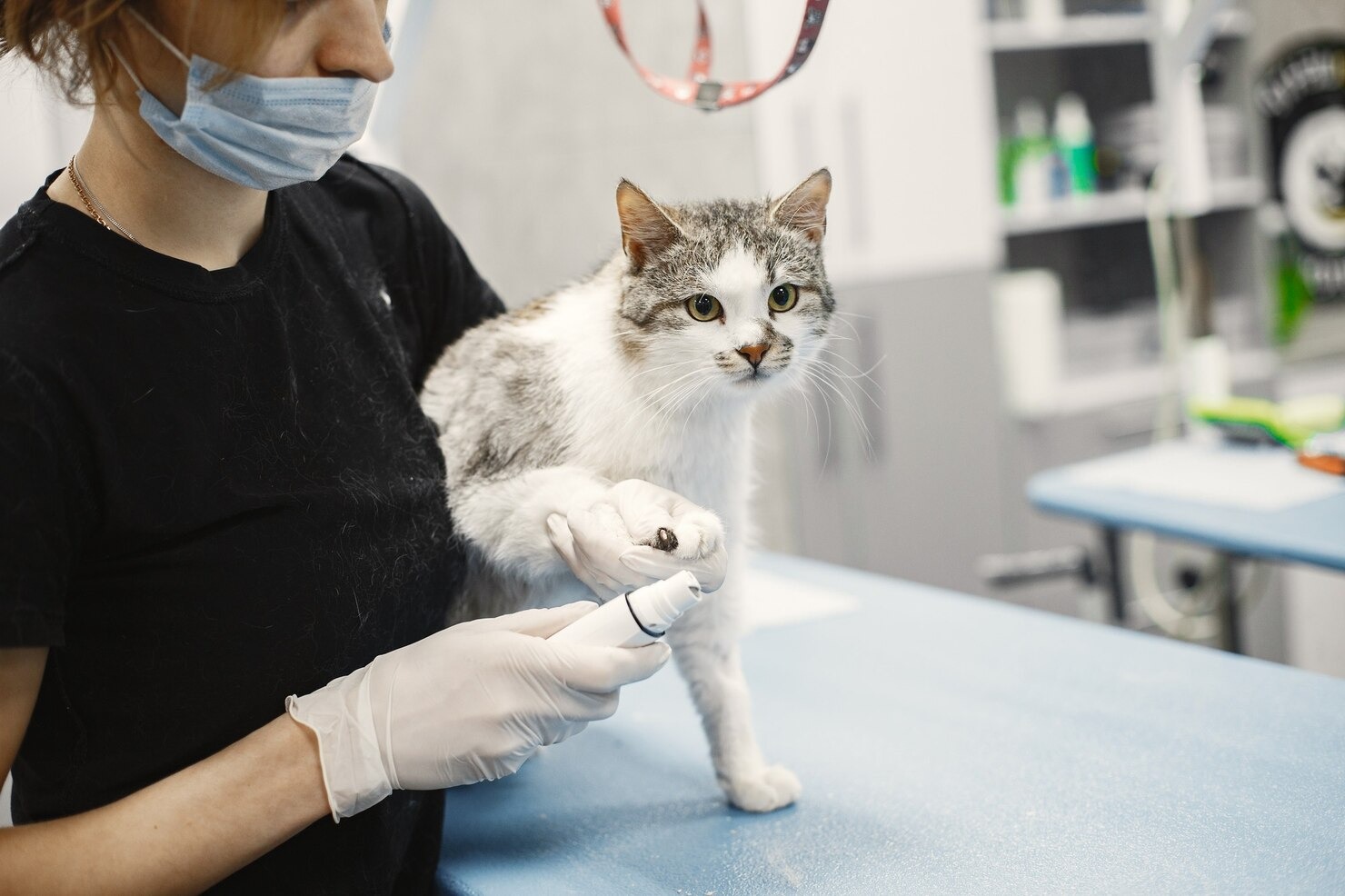
What are Gastrointestinal Parasites in Cats?
This content was prepared with AI assistance and reviewed by a licensed professional for accuracy.
Introduction
When pet owners notice their feline companions losing weight, developing a dull coat, or experiencing chronic digestive upset, the underlying culprit is often microscopic. Gastrointestinal parasites in cats are a pervasive veterinary issue, impacting millions of felines worldwide across all breeds, lifestyles, and life stages. While it is a common misconception that an indoor-only lifestyle guarantees safety from parasitic threats, these resilient organisms can easily infiltrate the home via potting soil, insects, rodents, or even on the soles of human shoes. Understanding the wide array of intestinal worms found in cats, alongside protozoan invaders, is essential for maintaining optimal feline wellness and safeguarding public health.[1]
Gastrointestinal parasites encompass two primary categories: helminths (parasitic worms such as nematodes and cestodes) and protozoa (single-celled microscopic organisms). Regardless of their biological classification, these parasites establish residence within the feline gastrointestinal tract—typically the stomach, small intestine, or colon—where they siphon essential nutrients, damage the mucosal lining, and disrupt normal digestion. The clinical consequences of an infestation can range from asymptomatic shedding to severe, life-threatening malnutrition, anemia, and profound dehydration. While kittens are statistically more vulnerable due to their immature immune systems and high nutritional demands relative to their body weight, older felines and adult cats remain highly susceptible to infection, especially if they experience immunocompromise or environmental stress.[2]
Contrary to outdated assumptions, many feline gastrointestinal parasites pose a significant zoonotic risk, meaning they can be transmitted from animals to humans. The shedding of microscopic eggs or cysts in a cat’s feces contaminates the environment, creating a vector for human infection, particularly in young children or immunocompromised individuals. Conditions such as visceral larva migrans, cutaneous larva migrans, and human toxoplasmosis trace their origins directly to environmental contamination by infected felines.[3] Furthermore, while parasitic infections typically manifest as localized gastrointestinal distress, severe migrations or systemic protozoal infections can cross the blood-brain barrier, leading to profound neurological emergencies, including blindness, ataxia, and severe seizures. Recognizing the signs, understanding the life cycles of these parasites, and implementing rigorous preventive strategies are non-negotiable responsibilities for modern pet ownership.
Types of Gastrointestinal Parasites in Cats

Felines act as definitive or intermediate hosts to a remarkably diverse array of parasite species. Understanding the unique biological behaviors, transmission routes, and clinical manifestations of each specific organism is critical for effective veterinary intervention and environmental management. The following sections detail the most prevalent and clinically significant gastrointestinal parasites affecting domestic cats.
Roundworms
Roundworms, scientifically classified as ascarids, are arguably the most ubiquitous intestinal nematodes diagnosed in feline medicine. The two primary species affecting domestic cats are Toxocara cati and Toxascaris leonina. Visually, adult roundworms are large, robust, and cylindrical, closely resembling cooked spaghetti, and can reach lengths of up to four to four-and-a-half inches within the feline small intestine. These parasites do not attach to the intestinal wall; instead, they swim freely within the lumen, competing directly with the host for ingested nutrients, particularly amino acids and carbohydrates.[4]
The life cycle of Toxocara cati is exceptionally complex and highlights the parasite’s evolutionary success. Cats can become infected through multiple pathways: ingesting embryonated eggs from contaminated soil, consuming infected paratenic hosts (such as mice, birds, or beetles), or, most commonly in kittens, via transmammary transmission. When a queen is infected, dormant roundworm larvae encysted in her somatic tissues become reactivated during the late stages of pregnancy and lactation, passing directly into the kittens through her milk.[5] Once inside the feline host, the larvae undergo a dramatic migration, burrowing through the intestinal wall, traveling to the liver, and eventually reaching the lungs. Here, they are coughed up into the trachea and swallowed back down into the digestive tract to mature into reproductive adults.[4]
Clinical signs of a roundworm burden depend heavily on the age of the cat and the severity of the infestation. Infected kittens classically present with a distended, “pot-bellied” appearance, accompanied by poor growth, a dull and unkempt hair coat, vomiting, and chronic diarrhea. In extreme cases, a massive adult worm burden can physically occlude the narrow intestinal lumen of a kitten, resulting in a fatal bowel obstruction or intestinal rupture. From a public health perspective, Toxocara cati is highly zoonotic. Humans accidentally ingesting the microscopic eggs can develop visceral larva migrans or ocular larva migrans, conditions where the confused larvae migrate through human organs or the retina, causing severe inflammation and tissue damage.[3] Treatment involves the administration of specific anthelmintics (such as pyrantel pamoate or milbemycin oxime) tailored to paralyze and expel the adult worms, though repeated dosing is mandatory to eliminate migrating larvae as they return to the gut.
Hookworms
Hookworms are voracious, blood-feeding nematodes that pose a severe threat to feline health. The predominant species affecting cats is Ancylostoma tubaeforme, though Ancylostoma braziliense is also endemic in certain warm, humid climates. Unlike the large, free-swimming roundworms, hookworms are exceedingly small—often less than an inch in length—and equipped with specialized, hook-like mouthparts containing sharp cutting plates. They use these mouthparts to anchor themselves deeply into the mucosal lining of the cat’s small intestine, where they lacerate capillaries and secrete powerful anticoagulants to feed continuously on the host’s blood.[6]
Transmission of hookworms is highly efficient. Cats can ingest infective larvae present in contaminated environments or consume infected prey animals. However, hookworm larvae also possess the remarkable ability to actively penetrate intact mammalian skin. A cat simply walking across contaminated, damp soil can acquire an infection as the microscopic larvae burrow through the paw pads, enter the bloodstream, and migrate to the lungs before eventually being swallowed and reaching the intestines.[7]
Because they are active blood-feeders, the primary clinical consequence of a hookworm infestation is profound, often life-threatening anemia. As the worms feed and continuously detach to find new biting sites, the old wounds continue to bleed due to the secreted anticoagulants. Infected cats may exhibit severe lethargy, pale or white mucous membranes, weakness, and dark, tarry stools (melena) caused by digested blood passing through the gastrointestinal tract. In young kittens, the rapid blood loss can lead to acute hypovolemic shock and sudden death before the worms are even old enough to lay eggs.[6] Furthermore, hookworms present a notable zoonotic hazard; infective larvae in the soil can penetrate human skin, causing a highly pruritic (itchy) dermatological condition known as cutaneous larva migrans, or “creeping eruption.” Aggressive deworming protocols, alongside intensive supportive care such as blood transfusions and iron supplementation, are often required to save severely affected felines.[8]
Tapeworms
Tapeworms, or cestodes, are segmented flatworms that colonize the feline small intestine. The most frequently diagnosed species in domestic cats are Dipylidium caninum (the flea tapeworm) and Taenia taeniaeformis (the feline rodent tapeworm). Tapeworms have a unique morphology; they possess a specialized head structure called a scolex, equipped with suckers and hooks to permanently anchor to the intestinal wall. The rest of the parasite’s body, known as the strobila, is composed of individual reproductive segments called proglottids. As the tapeworm matures, the terminal proglottids—each packed with thousands of eggs—detach and pass through the cat’s digestive tract.[9]
A defining characteristic of tapeworms is their obligate indirect life cycle, meaning they absolutely require an intermediate host to complete their development; a cat cannot become infected simply by ingesting a tapeworm egg from the ground. For Dipylidium caninum, the intermediate host is the common flea. A cat grooming itself will inevitably ingest an infected adult flea, releasing the tapeworm cysticercoid into the cat’s digestive tract. For Taenia species, rodents serve as the intermediate host. Outdoor cats or adept indoor hunters acquire the parasite by consuming an infected mouse or rat.[10]
Clinical signs of a tapeworm burden are often mild compared to nematodes, typically limited to failure to gain weight, an unkempt coat, or mild gastrointestinal upset. The most common presenting complaint from pet owners is the visual discovery of the proglottids. These segments, which resemble small grains of white rice or sesame seeds, are frequently observed actively crawling around the cat’s perianal region, clinging to the fur under the tail, or resting on the surface of freshly passed feces. This migration often causes intense perianal pruritus, leading the cat to aggressively lick its hindquarters or “scoot” across carpets. Treatment requires specific cestocidal medications, predominantly praziquantel, which dissolves the tapeworm in situ.[9] Crucially, tapeworm treatment will inevitably fail if the underlying vector—the flea infestation or the rodent access—is not simultaneously eradicated.
Giardia
Moving away from parasitic worms, Giardia duodenalis (formerly Giardia intestinalis or Giardia lamblia) is a microscopic, single-celled protozoan flagellate that commonly infects the feline gastrointestinal tract. Unlike helminths, Giardia does not grow into a visible organism; instead, it exists in two distinct morphological phases: the fragile, actively feeding trophozoite stage that resides in the cat’s gut, and the resilient, environmentally hardy cystic stage that is shed in the feces.[11]
Transmission occurs strictly via the fecal-oral route. Cats ingest the infective cysts from contaminated water sources, communal food bowls, shared litter boxes, or by grooming cysts off their fur. Once the cyst reaches the acidic environment of the cat’s stomach, it undergoes excystation, releasing two motile trophozoites into the small intestine. These trophozoites use a specialized ventral adhesive disc to attach firmly to the microvilli of the intestinal enterocytes. This attachment, combined with rapid binary fission, causes significant blunting of the microvilli, massive inflammation, and apoptosis (cell death) of the intestinal lining, ultimately resulting in profound malabsorption of water, electrolytes, and nutrients.[12]
The hallmark clinical sign of feline giardiasis is acute, chronic, or intermittent diarrhea. The feces are typically pale, highly malodorous, mucoid, and often present with a greasy appearance (steatorrhea) due to unabsorbed fats. Affected cats may also exhibit flatulence, weight loss, and generalized lethargy, though subclinical carrier states are also extremely common in adult felines. The zoonotic potential of feline Giardia is a subject of ongoing scientific debate; while cats primarily harbor species-specific assemblages (Assemblage F), they can occasionally be infected by and shed Assemblage A, which is infective to humans.[13] Therefore, meticulous hygiene is paramount. Eradicating Giardia is notoriously difficult due to the cyst’s resistance to standard disinfectants. Treatment protocols typically involve multi-day courses of fenbendazole, metronidazole, or a combination of both, alongside rigorous daily bathing of the cat’s hindquarters and thorough environmental sanitization using quaternary ammonium compounds.[11]
Coccidia
Coccidia are another group of microscopic, single-celled protozoan parasites, with Cystoisospora felis and Cystoisospora rivolta being the most prevalent species affecting domestic cats. Coccidiosis is fundamentally a disease of the young, the stressed, and the immunocompromised, frequently devastating litters of kittens in high-density environments such as animal shelters, catteries, and rescue organizations. These parasites are highly host-specific, meaning feline coccidia cannot infect dogs or humans, effectively eliminating any zoonotic risk.[14]
The pathogenesis of feline coccidiosis involves the ingestion of sporulated oocysts from contaminated environments. Once ingested, the sporozoites invade the epithelial cells lining the cat’s small and large intestines. Inside these cells, the parasite undergoes explosive asexual and sexual reproduction, leading to the ultimate destruction and rupture of the host cells to release new oocysts. This massive cellular destruction ulcerates the intestinal mucosal lining, leading to severe fluid loss, hemorrhage, and secondary bacterial infections.[15] Furthermore, freshly shed oocysts are not immediately infective; they require a brief period of maturation (sporulation) in the environment, which is highly dependent on ambient temperature and oxygen levels.
Clinical manifestations in vulnerable kittens are often acute and severe. Symptoms include profuse, watery, and sometimes bloody diarrhea, anorexia, vomiting, severe abdominal pain, and rapid, life-threatening dehydration. In stark contrast, healthy adult felines may harbor the organism and shed oocysts entirely asymptomatically, acting as silent reservoirs of infection. Managing coccidiosis requires specific anti-protozoal medications, generally sulfa-based antibiotics like sulfadimethoxine or off-label use of ponazuril, which inhibit parasite reproduction and allow the feline immune system to mount an effective response.[14] Because environmental oocysts can survive for many months and are highly resistant to standard household cleaners, strict sanitation utilizing high-heat steam cleaning or high-concentration ammonia solutions is critical to break the cycle of reinfection.
Toxoplasma
Toxoplasma gondii is an obligate intracellular protozoan parasite of immense global medical and veterinary significance. While it is capable of infecting virtually all warm-blooded vertebrates—including humans, livestock, and marine mammals—domestic and wild felids hold the unique biological distinction of being the parasite’s only definitive hosts. This means that the crucial sexual reproduction phase of Toxoplasma gondii, which results in the shedding of environmentally resilient oocysts, can only occur within the epithelial cells of the feline small intestine.[16]
Cats typically acquire a Toxoplasma infection by engaging in normal predatory behaviors, specifically consuming the raw tissue of infected intermediate hosts (like rodents or birds) that harbor dormant parasitic cysts, known as bradyzoites, within their muscle and brain tissue. Following ingestion, the parasite undergoes sexual replication in the cat’s gut, and the cat begins shedding millions of unsporulated oocysts in its feces. Crucially, this shedding period is remarkably brief, typically lasting only one to two weeks during the cat’s entire lifetime. Once in the environment, these oocysts sporulate over 24 to 48 hours, becoming highly infectious and capable of surviving in soil or water for over a year.[17]
Most cats mount a robust immune response to toxoplasmosis and show absolutely no clinical signs of illness. However, if the parasite spreads systemically beyond the gastrointestinal tract (a phase involving rapidly dividing tachyzoites), it can cause severe clinical disease affecting multiple organ systems. Symptoms of clinical feline toxoplasmosis include anterior uveitis (eye inflammation), persistent fever, interstitial pneumonia, hepatic necrosis, and severe neurological deficits. Treatment generally relies on long-term administration of clindamycin or azithromycin. From a public health standpoint, Toxoplasma gondii poses a profound risk to immunocompromised individuals and pregnant women, as primary human infection during gestation can result in catastrophic congenital defects or miscarriage.[18] Proper litter box hygiene, specifically removing feces daily before oocysts have time to sporulate, drastically mitigates this risk.
Lungworms
While their primary site of adult infection is the lower respiratory tract rather than the intestines, feline lungworms—predominantly Aelurostrongylus abstrusus—are frequently categorized and diagnosed alongside gastrointestinal parasites due to their unique migration pathways and diagnostic methods. These delicate, thread-like nematodes embed themselves deeply into the terminal bronchioles and alveolar ducts of the feline lungs, causing chronic inflammatory nodules.[19]
The life cycle is complex and heavily reliant on intermediate and paratenic hosts. Adult female worms in the lungs lay eggs that quickly hatch into first-stage larvae. These microscopic larvae are coughed up by the cat into the pharynx, swallowed, pass entirely through the gastrointestinal tract, and are excreted in the feces. In the environment, these larvae must be ingested by specific species of snails or slugs to continue their development. Cats acquire the infection either by consuming these terrestrial mollusks directly, or, more commonly, by eating paratenic hosts such as rodents, frogs, or birds that have recently fed on infected snails.[20]
Clinical signs of aedurostrongylosis are inherently respiratory. Cats typically present with a dry, chronic cough, tachypnea (rapid breathing), wheezing, and progressive dyspnea (difficulty breathing). In severe, acute infections, the massive inflammatory response in the lungs can mimic feline asthma or severe bacterial pneumonia, leading to cyanosis and respiratory failure. Diagnosis is uniquely reliant on identifying the larvae in the feces, rather than the respiratory secretions. Treatment utilizes specific anthelmintic formulations, often requiring extended or repeated courses of fenbendazole, or targeted topical applications of macrocyclic lactones like moxidectin. Strict restriction of hunting behavior is the only reliable method of preventing feline lungworm infection.[19]
Diagnosis of Feline Gastrointestinal Parasites

Accurate identification of gastrointestinal parasites requires a multifaceted diagnostic approach. Clinical presentation alone is rarely sufficient, as many parasites cause overlapping symptoms such as vomiting, diarrhea, and weight loss. Veterinarians rely on a battery of specific laboratory techniques to isolate and identify the eggs, cysts, larvae, or adult organisms responsible for the disease pathology.[21]
Fecal Flotation Analysis
The fecal flotation is the cornerstone of veterinary parasitology and the primary diagnostic modality for identifying the ova (eggs) of nematodes and cestodes, as well as the oocysts of coccidia. This technique exploits the principles of specific gravity. By mixing a standardized sample of feline feces with a specialized hyperosmolar flotation solution (typically zinc sulfate, sodium nitrate, or sucrose solutions with a specific gravity between 1.18 and 1.27), heavy fecal debris is forced to sink to the bottom of the test tube, while the lighter parasite eggs buoy upward.[22]
Modern veterinary practices predominantly utilize centrifugal fecal flotation, a method that spins the sample in a centrifuge to forcefully drive the eggs to the surface meniscus. A glass coverslip is placed on top of the tube, capturing the concentrated eggs, which is then meticulously examined under a microscope. This centrifugal method significantly increases diagnostic sensitivity compared to older, passive flotation techniques, ensuring that even low-burden infections of roundworms, hookworms, or whipworms are accurately detected.[21]
Fecal Smear Test
Also known as a direct wet mount, the direct fecal smear is a rapid, fundamental diagnostic test utilized primarily to identify delicate, motile organisms that would be destroyed or severely distorted by the high osmotic pressure of flotation solutions. A microscopic quantity of extremely fresh feline feces is blended with a drop of warm isotonic saline directly on a microscope slide.[23]
This technique is absolutely critical for the detection of active protozoan trophozoites, particularly Giardia species and Tritrichomonas foetus, which exhibit distinctive “falling leaf” or erratic, jerky swimming motions under high magnification. However, the direct smear has significant limitations; because only an infinitesimal amount of feces is examined, the test suffers from very low sensitivity. A negative smear does not definitively rule out a parasitic infection, and this test is almost always performed in conjunction with more comprehensive concentration methods.
Baermann Technique
Standard fecal flotation solutions are ineffective for diagnosing feline lungworms, as the high specific gravity forces the heavy, dense Aelurostrongylus abstrusus larvae to sink rather than float. To identify these specific parasites, veterinarians employ the Baermann technique, a specialized diagnostic procedure designed to isolate live, motile nematode larvae from fecal matter.[24]
The test involves suspending a significant volume of feces wrapped in cheesecloth or gauze within a glass funnel filled with warm water. The warmth stimulates the live larvae, causing them to actively migrate out of the fecal mass. Unable to swim against gravity, the larvae sink to the bottom of the funnel stem over a period of 12 to 24 hours. The concentrated fluid at the very tip of the funnel is then collected and examined microscopically to identify the characteristic morphological features of the lungworm larvae.[24]
Direct Observation
While laboratory analysis provides definitive diagnosis, many parasitic infections are initially identified through the direct, gross observation of the macroscopic adult organisms by the pet owner or the veterinary team. Tapeworms are the most common example of this; the motile, rice-like proglottids are frequently visualized contracting on the cat’s perineal region or within freshly voided stool. Additionally, felines suffering from a severe ascarid burden may actively pass adult roundworms within the cat’s vomit, presenting a startling but diagnostically conclusive finding. Any expelled parasite segments or whole worms should be safely collected in a sealed container and brought to the veterinary clinic for formal identification, as different worm species require distinctly different pharmacological interventions.[21]
Blood Testing
Though gastrointestinal parasites reside primarily in the gut, their systemic impact often leaves distinct markers within the feline bloodstream. A Complete Blood Count (CBC) frequently reveals a marked eosinophilia—an elevation in a specific type of white blood cell explicitly tasked with combating multicellular parasitic invaders. While non-specific, high eosinophil counts serve as a massive red flag prompting deeper parasitological investigation. Furthermore, severe hookworm burdens will manifest on a CBC as profound microcytic, hypochromic anemia due to chronic blood loss.[25]
Advanced serological blood tests, such as Enzyme-Linked Immunosorbent Assays (ELISA) or Polymerase Chain Reaction (PCR) panels, are critical for identifying elusive protozoa or systemic parasites. For example, testing for Toxoplasma-specific Immunoglobulin G (IgG) and Immunoglobulin M (IgM) antibodies helps determine if a cat has an active, recent infection versus historical exposure. Blood testing is also the primary diagnostic tool for identifying unrelated but concurrent parasitic infections that can diseases or affect the heart, such as Dirofilaria immitis (heartworm disease), which requires specific antigen and antibody screening.[26]
Prevention of Gastrointestinal Parasites in Cats

Given the ubiquitous nature of parasitic eggs in the environment and the diverse transmission pathways, true eradication is virtually impossible. Instead, veterinary medicine focuses on proactive, multi-tiered preventive strategies designed to minimize exposure, interrupt the parasite life cycle, and prevent the establishment of heavy, pathogenic burdens. Effective prevention requires unwavering commitment from the pet owner and a close partnership with veterinary professionals.
- Rigorous Environmental Hygiene: The absolute most effective method of preventing protozoan infections, particularly toxoplasmosis and giardiasis, is meticulous litter box management. Feces must be removed daily to prevent Toxoplasma oocysts from sporulating and becoming infectious to humans and other pets. Litter boxes should be periodically emptied completely, scrubbed with hot, soapy water, and thoroughly dried.[20]
- Strict Flea Control: Because fleas serve as the mandatory intermediate host for Dipylidium caninum, tapeworm prevention is synonymous with aggressive, year-round flea control. Every animal in the household must be maintained on a highly efficacious adulticide to break the flea life cycle and prevent the accidental ingestion of infected vectors.
- Dietary Management: Cats should be fed commercial, high-quality, balanced diets. Raw meat diets, particularly those incorporating undercooked pork, lamb, or wild game, pose a catastrophic risk for transmitting Toxoplasma gondii, Taenia tapeworms, and various bacterial pathogens. If hunting behavior cannot be curtailed, the risk of acquiring roundworms, hookworms, and lungworms remains persistently high.
- Routine Veterinary Surveillance: The Companion Animal Parasite Council (CAPC) mandates that all adult cats receive a minimum of one to two centrifugal fecal flotations annually, regardless of their indoor/outdoor status. Regular screening allows veterinarians to identify and treat asymptomatic shedders before environmental contamination becomes severe.
Preventive Medications Needed After Treatment
Treating an active parasitic infection is only the first phase of clinical management. Because anthelmintic medications possess little to no residual activity, a cat can easily become reinfected the very next day if exposed to contaminated environments. To combat this, veterinary pharmacology relies on the strategic, routine administration of broad-spectrum preventive medications. It is imperative to consult your veterinarian before making any changes to your pet’s care, as inappropriate use of dewormers can lead to toxicosis or parasite resistance.
- Macrocyclic Lactones: Topical and oral formulations containing drugs like moxidectin, selamectin, or milbemycin oxime represent the gold standard in feline parasite prevention. Applied or administered monthly, these medications achieve systemic distribution, providing highly effective prophylaxis against heartworms while simultaneously treating and controlling intestinal hookworms and roundworms.[27]
- Targeted Cestocides: For cats with persistent hunting habits or chronic flea exposure, routine prophylactic administration of praziquantel or epsiprantel may be required to continually purge the intestines of developing tapeworms before they can shed proglottids.
- Isoxazolines and Integrated Preventives: Modern veterinary medicine increasingly utilizes combination products that merge systemic flea and tick control (e.g., fluralaner, sarolaner) with broad-spectrum anthelmintics. These all-in-one monthly preventives dramatically increase owner compliance and ensure the cat is continuously protected from both the intestinal parasites and the ectoparasitic vectors that transmit them.[28]
How Often to Deworm a Cat After Treatment?
The ideal deworming schedule is not uniform; it is a dynamic protocol highly individualized to the specific cat’s geographic location, age, and lifestyle risk factors. Following the resolution of an active infection, a strategic, ongoing deworming calendar must be established to prevent disease recurrence.
For neonatal and pediatric felines, the risk of roundworm and hookworm infection is astronomically high due to transmammary transmission and immature immunity. The Companion Animal Parasite Council strongly recommends initiating prophylactic deworming in kittens at two weeks of age. This treatment should be repeated every two weeks until the kitten is eight weeks old, followed by monthly preventive administration. This aggressive early protocol ensures that migrating larvae are neutralized as soon as they mature and enter the gastrointestinal tract, preventing the shedding of eggs into the environment.[29]
For adult cats, the schedule hinges entirely on lifestyle risk assessment. Strictly indoor felines housed in single-pet homes without access to rodents or fleas may only require prophylactic, broad-spectrum deworming once or twice a year, accompanied by their annual fecal analysis. Conversely, outdoor felines, known hunters, or cats residing in multi-pet households where other animals venture outside demand a much more aggressive approach. These high-risk individuals should receive, at minimum, quarterly (four times a year) broad-spectrum deworming, or ideally, be maintained on a year-round, monthly comprehensive parasite preventive medication to ensure continuous, unbroken protection.[30]
Frequently Asked Questions
Can my strictly indoor cat still get gastrointestinal parasites?
Yes, indoor cats are completely capable of acquiring gastrointestinal parasites. Parasite eggs can be tracked into the home on human shoes, clothing, or equipment. Additionally, indoor cats can ingest infected fleas that hitch a ride inside, or consume indoor pests like mice and houseflies that act as intermediate or paratenic hosts for tapeworms and roundworms.
Are feline intestinal worms contagious to humans?
Many feline intestinal parasites are highly zoonotic, meaning they pose a significant health risk to humans. Roundworm eggs accidentally ingested from contaminated environments can cause visceral or ocular larva migrans in people. Hookworm larvae in soil can penetrate human skin, causing cutaneous larva migrans. Toxoplasma gondii also presents severe risks, particularly to pregnant women and immunocompromised individuals.
How quickly do deworming medications work in cats?
Most modern veterinary deworming medications begin paralyzing and killing adult intestinal parasites within 24 to 48 hours of administration. You may physically see dead worms passed in the cat’s feces during this time. However, because most medications only target the adult stages in the gut, a second or third dose is often required weeks later to kill maturing larvae that were migrating through tissues during the initial treatment.
References
- Companion Animal Parasite Council. General Guidelines for Parasite Control. CAPC, 2023.
- Merck Veterinary Manual. Gastrointestinal Parasites of Dogs and Cats. Merck & Co., 2022.
- Centers for Disease Control and Prevention. Toxocariasis (Roundworm Infection). CDC, 2020.
- Companion Animal Parasite Council. Ascarid Guidelines. CAPC, 2023.
- Lee, A. C., et al. “Clinical manifestations and treatment of Toxocara cati.” Journal of Feline Medicine and Surgery. SAGE, 2010.
- Companion Animal Parasite Council. Hookworm Guidelines. CAPC, 2023.
- Centers for Disease Control and Prevention. Zoonotic Hookworm. CDC, 2021.
- Merck Veterinary Manual. Hookworms in Small Animals. Merck & Co., 2022.
- Companion Animal Parasite Council. Tapeworms (Dipylidium caninum). CAPC, 2023.
- Cornell Feline Health Center. Gastrointestinal Parasites of Cats. Cornell University, 2021.
- Companion Animal Parasite Council. Giardia Guidelines. CAPC, 2023.
- Merck Veterinary Manual. Giardiasis in Dogs and Cats. Merck & Co., 2022.
- Centers for Disease Control and Prevention. Giardia Information. CDC, 2021.
- Companion Animal Parasite Council. Coccidia Guidelines. CAPC, 2023.
- Merck Veterinary Manual. Coccidiosis of Cats and Dogs. Merck & Co., 2022.
- Companion Animal Parasite Council. Toxoplasma gondii Guidelines. CAPC, 2023.
- Centers for Disease Control and Prevention. Toxoplasmosis FAQs. CDC, 2020.
- World Health Organization. Toxoplasmosis Information. WHO, 2022.
- Companion Animal Parasite Council. Feline Lungworm (Aelurostrongylus abstrusus). CAPC, 2023.
- Merck Veterinary Manual. Lungworm Infection in Dogs and Cats. Merck & Co., 2022.
- American Veterinary Medical Association. Internal Parasites in Cats and Dogs. AVMA, 2023.
- Companion Animal Parasite Council. Fecal Flotation Procedures. CAPC, 2023.
- Merck Veterinary Manual. Fecal Examination Procedures. Merck & Co., 2022.
- Companion Animal Parasite Council. The Baermann Technique. CAPC, 2023.
- Glaub, M., et al. “Feline Eosinophilia and Parasitism.” Veterinary Clinics of North America: Small Animal Practice. Elsevier, 2011.
- American Heartworm Society. Feline Heartworm Guidelines. AHS, 2020.
- Merck Veterinary Manual. Macrocyclic Lactones. Merck & Co., 2022.
- VCA Hospitals. Parasite Prevention for Cats. VCA, 2022.
- Cornell Feline Health Center. Kitten Care and Deworming. Cornell University, 2021.
- Companion Animal Parasite Council. Strategic Parasite Prevention Strategies. CAPC, 2023.


