Corneal Ulcers in Dogs: Causes, Symptoms, Treatment
What is it?
How is it Treated?
Breed Predispositions
Pekingese Shih Tzu Bulldog Boston Terrier Pug
Introduction
For as long as she could remember, Sarah and her faithful Border Collie, Rex, had been inseparable. Recently, though, she noticed that Rex was constantly rubbing his eye and seemed more sensitive to light than usual. Concerned about her beloved pet’s well-being, Sarah took Rex to the veterinarian for a thorough assessment. After a careful examination, the vet revealed that Rex was suffering from a corneal ulcer, a painful and potentially serious eye condition in dogs.
Corneal ulcer in dogs indicate where the cornea’s outermost layer, a transparent, safeguarding layer enveloping the iris, pupil, and anterior chamber of the eye, undergoes damage or erosion. This can result in considerable pain and may lead to vision loss if not treated promptly. These wounds, sometimes called superficial ulcers, particularly affect the eye’s surface. Due to their nature, these ulcers can be persistent, earning them the term ‘indolent ulcers.’
Given the potential severity of this condition, it is paramount to seek veterinary care if you suspect your dog may have a corneal ulcer. Timely and appropriate treatment can help avert further complications and maintain your dog’s sight.
What Causes Corneal Ulcers in Canine?
Different elements can trigger the formation of corneal ulcers in dogs, with factors falling into categories such as traumatic incidents, infections, or other underlying conditions. These ulcers are a substantial cause of discomfort in pets, representing nearly 0.80% of diagnosed conditions in primary care practices in the UK. Some prevalent sources of these corneal erosions include:
- Physical Trauma: Damages to the dog’s eye can lead to the development of superficial corneal ulcers such as:
- scratches
- punctures,
- foreign bodies like grass seeds, sand, or debris

- Infections: Bacterial or viral incursions can foster corneal ulcers. Occasionally, an existing ulcer can succumb to secondary infection.
- Insufficient Tear Production (Keratoconjunctivitis Sicca): When tear production falls short, it leads to a dry cornea that, due to the absence of natural lubrication and shielding, can result in corneal ulcers.
- Eyelid Conformation Abnormalities (Entropion or Ectropion): An eyelid that folds inward (entropion) or outward (ectropion) can provoke irritation and harm the cornea, triggering ulcers.
- Exposure Keratitis: In dogs with prominent eyes or nerve damage, the cornea’s prolonged exposure to air can incite corneal ulcers.
- Autoimmune Disorders: Some autoimmune conditions can spur inflammation and damage to the cornea, inducing corneal ulcers.
- Corneal Dystrophy or Degeneration: Dogs can inherit a predisposition to corneal disorders, making them more susceptible to ulcers. These predispositions can often lead to what’s known as indolent corneal ulcers or spontaneous chronic corneal epithelial defects.
Understanding the fundamental cause of a corneal ulcer is critical to devising an effective treatment strategy and avoiding a recurrence. In addition, quick access to veterinary care is crucial for diagnosing and treating corneal ulcers in dogs, as it dramatically aids corneal healing and helps to prevent severe complications like corneal perforation, which occurs when an ulcer penetrates the cornea’s deeper layer, the stroma, leading to refractory corneal ulcers.
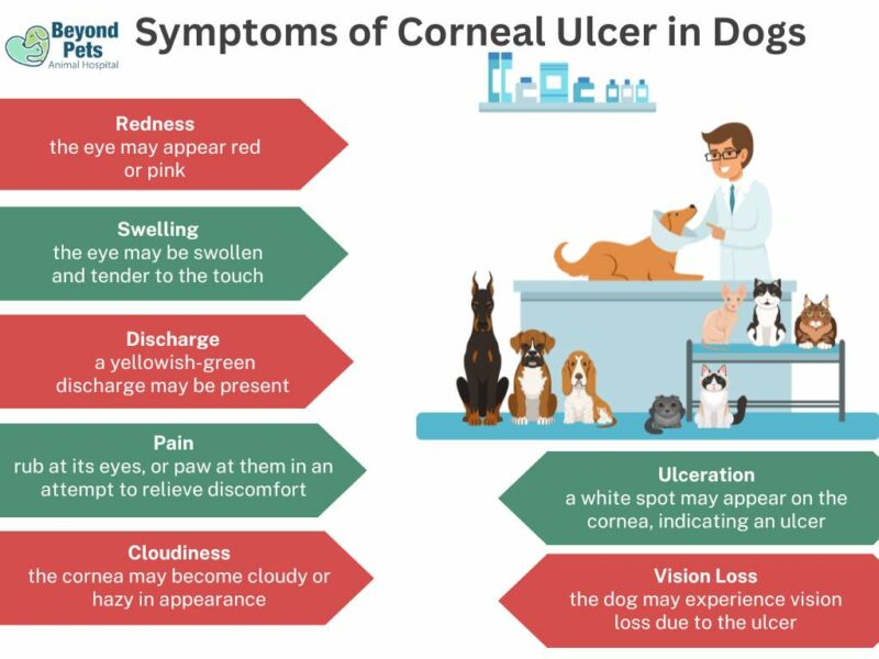
Symptoms of Canine Corneal Ulceration
The indicators of corneal ulcers in dogs might differ depending on the depth of the ulcer and its cause. The corneal surface might show various signs of discomfort and distress in your canine friend, including:
Diagnosis of Corneal Ulcer in Dogs
Corneal ulcers in dogs are a prevalent, intense ocular condition requiring precise diagnosis and immediate intervention. Several diagnostic methods are employed in veterinary medicine, each offering distinct benefits and features.
Fluorescein Staining
This is the principal method for detecting corneal ulcers. First, a harmless fluorescent dye is applied to the corneal surface. This dye sticks to any region where the cornea’s outer layer or epithelium is damaged or missing, such as in a melting ulcer, causing the affected area to glow bright green under blue light. This enables the vet to identify the size and position of the ulcer.
Physical Examination
The vet may conduct an exhaustive physical assessment, including a meticulous eye examination for indications of injury, ectopic cilia, or visible irregularities in the cornea.
Slit Lamp Biomicroscopy
This device permits the vet to scrutinize the cornea under magnification. It provides a more detailed perspective of the cornea, facilitating an evaluation of the ulcer depth, the detection of foreign bodies, and an assessment of the overall ocular health.
Tonometry
This test measures the pressure inside the eye. Since corneal ulcers can precipitate secondary conditions like glaucoma (increased eye pressure), tonometry is a vital component of the diagnostic process.
Schirmer Tear Test
Should the vet suspect that dry eye (keratoconjunctivitis sicca) could contribute to the corneal ulcer, they may execute a Schirmer tear test, which quantifies the eye’s tear production.
Cytology
If an ulcer does not show signs of routine ulcer healing or if there are worries about a secondary bacterial or fungal infection, the vet may collect cells from the eye for microscopic evaluation.
Biopsy
In rare cases, mainly if there’s concern about corneal cancer, the vet might extract a small sample of corneal tissue for biopsy.
Advanced Imaging
Imaging examinations such as ultrasound or a CT scan can occasionally be used to better view the eye and surrounding structures, especially in complicated corneal ulcers.
The chosen methods depend on the ulcer’s characteristics, the dog’s health status, and the available veterinary equipment. However, it’s always crucial to secure an accurate diagnosis to implement the correct treatment plan, which could even involve an eyelid flap in certain severe cases.
Treatment of Corneal Ulcers in Dogs
The treatment strategies for canine corneal ulcers are contingent on the ulcer’s severity, cause, and nature. A veterinarian will devise a fitting treatment protocol following an exhaustive examination and diagnosis. Several standard treatment options encompass:

Medical Intervention
Primarily, corneal ulcers are medically managed to facilitate healing, thwart infection, and provide pain relief.
- Antibiotics: Topical antibiotic eye drops are employed to forestall or manage secondary bacterial infections. Popular options encompass aminoglycosides (gentamicin, tobramycin), fluoroquinolones (ciprofloxacin, ofloxacin), and polymyxin B combinations.
- Antivirals: For ulcers provoked by viruses, particularly those induced by the herpes virus, antiviral eye medications like idoxuridine or trifluridine might be utilized.
- Antifungals: In the event of a suspected or confirmed fungal infection, antifungal eye drops such as natamycin or voriconazole are essential.
- Cycloplegics: Drugs like atropine can soothe eye discomfort by paralyzing specific eye muscles to diminish spasms.
- Pain Relievers: Intense discomfort may be managed with systemically and topically administered non-steroidal anti-inflammatory drugs (NSAIDs).
- Collagenase Inhibitors: In ‘melting’ ulcers where the cornea’s collagen matrix is swiftly deteriorating, medications such as acetylcysteine are used to impede this process.
- Autologous Serum: Eye drops derived from the dog’s serum can be utilized for stubborn or non-healing ulcers due to their innate healing properties.
- Lubricants: Lubricants like artificial tears or lubricant ointments aid in maintaining a moist environment for the cornea, thereby promoting healing and comfort.
Surgical Intervention
Surgical intervention may be necessary for certain circumstances, such as when medical management proves ineffective, the ulcer is deep or perforated, or when there’s a ‘descemetocele’ – a severe form of ulcer on the verge of perforation.
- Keratectomy: To foster healing, a superficial keratectomy may remove loose, unhealthy corneal tissue.
- Conjunctival Pedicle Graft: This procedure involves suturing a portion of the conjunctiva (the pink tissue overlaying the white of the eye) over the ulcer to provide additional support and blood supply.
- Corneal Transplant: A corneal transplant may be considered in severe cases, though this is less frequently performed in dogs within veterinary ophthalmology.
- Cyanoacrylate Glue and Contact Lens: For impending or small perforations, veterinary ophthalmologists may apply medical-grade glue and a soft contact lens to seal the area and facilitate healing.
All treatments should be conducted under the supervision of a veterinarian, and owners should meticulously follow the prescribed treatment plan. The administration of eye medication, especially in cases of an infected corneal ulcer, is paramount. Corneal surgery may be an unavoidable course of action in the context of deep ulcers.
How Can I Prevent My Dog From Developing Corneal Ulcers?
Preventing corneal ulcers in dogs involves taking several steps to protect and maintain the overall health of your dog’s eyes. Here are some tips to help prevent corneal ulcers in dogs:
- Regular veterinary check-ups: Schedule routine veterinary visits for your dog, including eye exams. This will allow your veterinarian to detect any early signs of eye problems and address them before they progress into more severe conditions.
- Maintain proper eye hygiene: Clean your dog’s eyes by gently wiping away any discharge or debris with a damp cloth or a pet-safe eye wipe. Use a separate cloth for each eye to avoid spreading infections.
Grooming and hair management: Keep the hair around your dog’s eyes trimmed to prevent irritation and reduce the risk of foreign bodies entering the eye. Regular grooming also helps avoid excessive tearing and the development of infections. - Eye protection: If your dog is prone to eye injuries or spends much time outdoors, consider using protective eyewear, such as dog goggles, to shield their eyes from foreign objects, debris, and UV radiation.
- Be cautious with chemicals and irritants: Protect your dog from household cleaning products, pesticides, and other chemicals that may cause eye irritation or injury. If your dog comes into contact with an irritant, flush their eyes with clean water and consult your veterinarian.
- Monitor playtime: Supervise your dog during playtime, especially with other dogs or in areas with sharp objects or debris that could injure their eyes.
- Address underlying health issues: If your dog has a health condition that could contribute to corneal ulcers, such as dry eye, eyelid abnormalities, or allergies, work closely with your veterinarian to manage these conditions and minimize the risk of corneal ulcers.
Following these preventive measures and being vigilant about your dog’s eye health can significantly reduce the risk of corneal ulcers and help maintain your dog’s vision and overall well-being.
Frequently Asked Questions
Disclaimer: The information provided on this veterinary website is intended for general educational purposes only and should not be considered as a substitute for professional veterinary advice, diagnosis, or treatment. Always consult a licensed veterinarian for any concerns or questions regarding the health and well-being of your pet. This website does not claim to cover every possible situation or provide exhaustive knowledge on the subjects presented. The owners and contributors of this website are not responsible for any harm or loss that may result from the use or misuse of the information provided herein.

