


 March 1, 2023
 Phil Good, DVM
Understanding Brachycephalic Syndrome in Dogs
This content was prepared with AI assistance and reviewed by a licensed professional for accuracy.
Introduction
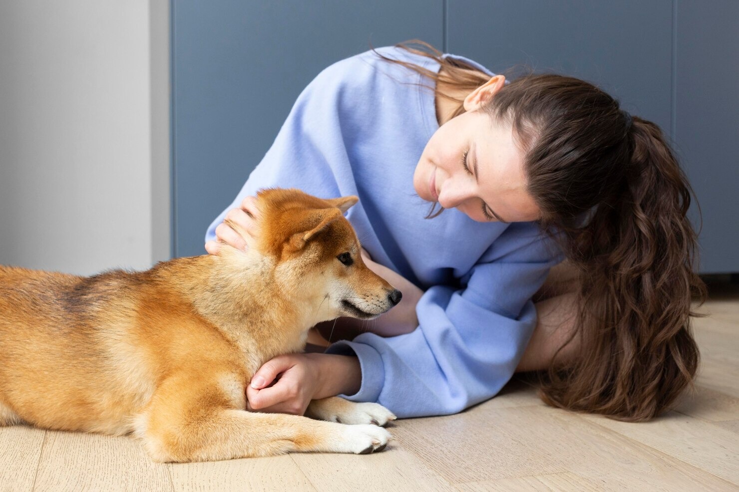
When you look at the beloved faces of Pugs, French Bulldogs, English Bulldogs, and Boston Terriers, it is easy to understand why these distinctive breeds have captured the hearts of millions of pet owners around the world. However, beneath their charming, pushed-in faces and expressive eyes lies a complex anatomical reality. The focus of this reality is Brachycephalic Syndrome, a comprehensive medical classification for the significant breathing difficulties that these dogs frequently endure. Because of their uniquely squished facial structure and shallow cranial cavity, it is routinely difficult for the dog to breathe, particularly during exercise, stress, or exposure to warm weather. This intricate condition, which veterinarians often refer to as Brachycephalic Obstructive Airway Syndrome (BOAS), is a lifelong health concern that requires diligent management, veterinary oversight, and a deep understanding from pet owners.[1]
To truly grasp the impact of this condition, we must first look at the terminology. The word “brachycephalic” is derived from Greek roots, where “brachy” means short and “cephalic” refers to the head. Dogs belonging to this category have been intentionally bred over generations to possess a noticeably shortened upper jaw and snout, all while retaining a relatively normal-sized lower jaw. This severe alteration in skull shape directly results in a compressed respiratory tract. Unlike dogs with long snouts, such as Greyhounds or German Shepherds, a brachycephalic dog must force the same volume of inhaled air through a significantly narrower and more convoluted space.[2] This physical limitation is the hallmark of Brachycephalic Syndrome, which is an unfortunate condition common in dogs that have been bred specifically for flat faces.
The syndrome itself is not simply a single physical defect; rather, it is a combination of multiple upper airway abnormalities that compound upon one another. The primary components of this syndrome typically include narrowed nostrils, an excessively long soft palate, and a dangerously narrow windpipe. When these anatomical flaws occur together, they create an immense amount of resistance within the dog’s airway. Just as trying to breathe through a tiny cocktail straw requires significantly more effort than breathing through a wide tube, a dog suffering from Brachycephalic Syndrome must exert tremendous muscular effort just to pull a normal breath into their lungs.[3] Over time, this constant, forceful inhalation causes profound secondary changes to the tissues of the throat and larynx, worsening the breathing difficulties as the dog ages.
For decades, many pet owners mistakenly believed that the loud snoring, snorting, and heavy panting exhibited by these breeds were simply “normal” or “cute” breed quirks. Today, the veterinary community emphasizes that these sounds are actually clinical indicators of respiratory distress. A dog that snores loudly while awake is not merely demonstrating a funny breed trait; they are actively fighting against a compromised airway to deliver life-sustaining oxygen to their brain and body.[4] Because of the systemic nature of this oxygen deprivation, Brachycephalic Syndrome impacts almost every aspect of a dog’s life, from their ability to play and regulate their body temperature to their sleeping patterns and digestive health.
Managing Brachycephalic Syndrome requires a multifaceted approach that includes lifestyle modifications, environmental control, and, in many cases, corrective surgical intervention. By gaining a comprehensive understanding of the anatomy, pathophysiology, symptoms, and modern treatment options associated with this syndrome, pet owners can take proactive steps to dramatically improve their brachycephalic dog’s quality of life. The goal of veterinary medicine is not to simply manage the inevitable decline of these dogs, but to intervene appropriately so that flat-faced breeds can lead comfortable, active, and fulfilling lives without the constant burden of respiratory struggle.[5]
Causes of Brachycephalic Syndrome in Dogs

The root causes of Brachycephalic Syndrome are inherently tied to the history of canine domestication, selective breeding practices, and genetics. To fully understand why these dogs suffer from such severe airway obstruction, it is necessary to examine how human preference has physically shaped the canine skull over the last two centuries. Unlike many diseases that are caused by environmental pathogens, viruses, or sudden trauma, Brachycephalic Syndrome is an entirely man-made condition, driven by the desire for specific aesthetic traits.[6]
The deliberate breeding for neotenous, or “baby-like,” features has been the primary driving force behind the development of brachycephalic breeds. Humans are naturally drawn to large, forward-facing eyes, flat faces, and rounded heads, as these features trigger a caregiving response. Over generations, breeders selectively mated dogs that displayed these exaggerated physical characteristics. As a result, the facial bones of these dogs became progressively shorter. However, while the skeletal structure of the skull was dramatically compressed, the soft tissues of the face and respiratory tract were not genetically downsized at the same rate.[7] This mismatch between a shortened bony framework and an excessive volume of soft tissue is the fundamental architectural flaw of the brachycephalic airway.
Recent advancements in canine genomics have allowed researchers to pinpoint specific genetic mutations responsible for this severe facial flattening. One notable discovery is the mutation in the SMOC2 gene, which heavily influences facial development in the embryo. Dogs carrying specific variations of this gene exhibit extreme shortening of the muzzle.[8] Because these genetic traits are recessive or intentionally fixed within the breed populations, every purebred English Bulldog, French Bulldog, or Pug is essentially guaranteed to inherit the anatomical blueprint for a compromised respiratory system. Genetics dictate that the nasal cavity is severely truncated, forcing the intricate network of internal nasal structures into a space that is far too small to accommodate them.
Furthermore, the strict breed standards maintained by various kennel clubs historically rewarded these extreme physical exaggerations. Dogs with the flattest profiles were frequently awarded top honors in conformation shows, cementing the short-muzzled appearance as the ultimate standard of perfection for the breed. Consequently, breeding programs prioritized this look over the functional health and athletic ability of the animals. While modern veterinary advocacy has begun to influence kennel clubs to amend their standards and penalize dogs displaying overt respiratory distress, the genetic legacy of these breeding practices continues to heavily impact the millions of brachycephalic dogs alive today.[9]
Causes of Brachycephalic Syndrome in Dogs
While the first aspect of causation is historical and genetic, the direct physiological and anatomical causes of Brachycephalic Syndrome are found in the precise structural abnormalities present within the dog’s respiratory tract. When a veterinarian evaluates a flat-faced dog, they are looking for a specific collection of primary and secondary defects. The primary abnormalities are the structural flaws the dog is born with, while the secondary abnormalities are the progressive, degenerative changes that occur over time due to the constant trauma of abnormal breathing.[10]
The first primary anatomical cause is Stenotic Nares, which is the medical term for severely narrowed or collapsed nostrils. In a healthy dog, the nostrils are wide, open, and rigid, allowing for effortless inhalation. In a brachycephalic dog, the cartilage that forms the outer wall of the nostril is often weak, malformed, and folded inward. When the dog attempts to breathe in, the negative pressure of the inhalation literally sucks the nostrils shut, pinching off the vital flow of air at the very entrance of the respiratory tract.[11] This forces the dog to rely heavily on open-mouth breathing, which is less efficient and drastically limits their ability to cool themselves.
The second major primary abnormality is an Elongated Soft Palate. The soft palate is the fleshy tissue that forms the roof of the back of the mouth, separating the oral cavity from the nasal passages. In brachycephalic dogs, the skeletal jaw has been shortened, but the soft palate remains the length of a normal-snouted dog. Because there is nowhere else for this excess tissue to go, the soft palate extends too far back, dangling loosely over the epiglottis and the opening of the windpipe. Every time the dog breathes, this long flap of tissue is drawn into the airway, creating a physical blockage and producing the loud, rattling snoring sounds characteristic of these breeds.[12]
Another primary anatomical cause is a Hypoplastic Trachea, which is particularly prevalent in the English Bulldog. The trachea, or windpipe, is a tube composed of rigid cartilaginous rings that carry air to the lungs. A hypoplastic trachea has rings that overlap or are abnormally small in diameter, resulting in a windpipe that is dramatically narrower than it should be for the dog’s body size. This is equivalent to forcing a large volume of air through a cocktail straw. The resulting air resistance places an immense workload on the dog’s diaphragm and chest muscles.[13]
Secondary anatomical causes develop as a direct consequence of the immense negative pressure generated by the dog struggling to breathe against these primary blockages. The most common secondary defect is Everted Laryngeal Saccules. These saccules are tiny, pouch-like structures located just inside the larynx (voice box). Due to the powerful vacuum created during a difficult inhalation, the mucosal lining of these saccules is sucked outward, flipping inside out. These swollen, everted tissues protrude directly into the already-crowded airway, significantly worsening the obstruction. If left untreated, the chronic stress on the airway cartilage eventually leads to Laryngeal Collapse, a devastating, end-stage condition where the rigid cartilages of the voice box completely lose their structural integrity and fold inward, completely occluding the airway.[14]
Symptoms of Brachycephalic Syndrome in Dogs
The clinical signs and symptoms of Brachycephalic Syndrome exist on a spectrum, ranging from mild respiratory noise to life-threatening respiratory collapse. Understanding these symptoms is critical for early intervention, as pet owners are the first line of defense in recognizing when their dog is in distress. One of the most ubiquitous symptoms is noisy breathing. Veterinarians classify these noises into two distinct categories: stertor and stridor. Stertor is a low-pitched, snoring, or snorting sound that originates from the vibrations of the excessively long soft palate and redundant tissues in the back of the throat. Stridor is a much higher-pitched, wheezing sound that indicates a severe narrowing or restriction within the larynx or windpipe itself.[15]
Exercise intolerance is another profound symptom. Because a brachycephalic dog must expend significant energy just to breathe, engaging in physical activity quickly overwhelms their cardiovascular and respiratory systems. A normal dog can run for miles and recover rapidly; a brachycephalic dog may become profoundly exhausted, begin to heave heavily, and refuse to walk after just a short stroll around the block. When their muscles demand more oxygen during exercise, their anatomically compromised airways simply cannot deliver the necessary volume of air, leading to a buildup of carbon dioxide and rapid fatigue.[16]
Heat intolerance and extreme susceptibility to heatstroke are arguably the most dangerous symptoms of this syndrome. Dogs do not sweat through their skin like humans; their primary method of thermoregulation is panting. Panting rapidly moves cool air over the moist surfaces of the tongue, mouth, and internal nasal passages, allowing heat to evaporate. In a brachycephalic dog, the nasal passages are crushed and the airway is obstructed, rendering their panting highly inefficient. Furthermore, the massive physical effort required to breathe through the narrow airway generates additional internal body heat. Because they cannot cool down effectively, these dogs can quickly spiral into fatal heatstroke even in moderately warm temperatures or mildly stressful situations.[17]
Additionally, brachycephalic dogs frequently suffer from severe sleep disturbances and obstructive sleep apnea. When the dog falls asleep, the muscles of the throat relax. The heavy, redundant tissue of the elongated soft palate sags into the airway, temporarily cutting off the flow of oxygen. The dog will suddenly awaken, often gasping or choking, in order to restore their breath. This chronic sleep disruption leads to severe daytime lethargy and prevents the dog from achieving the deep, restorative sleep necessary for overall cellular health.[18]
Gastrointestinal symptoms are surprisingly common but often overlooked signs of Brachycephalic Syndrome. Due to the intense negative pressure in the chest cavity required to pull air past the airway blockages, the dog’s stomach is chronically subjected to a powerful suction effect. This mechanical force can literally pull the stomach forward through the diaphragm, creating a hiatal hernia. It also prevents the esophageal sphincter from closing properly, leading to chronic acid reflux, frequent regurgitation, gagging, and vomiting of white foam.[19]
Finally, because of their shallow eye sockets, these breeds are more prone to specific health issues involving their vision and ocular health. The prominent, bulging nature of their eyes means their corneas are highly exposed and vulnerable to trauma, dryness, and severe Eye problems: which can develop concurrently with their respiratory distress. If a brachycephalic dog strains heavily to breathe or is tightly restrained around the neck, the increased venous pressure in the head can even exacerbate issues within the eyes, highlighting the complex, systemic nature of this syndrome.[20]
Diagnosing Brachycephalic Syndrome in Dogs

Diagnosing Brachycephalic Syndrome requires a comprehensive, step-by-step veterinary evaluation, utilizing both physical examinations and advanced diagnostic imaging. Because the syndrome is a complex interplay of multiple anatomical defects, a veterinarian must precisely identify which specific structures are contributing to the airway obstruction before formulating an effective treatment plan. The diagnostic process begins with a detailed clinical history and a thorough physical examination in the exam room. The veterinarian will carefully observe the dog’s breathing patterns at rest, noting any audible stertor or stridor, the degree of effort required to inhale, and whether the dog relies heavily on open-mouth breathing.[21]
During the physical exam, the veterinarian will closely inspect the nose to evaluate the presence and severity of stenotic nares. By gently manipulating the alar folds (the outer wings of the nostrils), the vet can determine how much of the nostril collapses inward during a forced breath. The doctor will also listen to the heart and lungs with a stethoscope to check for signs of secondary complications, such as aspiration pneumonia or heart murmurs. However, a standard awake physical examination is limited because the internal structures of the throat, such as the soft palate and the laryngeal saccules, cannot be safely or accurately visualized while the dog is conscious and panting.[22]
Therefore, a definitive evaluation of the upper airway necessitates an upper airway exam under light, carefully monitored sedation or general anesthesia. Using a specialized tool called a laryngoscope, the veterinarian can peer directly into the back of the throat. This allows for a clear visual assessment of the length and thickness of the soft palate, checking if it extends past the tip of the epiglottis. The vet will also look for the presence of everted laryngeal saccules, appearing as fleshy, pale blue or pink masses obstructing the vocal folds. Importantly, the airway exam is the only definitive way to evaluate the rigidity of the laryngeal cartilages and determine if life-threatening laryngeal collapse has begun to develop.[23]
Radiography, or X-ray imaging, plays a crucial role in diagnosing the lower components of the respiratory tract that cannot be seen with a laryngoscope. Cervical and thoracic radiographs are taken to evaluate the diameter of the trachea and determine the severity of a hypoplastic (undersized) trachea. The veterinarian will compare the width of the tracheal lumen to the width of the thoracic inlet to calculate a standardized ratio. Furthermore, chest X-rays are vital for evaluating the lung fields to rule out aspiration pneumonia—a very common complication in brachycephalic dogs caused by inhaling regurgitated stomach contents—and to assess the size and shape of the heart, as chronic oxygen deprivation can lead to right-sided heart enlargement (cor pulmonale).[24]
In modern, advanced veterinary hospitals, additional modalities such as fluoroscopy or Computed Tomography (CT) scans may be utilized. Fluoroscopy provides a continuous, real-time “moving X-ray” of the dog’s chest and airway, allowing the veterinarian to see dynamic issues like tracheal collapse or bronchial compression as the dog actually breathes in and out. CT scans have become increasingly valuable for surgical planning in brachycephalic breeds. A 3D CT reconstruction provides an incredibly detailed map of the skull, revealing hidden obstructions such as aberrant, thickened nasal turbinates inside the nasal cavity that may be completely blocking airflow. Additionally, a full blood panel and electrocardiogram (ECG) are routinely performed to ensure the dog’s internal organs are healthy enough to safely process anesthetic medications before any corrective surgery is scheduled.[25]
Treatment for Dogs with Brachycephalic Syndrome

The treatment of Brachycephalic Syndrome is highly tailored to the individual dog, taking into account their age, overall health, and the specific anatomical defects present. Broadly, treatment strategies are divided into medical management and surgical intervention. It is important to recognize that while medical management can alleviate symptoms and improve comfort, it cannot cure the underlying structural deformities. Therefore, the most effective way to successfully manage the respiratory issues linked with Brachycephalic Syndrome and significantly improve the affected dog’s long-term quality of life is typically a combination of precise surgical corrections followed by lifelong medical and environmental care.[26]
Medical management is primarily utilized for dogs with very mild symptoms, dogs that are not candidates for surgery due to severe underlying health issues, or to stabilize a dog presenting in an acute respiratory crisis. During an emergency crisis—where a dog is overheating, cyanotic (blue gums), and gasping for air—immediate veterinary intervention is required. The dog is placed in an oxygen-rich environment, actively cooled using fans and cool water, and administered fast-acting intravenous sedatives to calm their panic and slow their breathing rate. Injections of an anti-inflammatory steroid are often given to rapidly reduce the life-threatening swelling of the throat tissues. Once stabilized, long-term medical management focuses on strict weight control, strictly limiting exercise to cool times of the day, using a properly fitted body harness instead of a neck collar, and administering medications to treat concurrent gastrointestinal issues like acid reflux.[27]
Surgical intervention remains the gold standard for treating Brachycephalic Syndrome, as it physically removes the anatomical barriers preventing normal airflow. Surgery is most successful when performed while the dog is young (typically between 6 to 12 months of age), before the chronic negative airway pressures have caused irreversible secondary damage such as laryngeal collapse. The most common surgical procedure is an Alarplasty (or stenotic nares resection), which involves removing a wedge of cartilage and tissue from the nostrils to permanently widen the nasal openings. This immediately relieves the vacuum effect in the nose, allowing air to flow freely.[28]
The second critical procedure is a Staphylectomy, or soft palate resection. During this surgery, the veterinarian carefully measures and trims away the excess length of the elongated soft palate, ensuring it just brushes the tip of the epiglottis. This prevents the tissue from being sucked into the airway opening. Advanced techniques, such as the folded flap palatoplasty, may be used to not only shorten the palate but also thin it out, further increasing the space in the back of the throat. Many veterinary surgeons now utilize a CO2 laser or advanced bipolar sealing devices to perform palate surgeries, as these tools cut and cauterize simultaneously, dramatically reducing bleeding and postoperative swelling.[29]
If the dog has already developed secondary complications, additional procedures may be necessary. If everted laryngeal saccules are present, they are carefully amputated using long-handled scissors or a surgical snare to clear the glottis. In extreme cases where laryngeal collapse has occurred, more invasive salvage procedures, such as a permanent tracheostomy (creating a permanent breathing hole in the dog’s neck), may be the only option to bypass the totally obstructed upper airway and save the dog’s life.[30]
Anesthesia and postoperative care are the most critical, high-risk components of brachycephalic surgery. Because their airways are so compromised, these dogs require intensely specialized anesthetic protocols, preemptive anti-nausea medications, and incredibly close monitoring. During recovery, they must be watched continuously in a dedicated intensive care unit. The breathing tube (endotracheal tube) is left in place until the dog is fully awake, swallowing, and actively protesting its presence, ensuring they can protect their own airway as they recover. Swelling of the surgical sites in the throat is a major risk in the first 24 to 48 hours, so dogs are kept calm, given potent pain relief, and monitored for any signs of respiratory distress before they are cleared to return home. Always consult your veterinarian before making any changes to your pet’s care, especially regarding postoperative restrictions and medication schedules.[31]
Prevention of Brachycephalic Syndrome in Dogs
Preventing Brachycephalic Syndrome is a complex challenge because the disease is deeply embedded in the genetic blueprint of these specific breeds. Complete eradication of the syndrome is virtually impossible without drastically changing the physical appearance and breed standards of French Bulldogs, Pugs, and English Bulldogs. However, the severity of the disease can be vastly mitigated through highly responsible, ethical breeding practices and vigilant, preventative care by the pet owner. The ultimate goal of prevention is to reduce the incidence of severe anatomical defects in future generations while actively managing the lifestyle of current dogs to delay or prevent the onset of secondary airway damage.[32]
The most profound method of prevention begins with breeders. Ethical breeders are now prioritizing respiratory health over extreme aesthetic traits. They achieve this by utilizing pre-breeding screening tools, such as the Respiratory Function Grading (RFG) Scheme, a program developed to objectively score a dog’s breathing ability before and after exercise. By exclusively selecting breeding pairs that score highly for clear, unobstructed breathing, breeders can progressively lengthen the muzzle and widen the nares in successive generations, breeding the severe forms of BOAS out of the bloodline. Potential puppy buyers play a vital role here; by demanding health clearances and refusing to purchase puppies from breeders who produce dogs with obvious respiratory distress, consumers drive the market toward healthier animals.[33]
For the individual dog owner, prevention involves strict environmental and nutritional management. Weight management: Keeping a healthy weight is critical for all dogs, but it is an absolute medical necessity for a brachycephalic dog. Even a single pound of excess body fat significantly increases the deposition of fatty tissue in the neck and chest, further compressing the already restricted airway and increasing the body’s baseline demand for oxygen. Maintaining a lean body condition score is one of the most effective, non-surgical ways an owner can prevent the exacerbation of breathing difficulties.[34]
Furthermore, preventing respiratory crises requires environmental awareness. Owners must prevent their dogs from overexerting themselves, completely avoid walking them during the hottest parts of the day, and ensure they have constant access to air conditioning and fresh, cool water during the summer months. Using a well-fitted chest harness instead of a traditional neck collar prevents pressure on the delicate windpipe during walks. By anticipating the physical limitations of their dog, owners can effectively prevent the traumatic, acute respiratory emergencies that so often plague these endearing breeds.[35]
What is the Prognosis For Dogs With Brachycephalic Airway Syndrome?
The prognosis for a dog diagnosed with Brachycephalic Syndrome is highly variable and depends on a multitude of clinical factors. The primary determinants of long-term success are the severity of the anatomical defects, the dog’s age at the time of surgical intervention, and the presence or absence of irreversible secondary changes. When a young dog (typically under two years of age) with moderate symptoms undergoes corrective airway surgery, the prognosis is generally considered excellent. Surgical interventions like alarplasty and staphylectomy have a remarkably high success rate, with numerous clinical studies indicating that over 85% to 90% of dogs show significant, permanent improvement in their breathing, exercise tolerance, and overall vitality following recovery.[36]
For these early-intervention patients, the surgery not only relieves their immediate distress but serves as a crucial preventative measure. By widening the airway early in life, the immense negative pressure inside the throat is neutralized, effectively halting the progression of laryngeal saccule eversion and preventing the catastrophic collapse of the laryngeal cartilages. These dogs frequently go on to live long, active, and happy lives, though they will always require slightly more care in extreme heat compared to their long-snouted canine counterparts.[37]
Conversely, the prognosis becomes guarded to poor for older dogs that have been suffering from untreated airway obstruction for years. By the time a senior brachycephalic dog reaches the surgical table, the chronic strain of labored breathing has often led to severe, irreversible laryngeal collapse and right-sided heart failure. While surgery may still be attempted to provide palliative relief, the cartilages of the airway may be too weak to support themselves, meaning the dog will continue to experience significant respiratory limitations. In these severe, late-stage cases, the risk of anesthetic death is elevated, and the surgical outcomes are far less predictable.[38]
Ultimately, Brachycephalic Syndrome is a condition that is managed, not perfectly cured. Even after the most successful surgical procedures, a brachycephalic dog will never have the pristine, highly efficient airway of a Greyhound or a Labrador Retriever. They will always possess the shortened skull genetics that predispose them to respiratory vulnerability. However, with prompt veterinary diagnosis, appropriate surgical intervention, strict weight management, and dedicated, informed at-home care, the vast majority of these dogs can overcome their anatomical challenges and enjoy a fantastic, comfortable quality of life alongside their families.[39]
Frequently Asked Questions
At what age should a brachycephalic dog undergo airway surgery?
Veterinary surgeons generally recommend that corrective airway surgery for brachycephalic dogs be performed when the dog is young, ideally between 6 to 12 months of age. Early intervention is crucial because it corrects the primary anatomical defects (like narrow nostrils and an elongated palate) before the chronic, forceful effort of breathing causes irreversible, secondary damage to the delicate cartilages of the larynx. Operating on a young, otherwise healthy dog drastically improves the long-term prognosis and minimizes surgical complications.
Are the gastrointestinal issues in my French Bulldog related to their breathing?
Yes, gastrointestinal symptoms are intricately linked to Brachycephalic Syndrome. When a flat-faced dog struggles to inhale air through a narrow, obstructed airway, it creates a powerful vacuum effect in the chest. Over time, this intense negative pressure can literally pull the stomach forward into the chest cavity (causing a hiatal hernia) and draw stomach acid up into the esophagus. This results in frequent regurgitation, gagging, acid reflux, and vomiting of white foam. Treating the airway obstruction surgically often resolves or significantly improves these gastrointestinal issues.
Is it safe for a dog with Brachycephalic Syndrome to fly on a commercial airplane?
Flying presents an extreme risk for dogs with Brachycephalic Syndrome, and many major airlines strictly ban brachycephalic breeds from traveling in the cargo hold. The stress of travel, fluctuating temperatures, and changes in cabin air pressure can quickly trigger a severe respiratory crisis or heatstroke. Because their compromised airways cannot effectively regulate body temperature or handle panic-induced rapid breathing, commercial flying is considered highly dangerous. If travel is absolutely necessary, consulting a veterinarian for specific guidance and considering specialized, climate-controlled pet transport services or in-cabin travel (if the dog is small enough) is essential.
References
- American College of Veterinary Surgeons. Brachycephalic Syndrome. ACVS, 2023.
- American Veterinary Medical Association. Brachycephalic Dogs. AVMA, 2023.
- Packer, R. M. A., et al. “Impact of Facial Conformation on Canine Health.” PLoS One, 2015.
- Merck Veterinary Manual. Brachycephalic Airway Syndrome in Dogs and Cats. Merck & Co., 2022.
- Fasanella, F. J., et al. “Brachycephalic Airway Obstructive Syndrome in Dogs: 90 cases.” JAVMA, 2010.
- VCA Animal Hospitals. Brachycephalic Airway Syndrome in Dogs. VCA Hospitals, 2021.
- Ekenstedt, K. J., et al. “Canine Genetics and Breeding for Healthy Traits.” Vet Clin North Am Small Anim Pract, 2017.
- Marchant, T. W., et al. “Canine Brachycephaly is Associated with a Retrotransposon Splicing Mutation.” Current Biology, 2017.
- University of Cambridge. The BOAS Research Group. Cambridge Veterinary School, 2023.
- Riecks, T. W., et al. “Surgical Correction of Brachycephalic Syndrome in Dogs: 62 Cases.” JAVMA, 2007.
- Veterinary Information Network. “Anatomical Considerations of Brachycephalic Breeds.” VIN, 2015.
- Meola, S. D. “Brachycephalic Airway Syndrome.” Top Companion Anim Med, 2013.
- Kaye, B. M., et al. “Relationship Between Brachycephalic Airway Syndrome and Tracheal Hypoplasia.” J Small Anim Pract, 2018.
- Pink, J. J., et al. “Laryngeal Collapse in Seven Brachycephalic Puppies.” J Small Anim Pract, 2006.
- ASPCA. Common Dog Health Conditions and Symptoms. ASPCA, 2023.
- Roedler, F. S., et al. “How does severe brachycephaly affect dog’s lives? Results of a structured preoperative owner questionnaire.” Vet J, 2013.
- Cornell University College of Veterinary Medicine. Heat Stroke in Dogs. Cornell CVM, 2022.
- Hendricks, J. C., et al. “The English Bulldog: A Natural Model of Sleep-Disordered Breathing.” J Appl Physiol, 1987.
- Poncet, C. M., et al. “Prevalence of Gastrointestinal Tract Lesions in 73 Brachycephalic Dogs.” J Small Anim Pract, 2005.
- Maggs, D. J., et al. “Brachycephalic Ocular Syndrome.” Vet Clin North Am Small Anim Pract, 2015.
- Merck Veterinary Manual. Clinical Evaluation of the Respiratory Tract. Merck & Co., 2022.
- American College of Veterinary Surgeons. Laryngeal Anatomy and Disease. ACVS, 2023.
- De Lorenzi, D., et al. “Bronchial abnormalities found in a consecutive series of 40 brachycephalic dogs.” JAVMA, 2009.
- Downing, F., et al. “Complications associated with surgical treatment of brachycephalic obstructive airway syndrome.” Vet Rec, 2018.
- Oechtering, G. U., et al. “Computed Tomographic Evaluation of the Brachycephalic Nose.” Vet Surg, 2016.
- Schunck, N., et al. “Perioperative outcomes in brachycephalic dogs.” Vet Surg, 2018.
- Veterinary Information Network. “Emergency Management of Respiratory Distress.” VIN, 2017.
- Torrez, C. V., et al. “Results of surgical correction of abnormalities associated with brachycephalic airway obstruction syndrome.” JAVMA, 2006.
- Findji, L., et al. “Folded flap palatoplasty for treatment of elongated soft palates.” Vet Surg, 2008.
- MacPhail, C. M. “Laryngeal Disease in Dogs and Cats.” Vet Clin North Am Small Anim Pract, 2010.
- Gruenheid, M., et al. “Risk of anesthesia-related complications in brachycephalic dogs.” JAVMA, 2013.
- American Veterinary Medical Association. UK vets take on brachycephalic dogs. AVMA News, 2017.
- The Kennel Club. Respiratory Function Grading Scheme. Kennel Club UK, 2023.
- Liu, N. C., et al. “Conformational risk factors of brachycephalic obstructive airway syndrome.” PLoS One, 2017.
- ASPCA. Hot Weather Safety Tips for Pets. ASPCA, 2022.
- Haimel, G., et al. “Long-term outcome of surgical management of brachycephalic airway syndrome.” J Small Anim Pract, 2017.
- Fawcett, A., et al. “Consequences and Management of Canine Brachycephaly.” Vet Med (Auckl), 2018.
- Bernaerts, F., et al. “Description of original endoscopic findings and respiratory functional assessment in brachycephalic dogs.” Vet Rec, 2010.
- UC Davis Veterinary Medicine. Brachycephalic Syndrome. UC Davis Soft Tissue Surgery, 2023.


