


 March 3, 2023
 Phil Good, DVM
What is Medial Luxating Patella in Dogs?
This content was prepared with AI assistance and reviewed by a licensed professional for accuracy.
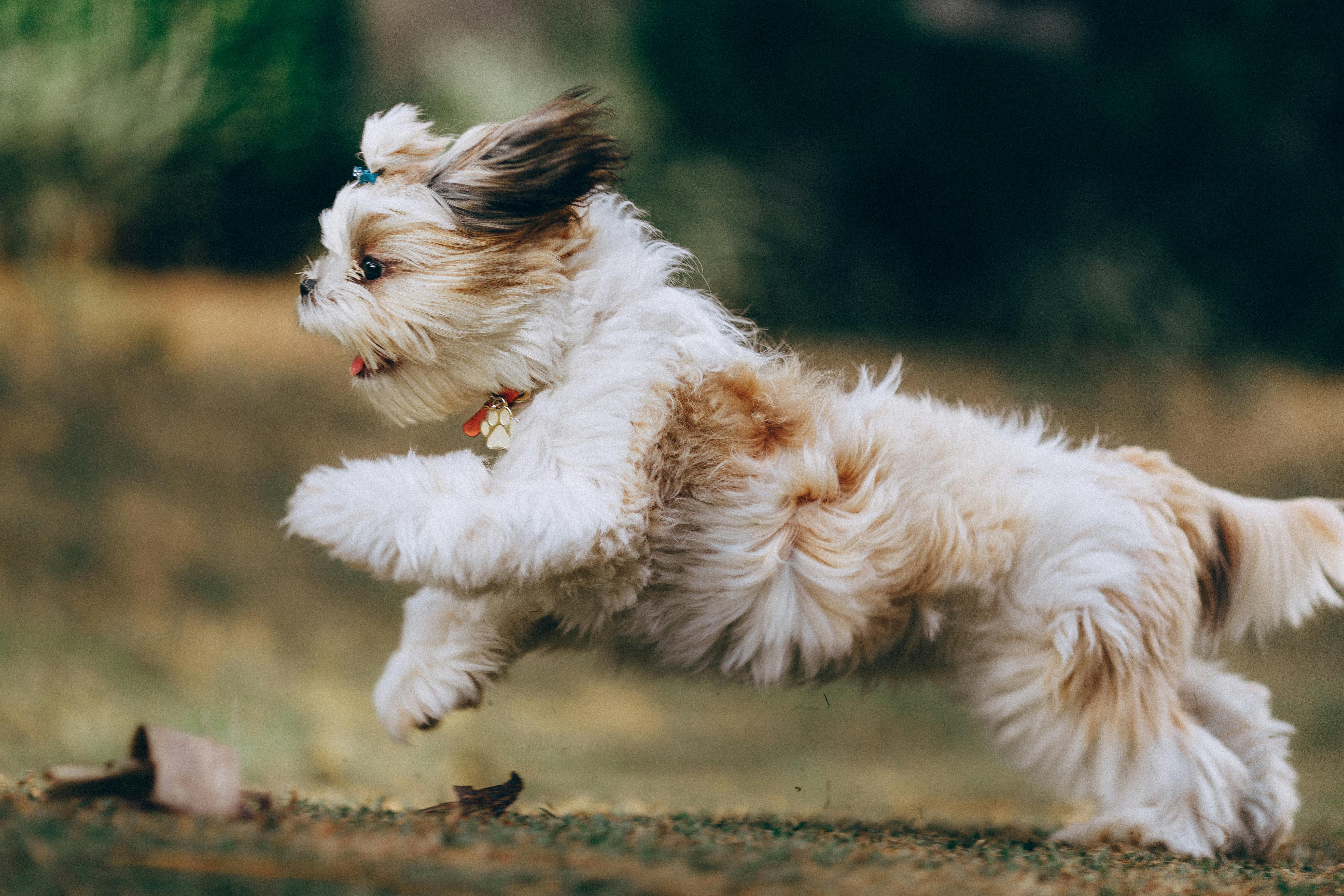
When you observe your spirited dog suddenly stop to hold up a hind leg, only to resume running a few moments later as if nothing happened, you may be witnessing the telltale signs of a medial luxating patella. A medial luxating patella, commonly referred to as a dislocated kneecap, is one of the most frequently diagnosed orthopedic conditions in veterinary medicine. It occurs when the dog’s kneecap (patella) slips out of the natural groove located at the base of the thigh bone (femur), shifting toward the inside (medial aspect) of the leg. This abnormal biomechanical movement often leads to a sudden skipping gait, localized discomfort, and noticeable swelling dog’s knee. While medial patellar luxation can affect any breed, it is overwhelmingly prevalent in small and toy-breed dogs. Patellar luxation disrupts the normal function of the entire hind limb, leading to progressive wear on the joint cartilage and the inevitable onset of osteoarthritis. Understanding the mechanics of this condition, the subtle early warning signs, and the comprehensive treatment options available is absolutely essential for preserving your dog’s mobility and long-term joint health.
To fully grasp the impact of a medial luxating patella, it is crucial to understand the anatomy of the normal canine stifle (knee) joint. The stifle is a complex hinge joint connecting the femur (thigh bone) to the tibia (shin bone). At the front of this joint lies the patella, a small bone embedded within the robust tendon of the quadriceps muscle group. When a healthy dog bends and straightens its leg, the quadriceps muscle contracts, pulling the patella in a straight line up and down within a deep, cartilaginous channel at the end of the femur known as the trochlear groove. This mechanism, often referred to as the quadriceps mechanism, acts as a biological pulley system, generating the immense leverage required for running, jumping, and normal ambulation. [1]
However, when a dog suffers from medial patellar luxation, this pulley system is fundamentally misaligned. Instead of gliding smoothly within the trochlear groove, the quadriceps muscle pulls the kneecap out of its track and over the inner ridge of the femur. This dislocation forces the dog’s knee to lock in an abnormal position, preventing weight-bearing and causing the characteristic “skip” or three-legged hop. As the kneecap snaps in and out of place, it scrapes away the smooth, protective hyaline cartilage that coats the joint surfaces. Over time, this repetitive mechanical friction leads to cartilage erosion, exposing the sensitive nerve endings in the underlying bone and resulting in chronic inflammation and pain. [2]
The consequences of a dislocated kneecap extend far beyond the patella itself. The instability within the stifle joint places an immense, unnatural strain on the surrounding ligaments and supporting structures. It is highly common for dogs with a chronic medial luxating patella to subsequently develop secondary complications, such as tears to the cranial cruciate ligament. The cranial cruciate ligament (CCL) is the primary stabilizing ligament of the canine knee, and the rotational instability caused by a luxating patella drastically increases the risk of a complete or partial CCL rupture. [3] Small-breed dogs, such as Yorkshire Terriers, Chihuahuas, Pomeranians, and Miniature Poodles, are genetically predisposed to these complex anatomical abnormalities, making early detection and proactive veterinary management critical to preventing lifelong disability.
Classifications of Patellar Luxation in Dogs
Because the severity of a medial luxating patella can vary drastically from a mild, almost unnoticeable quirk to a severe, crippling deformity, the veterinary community utilizes a standardized grading system to classify the condition. This system, originally developed by Dr. R.W. Putnam, allows veterinarians to accurately describe the extent of the luxation, track the progression of the disease over time, and formulate highly specific, individualized treatment plans. The classification is based on a meticulous physical examination of the awake patient, evaluating how easily the kneecap slips out of the trochlear groove and, more importantly, whether it can return to its normal anatomical position. [4]
During the orthopedic evaluation, the veterinarian will gently manipulate the dog’s stifle joint, putting it through a full range of motion while applying slight pressure to the patella. This hands-on assessment helps determine the degree of soft tissue laxity, the depth of the femoral groove, and the presence of any grating or crunching sensations (crepitus) that indicate cartilage damage. Based on these findings, dogs with patellar luxation are categorized into one of four distinct grades:
- Grade I: This represents the mildest form of the condition. The kneecap rests in its normal position within the trochlear groove during regular movement. However, when the veterinarian applies direct manual pressure to the patella while the dog’s leg is fully extended, the kneecap can be luxated (pushed out of place). The critical defining feature of Grade I is that the moment the manual pressure is released, the patella immediately and spontaneously pops back into its proper place. Dogs with Grade I luxation rarely exhibit noticeable clinical signs, though they may display an occasional, fleeting skip while running. Structural deformities of the bones are typically absent at this stage, and the joint cartilage remains largely intact. [5]
- Grade II: At this stage, the condition becomes more pronounced and clinically significant. The patella rests in its normal position most of the time but will spontaneously luxate during routine physical activity or when the stifle is flexed. Unlike Grade I, the kneecap in a Grade II patient will often remain dislocated until the dog actively extends the leg and internally rotates the tibia to snap it back into place, or until the veterinarian manually reduces it. This intermittent luxation is the primary cause of the classic “skipping” gait, where the dog holds the leg up for several strides before the kneecap returns to the groove and normal weight-bearing resumes. Over months and years, the repetitive friction of the patella riding over the medial femoral ridge causes significant cartilage wear, leading to mild to moderate bone deformities and the early onset of osteoarthritis. [6]
- Grade III: This represents a severe progression of the disease. In a Grade III luxation, the kneecap is perpetually dislocated—it lives outside of the trochlear groove, resting permanently on the medial aspect of the femur. The veterinarian can manually push the patella back into the correct position when the leg is extended, but the instant the leg is flexed or the manual pressure is removed, the kneecap immediately luxates again. Because the patella is constantly out of alignment, the biomechanics of the entire hind limb are compromised. Dogs with Grade III luxation typically endure consistent lameness, a noticeable crouching or bow-legged stance, and significant loss of muscle mass (atrophy) in the affected leg. The constant malpositioning forces the bones to grow abnormally, resulting in moderate to severe skeletal deformities, such as twisting of the tibia and bowing of the femur. [7]
- Grade IV: This is the most extreme and debilitating classification. The patella is permanently and rigidly luxated to the medial side of the joint and cannot be manually repositioned into the trochlear groove, even when the dog is under heavy sedation or general anesthesia. The soft tissues on the medial side of the knee have become severely contracted and scarred, while the lateral tissues are overstretched. The trochlear groove itself is often completely flat or even convex because the patella has never articulated with it to stimulate proper depth development. Dogs with Grade IV medial patellar luxation exhibit constant, severe lameness. They often hold the affected limb permanently off the ground or walk with a severe, crippling crouch, transferring their body weight to their front legs. Extreme skeletal deformities are always present, and surgical intervention is exceptionally complex, often requiring the breaking and realigning of the long bones (osteotomies) to restore any semblance of normal function. [8]
Furthermore, it is essential to recognize that patellar luxation can be either medial or lateral, dictating the specific direction in which the kneecap dislocates. Medial patellar luxation, where the kneecap slips toward the inside of the leg (the body’s midline), is overwhelmingly the most prevalent form, accounting for roughly 75% to 80% of all cases. [9] It is the hallmark orthopedic condition of small and toy breeds. Conversely, lateral patellar luxation transpires when the kneecap dislocates toward the outside of the leg. While less common overall, lateral luxation is more frequently diagnosed in large and giant breed dogs, such as Great Danes, St. Bernards, and Irish Wolfhounds, and is often associated with a distinct set of anatomical abnormalities known as a “knock-kneed” (genu valgum) conformation.
Certain breeds are exponentially more prone to this condition due to deeply ingrained genetic traits. Small dogs like the Yorkshire Terrier, Boston Terrier, Chihuahua, Pomeranian, and Miniature Poodle carry a heavily documented genetic predisposition for medial luxation. The patellar tendon, which securely connects the bottom of the kneecap to the bony prominence on the shin bone (the tibial tuberosity), plays a crucial role in maintaining knee stability. If the tibial tuberosity is positioned too far to the inside of the leg—a common anatomical flaw in these breeds—the entire quadriceps mechanism is pulled off-center, virtually guaranteeing that the patella will dislocate and severely affect the dog’s mobility and quality of life over time. [10]
Two Categories of Patellar Luxation
In addition to the classifications based strictly on the severity and direction of the dislocation, veterinary specialists further categorize patellar luxation by its origin. Understanding whether the condition is congenital (present from birth) or acquired (developing later in life) is vital for determining the underlying cause, predicting the long-term prognosis, and establishing appropriate preventive measures for future generations of dogs.
Congenital Patellar Luxation
Congenital patellar luxation is by far the most common category, accounting for the vast majority of cases seen in veterinary practices. This form of the disease is present at birth or develops very shortly thereafter as the puppy begins to grow and bear weight. It is not caused by a single isolated defect, but rather by a complex, cascading series of anatomical abnormalities that affect the entire alignment of the hind limb. These structural flaws are heavily driven by genetic factors, and specific breeds, including Toy and Miniature Poodles, Chihuahuas, Yorkshire Terriers, and Pomeranians, have a significantly elevated risk of inheriting these traits. [11]
In congenital cases, the skeletal architecture of the dog is fundamentally misaligned from the very beginning. Common developmental abnormalities include a decreased angle of the femoral neck (coxa vara), a bowing outward of the lower femur (distal femoral varus), a pathologically shallow or absent trochlear groove, and an inward twisting of the shin bone (internal tibial torsion). Because the bones are growing in an abnormal shape, the muscles and connective tissues of the quadriceps mechanism are pulled off their natural axis. As the puppy grows, the constant abnormal pull of the muscles exacerbates the skeletal deformities, creating a vicious cycle. The patella fails to sit in the groove properly, which means the groove never develops the necessary depth, further ensuring that the luxation will persist and worsen as the dog reaches adulthood. [12]
Acquired Patellar Luxation
Acquired patellar luxation, on the other hand, occurs later in a dog’s life and is typically the direct result of a specific traumatic event, a severe injury, or the gradual, chronic degeneration of the joint’s supporting structures. Unlike congenital cases, where the dog’s skeletal alignment is flawed from birth, dogs with acquired patellar luxation usually possess a normal, healthy leg structure prior to the inciting incident. Traumatic luxation happens when a massive external force—such as being struck by a vehicle, a severe fall, or a high-speed collision with another dog—ruptures the fibrous joint capsule and the strong fascial tissues (retinaculum) that hold the kneecap in its correct position. [13]
Additionally, overweight dogs are more prone to acquired patellar luxation because the excess weight places relentless, crushing stress on the stifle joints day after day. Obesity is not merely a passive issue; adipose (fat) tissue is an active endocrine organ that releases inflammatory chemicals (adipokines) into the bloodstream, accelerating the degradation of joint cartilage and weakening the supporting ligaments. As the joint tissues degenerate from chronic overload and inflammation, the structural integrity of the knee fails, eventually allowing the patella to slip out of the trochlear groove. [14]
Causes of Luxating Patella in Dogs

A medial luxating patella, also known widely in lay terms as a slipped kneecap, is a multifactorial orthopedic disease. It is rarely caused by a single isolated issue; rather, it is the culmination of several overlapping biomechanical, genetic, and environmental factors. Understanding the deep root causes of this complex condition can empower dog owners to identify risk factors early, seek prompt veterinary intervention, and take meaningful preventive measures to protect their pet’s mobility. The primary causes of a luxating patella include:
- Genetics and Hereditary Predisposition: Genetic makeup plays an overwhelmingly significant role in the development of a medial luxating patella. The condition is considered polygenic, meaning it is influenced by multiple different genes passed down from the dog’s parents. Certain small and toy breeds, such as Toy and Miniature Poodles, Chihuahuas, Yorkshire Terriers, and Pomeranians, have been heavily bred for specific physical characteristics (like short stature and fine bone structure) that inadvertently carry the genetic coding for these joint abnormalities. [15] In these highly predisposed breeds, the complex structural abnormalities in the bones, the exact attachment points of the muscles, and the elasticity of the connective tissues contribute directly to the failure of the patellar mechanism. Breeding affected dogs drastically increases the likelihood of passing the debilitating condition to the next generation.
- Trauma or Acute Injury: Acquired patellar luxation can occur in an otherwise structurally perfect dog due to an overwhelming external force. This trauma might involve a severe fall from a significant height, a forceful collision at the dog park, or an awkward landing after jumping to catch a frisbee. Injuries to the ligaments and joint structures, specifically the tearing or severe stretching of the lateral retinaculum (the tough fibrous tissue on the outside of the knee that counterbalances the inward pull of the muscles), can lead to immediate and profound instability. When the lateral restraints are compromised, the medial muscles overpower the joint, resulting in a sudden and painful dislocation of the kneecap. [16]
- Degenerative Joint Disease and Osteoarthritis: Over time, the daily wear and tear on the joints can cause the gradual development of a medial luxating patella. This degenerative progression is especially true for senior dogs or clinically overweight and obese pets. The additional mechanical stress on the stifle joint compresses the cartilage and strains the joint capsule. Chronic osteoarthritis leads to the formation of bone spurs (osteophytes) and the remodeling of the joint surfaces, which can physically alter the depth and shape of the trochlear groove. As the joint architecture slowly degrades, the patella loses its secure track, leading to an acquired luxation later in the dog’s life. [17]
- Severe Muscle Imbalances: The stability of the canine knee relies heavily on a perfect balance of tension between the various muscles of the thigh and lower leg. In some cases, uneven muscle development, neurological deficits, or profound weakness in the specific muscle groups surrounding the knee joint can lead directly to instability and dislocation of the patella. For example, if the vastus medialis (the inner thigh muscle) is abnormally tight or overdeveloped compared to the vastus lateralis (the outer thigh muscle), it will constantly pull the kneecap inward. This dangerous imbalance may be due to poor nutrition during critical growth phases, inadequate or inappropriate exercise regimens, or other underlying neurological or muscular health issues.
- Congenital Skeletal Abnormalities: Many dogs affected by this condition are born with profound structural abnormalities that guarantee the development of a medial luxating patella. These deep-seated orthopedic flaws can include misaligned or severely bowed femur bones (femoral varus), a severely twisted tibia (tibial torsion) that rotates the attachment point of the patellar tendon inward, or a pathologically shallow—or completely absent—femoral trochlear groove. Furthermore, some puppies are born with excessively lax or stretchy ligaments that fail to hold the kneecap in place tightly against the bone, allowing it to float freely out of its intended anatomical position. [18]
Prevention and early, aggressive intervention are absolutely crucial in managing a medial luxating patella. Identifying and addressing the complex underlying causes through careful physical examinations and advanced diagnostic imaging can help reduce the risk of secondary complications, such as debilitating cruciate ligament tears, and ensure a dramatically better quality of life for affected dogs.
Symptoms of Medial Patellar Luxation in Dogs

The clinical presentation of a medial patellar luxation can vary wildly depending on the individual dog, their pain tolerance, and the exact anatomical nature of the deformity. Because the condition is characterized by a displaced kneecap that disrupts the mechanics of the entire leg, the symptoms are deeply tied to the four established grades of severity. Understanding these specific symptoms can help pet owners recognize the issue early and seek veterinary care before permanent joint damage occurs. [19]
Grade 1 Symptoms
In Grade 1, the kneecap can be manually dislocated during a physical exam but readily pops back into its rightful position the moment it is let go. Because the patella spends the vast majority of its time securely within the normal trochlear groove, the affected dog may show absolutely no signs of pain or discomfort most of the time. However, astute owners may occasionally notice very mild, intermittent signs. The dog might exhibit occasional lameness, a fleeting skipping motion where they carry the back leg for a single step, or a very slightly abnormal gait after vigorous exercise. These symptoms are often so brief and self-resolving that many owners mistakenly assume the dog simply stepped on a sharp rock or experienced a temporary muscle cramp. At this early stage, cartilage damage is minimal, and pain is generally absent.
Grade 2 Symptoms
As the condition progresses to Grade 2, the kneecap may spontaneously luxate with normal, everyday movement. It frequently slips out of the groove during activity but can still be manually repositioned or may snap back into place when the dog kicks its leg backward. This is the stage where clinical signs become obvious to most pet owners. The classic presentation is a sudden, intermittent lameness that appears out of nowhere and resolves just as quickly. Signs that an affected dog may exhibit include:
- More frequent and noticeable limping, especially after resting or upon waking up.
- The hallmark “skipping” gait, where the dog runs on three legs for several strides before the kneecap pops back into place, allowing them to instantly resume walking on all fours.
- An overall abnormal, stiff, or slightly bow-legged gait in the hindquarters.
- Noticeable difficulty or hesitation when jumping onto furniture, running up steep hills, or climbing stairs, as these actions require profound contraction of the quadriceps muscle which exacerbates the luxation.
- Occasional yelping or crying out if the patella luxates awkwardly and pinches the joint capsule or surrounding soft tissues. [20]
Grade 3 Symptoms
When a dog reaches a Grade 3 luxation, the kneecap remains permanently displaced out of the groove. While a veterinarian can manually relocate it, the patella will instantly luxate again the moment the physical pressure is removed or the leg is bent. Because the knee joint is now fundamentally functioning out of alignment 100% of the time, the biomechanics of the leg are severely compromised. The cartilage on the medial aspect of the femur is subjected to constant, grinding friction, leading to severe inflammation and the rapid onset of osteoarthritis. Dogs at this more serious level may suffer from the following:
- Persistent, chronic limping that does not resolve with rest.
- A continuously abnormal, crouching, or heavily bow-legged gait, as the dog tries to balance its weight on a structurally compromised limb.
- Chronic pain and joint stiffness, particularly noticeable in colder weather or after periods of inactivity.
- Dramatically reduced activity levels; the dog may tire easily on walks, refuse to play fetch, or become reluctant to participate in normal daily activities.
- Noticeable muscle atrophy (shrinking of the muscle mass) over the thigh and hip, as the dog subconsciously shifts its weight off the painful leg and relies heavily on the front limbs.
- Significant difficulty bearing full weight on the affected leg, often standing with the toes of the affected limb only lightly touching the ground. [21]
Grade 4 Symptoms
Grade 4 represents the most extreme and severe form of the disease, where the kneecap is permanently, rigidly luxated and cannot be manually repositioned into the groove under any circumstances. The soft tissues on the inside of the leg have contracted and scarred, essentially cementing the patella in its dislocated position. The underlying bone deformities are profound. Dogs with Grade 4 medial patellar luxation often display:
- Severe, constant, and highly debilitating lameness.
- Chronic, severe pain that significantly impacts their overall quality of life.
- The dog may be entirely unable to bear any weight on the affected leg, carrying it completely off the ground tucked up against the abdomen.
- Noticeable joint swelling, thickening of the joint capsule, and severe inflammation around the knee.
- Profound, visually striking muscle atrophy of the entire hind limb due to prolonged non-use.
- A severely reduced range of motion in the affected joint; the knee may be physically locked in a semi-flexed position, unable to fully extend or bend. [22]
The intensity of clinical symptoms can fluctuate significantly from one dog to another, even within the exact same grade, and the condition will almost certainly escalate over time without appropriate, proactive veterinary medicine intervention. Therefore, it is absolutely crucial to consult a veterinarian immediately if you suspect your dog may be experiencing even intermittent luxation of the patella. This condition can occur particularly in breeds with disproportionate long bones, affecting the fundamental alignment and stability of the stifle joint in the affected leg. Delaying veterinary evaluation allows the irreversible process of cartilage destruction and osteoarthritis to take hold, drastically complicating future treatment efforts.
Diagnosing Medial Luxating Patella in Dogs

Diagnosing a medial luxating patella in dogs calls for a highly structured, integrative approach that relies heavily on the veterinarian’s clinical expertise. It includes a comprehensive physical examination, meticulous palpation of the joint in question, and advanced diagnostic imaging tests to verify the diagnosis, rule out concurrent injuries, and precisely gauge the condition’s severity. Because the signs of patellar luxation can mimic other severe orthopedic issues, a thorough workup is mandatory. Here is the rigorous methodology veterinary professionals employ to diagnose a medial luxating patella in dogs:
- Physical examination and Gait Analysis: The diagnostic process begins before the vet even touches the dog. The veterinarian’s first step involves carefully scrutinizing the dog’s walking style across the exam room or down a hallway. They are looking for specific biomechanical clues, noting any indications of limping, the classic intermittent skipping, abnormal weight shifting, or crouching gait patterns that may hint at patellar luxation. The vet will also observe how the dog sits and stands; dogs with severe luxation often sit with the affected leg splayed awkwardly out to the side to avoid flexing the painful knee. [23]
- Orthopedic Palpation: This is the cornerstone of the diagnosis. The vet carefully palpates the dog’s stifle joint using specific orthopedic maneuvers. By stabilizing the femur with one hand and gently manipulating the patella with the thumb of the other, the vet attempts to luxate the kneecap medially and laterally. They evaluate how easily the kneecap is displaced, whether it returns to the groove spontaneously, and the presence of any grinding or crunching (crepitus) that indicates cartilage erosion. This crucial step directly assists the veterinarian in determining the exact grade of the patellar luxation and identifying any localized swelling, heat, or associated discomfort. The vet will also perform the “cranial drawer test” and “tibial compression test” during this palpation to evaluate the integrity of the cranial cruciate ligament, as concurrent CCL tears are highly prevalent in these patients. [24]
- Radiographs (X-rays): While the diagnosis is primarily made through physical palpation, comprehensive X-rays are absolutely essential. Radiographs of the leg affected by the condition must be conducted to examine the overall bone structure, gauge the level of joint deformity, and conclusively exclude other potential causes of the dog’s symptoms, such as traumatic bone fractures, bone cancer (osteosarcoma), or severe joint infections. Standard views include the mediolateral (side) and craniocaudal (front-to-back) projections. A special “skyline view” of the femur may also be taken to directly visualize the depth and shape of the trochlear groove. The position of the tibial crest—specifically how far it has rotated inward—can be particularly informative for surgical planning. Radiographs also reveal the severity of secondary osteoarthritis, marked by joint effusion and osteophyte formation. [25]
- Specialized Orthopedic Evaluation: In some complex instances, or when the dog has bilateral (both knees) Grade 3 or Grade 4 luxation, a more in-depth orthopedic evaluation may be required. This could involve an immediate referral to a board-certified veterinary surgeon at a specialized animal hospital. A specialist can provide a highly detailed, quantitative assessment of the extent of the luxation, measure specific bone angulation using precise orthopedic software, and evaluate any associated joint capsule or ligament damage. This expert assessment is crucial to inform the most suitable, and often highly complex, surgical treatment plan, especially in cases of lateral patellar luxation or when severe bone twisting requires corrective cutting of the bones (osteotomies).
- Additional Diagnostic Tests: Depending on the individual case and the dog’s age, the vet may recommend further diagnostic tests like comprehensive blood work (complete blood count and serum biochemistry) to ensure the dog is a safe candidate for general anesthesia and heavy pain medications. In exceptionally complex cases involving severe bone deformities, advanced 3D imaging such as CT (Computed Tomography) scans or an MRI may be utilized. A CT scan allows the surgeon to create a 3D model of the dog’s bones, calculating the exact Center of Rotation of Angulation (CORA) to plan precise bone cuts. These tests can give a more holistic understanding of the dog’s overall health and spot any underlying health issues contributing to the repeated luxation of the patella.
An accurate, comprehensive diagnosis of a medial luxating patella is vital in determining the correct treatment strategy and effectively managing the condition over the long term. A thorough workup ensures that concurrent injuries are not missed, which can significantly enhance the dog’s quality of life, dictate the success of any surgical intervention, and profoundly reduce the risk of enduring complications and chronic pain.
Treatment Options for Medial Luxating Patella in Dogs

Treating a medial luxating patella in dogs is never a one-size-fits-all endeavor. The therapeutic approach is highly individualized and is carefully tailored to the specific grade of the severity of the condition, the dog’s age, body condition score, overall health, and the presence of any associated complications, such as a ruptured cranial cruciate ligament or severe osteoarthritis. Based on a comprehensive orthopedic assessment, here is exactly how veterinary professionals treat a medial luxating patella in dogs, ranging from medical management to advanced surgical correction.
Conservative Management
Conservative, non-surgical management may be recommended as the primary course of action for mild, largely asymptomatic cases (Grade I or occasionally mild Grade II luxations), or for older dogs with significant underlying health issues that make general anesthesia unacceptably risky. The goal of conservative therapy is not to cure the anatomical defect—as bones cannot be realigned without surgery—but rather to minimize pain, reduce joint inflammation, and strengthen the surrounding musculature to provide better dynamic support to the unstable knee. Conservative management for a medial luxating patella in dogs typically includes a multimodal approach combining the following vital components: [26]
- Aggressive Weight Management: This is arguably the single most important non-surgical intervention. Excess body weight places exponential biomechanical stress on the compromised stifle joint. Furthermore, adipose (fat) tissue releases pro-inflammatory hormones that actively degrade joint cartilage. Achieving and strictly maintaining a lean, ideal body condition score will dramatically reduce joint pain and slow the progression of osteoarthritis. [27]
- Targeted Physical Therapy: Formal physical rehabilitation with a certified canine rehabilitation practitioner (CCRP) is highly beneficial. Therapies often include passive range of motion (PROM) exercises, use of an underwater treadmill to build muscle mass without placing full concussive weight on the joints, and specific therapeutic exercises (like stepping over cavaletti poles or balancing on wobble boards) designed to strengthen the quadriceps and hamstring muscles, thereby improving active joint stabilization.
- Controlled Exercise Modification: Unrestricted running, explosive jumping on and off high furniture, and roughhousing with other dogs must be severely curtailed. Exercise should consist of multiple short, controlled leash walks on flat, even surfaces. Ramps or pet stairs should be placed around the home to prevent the dog from having to jump, which violently stresses the patellar mechanism.
- Anti-inflammatory and Pain Medications: To control the chronic pain and inflammation associated with cartilage wear, veterinarians frequently prescribe specific prescription anti-inflammatory medications formulated safely for dogs. In more painful cases, additional nerve-pain medication prescribed by your veterinarian may be added to the protocol to address nerve-related pain and chronic wind-up pain. [28]
- Advanced Joint Supplements and Nutraceuticals: High-quality, veterinary-approved joint supplements are a cornerstone of conservative care. These typically include therapeutic levels of glucosamine, chondroitin, and highly concentrated Omega-3 fatty acids (EPA and DHA from fish oil) to naturally reduce joint inflammation, alongside prescription joint-protectant therapies administered by your veterinarian, which actively help protect the remaining cartilage and improve joint fluid viscosity. [29]
Conservative management aims to control the clinical symptoms of the medial luxating patella without resorting to surgical intervention. It requires profound, lifelong commitment from the pet owner. However, if conservative measures don’t provide sufficient pain relief, if the dog continues to exhibit frequent skipping and lameness, or if follow-up examinations reveal that the condition is progressively worsening to a higher grade, surgical treatment will become absolutely necessary to prevent irreversible joint destruction.
Surgical Intervention
Surgery is universally recommended to correct the anatomical defects of a patellar luxation in more severe cases (symptomatic Grade II, Grade III, and Grade IV) or when conservative management is ultimately unsuccessful at maintaining the dog’s quality of life. The primary goals of surgery are to realign the quadriceps mechanism, permanently deepen the trochlear groove, and stabilize the joint to halt the progression of debilitating osteoarthritis. There are several advanced surgical techniques that board-certified veterinary surgeons or highly experienced veterinarians may use to treat a medial luxating patella in dogs. The surgeon rarely uses just one technique; rather, they combine multiple procedures based on the specific anatomical needs of the individual dog. [30]
Some of the most common and effective surgical interventions for a medial luxating patella include:
- Trochleoplasty (Deepening the Groove): In most luxation cases, the natural groove at the end of the femur is pathologically shallow. To provide a secure track for the kneecap, the surgeon performs a trochleoplasty. The most common and cartilage-sparing techniques are the Trochlear Block Recession or the Trochlear Wedge Recession. In these highly delicate procedures, the surgeon uses a specialized bone saw to cut a rectangular block or V-shaped wedge of bone and cartilage out of the femur. The underlying bone bed is then excavated and deepened. Finally, the original piece of bone containing the smooth, healthy hyaline cartilage is firmly pressed back into the newly deepened hole. This provides a deep, secure groove for the patella while preserving the dog’s original joint surface. [31]
- Tibial Tuberosity Transposition (TTT): This is often the most critical component of the surgery. If the bony prominence on the shin bone where the patellar tendon attaches (the tibial crest) is twisted too far inward, no amount of groove deepening will keep the kneecap in place. In a TTT, the surgeon uses an osteotome (bone chisel) or saw to carefully cut the tibial tuberosity free from the main shin bone. The bone fragment, with the tendon still firmly attached, is physically moved outward (laterally) to perfectly align the quadriceps mechanism into a straight line. The bone fragment is then permanently secured in its new, correct location using specialized stainless steel orthopedic pins (K-wires) and a tension band wire. This powerful biomechanical realignment is essential for long-term success. [32]
- Soft Tissue Reconstruction (Imbrication and Release): Because the kneecap has been luxated for a long time, the soft tissues surrounding the joint have adapted abnormally. The tissues on the inside (medial aspect) of the knee are severely tightened and contracted, pulling the kneecap inward. The surgeon performs a medial desmotomy (a releasing incision) to cut and free these tight tissues, allowing the patella to return to the center. Conversely, the tissues on the outside (lateral aspect) of the knee are overstretched and baggy. The surgeon performs a lateral imbrication, cutting out the excess loose tissue and suturing the capsule tightly together to provide a strong, permanent wall of tension that pulls the kneecap outward and holds it securely in the newly deepened groove. [33]
- Corrective Osteotomies (for Severe Grade IV Cases): In extreme cases, particularly Grade IV luxations with massive skeletal deformities, the femur or tibia bones themselves may be severely twisted or bowed. Simple soft tissue corrections and groove deepening will fail. In these complex scenarios, the surgeon must perform a Distal Femoral Osteotomy (DFO) or Tibial Osteotomy. This involves completely cutting the long bone in half, rotating or straightening the bone into proper anatomical alignment, and rigidly securing the bone segments together using heavy orthopedic bone plates and screws. This is a major orthopedic intervention requiring significant expertise. [34]
- Combination Surgeries: It is incredibly rare for a surgeon to perform only one of the above techniques. To effectively address the complex biomechanics of a dog’s medial luxating patella and provide the absolute best possible outcome with the lowest rate of recurrence, the surgeon will meticulously combine a trochleoplasty, a TTT, and soft tissue reconstruction all during the same anesthetic event. [35]
Medial luxating patellar surgeries in dogs can profoundly and permanently improve a dog’s quality of life, especially when conservative management is unsuccessful and the dog is suffering from chronic pain. However, as with any major orthopedic intervention, it is essential for pet owners to realistically consider the comprehensive pros and cons of these surgeries before deciding on a definitive treatment plan.
Pros of medial luxating patellar surgeries:
- Profound Pain Relief: Surgery addresses the mechanical root cause of the problem. By permanently stabilizing the patella within the groove and correcting any underlying bone deformities, the surgery eliminates the constant, painful grinding of bone on bone, offering profound relief from chronic pain.
- Dramatically Improved Mobility: Dogs with severe patellar luxation may have immense difficulty walking, running, or playing. Successful surgical correction restores the normal biomechanical function of the stifle joint, allowing the dog to move smoothly, comfortably, and confidently without the fear of their knee collapsing.
- Prevention of Further Joint Damage: This is a critical benefit. If left untreated, the constant friction of a medial luxating patella rapidly destroys the joint cartilage, leading to severe, irreversible osteoarthritis and drastically increasing the risk of a cranial cruciate ligament tear. Surgery halts this destructive mechanical process, helping to prevent or significantly slow down the development of these secondary, crippling complications. [36]
- Customized, Targeted Treatment: Because there are various highly specific surgical techniques available to address each patient’s exact anatomical deficits, the veterinary surgeon can provide a completely tailored treatment plan based on the precise severity of the condition and the individual dog’s unique skeletal anatomy.
Cons of medial luxating patellar surgeries:
- General Anesthesia Risks: As with any major surgical procedure, there are inherent risks associated with general anesthesia, including adverse cardiovascular events, allergic reactions, difficulty breathing, or in extremely rare cases, death. However, with pre-anesthetic blood work, modern monitoring equipment, and dedicated veterinary anesthesiologists, these risks are generally very low.
- Postoperative Complications: Potential complications from orthopedic surgery can occur and include surgical site infection, excessive bleeding, nerve damage, failure of the orthopedic implants (such as pins migrating or wires breaking), or, most notably, a recurrence of the patellar luxation if the anatomical correction was insufficient or if the dog was allowed to be overly active during the healing phase. [37]
- Intensive Recovery and Rehabilitation: The recovery period after stifle surgery is rigorous and lengthy, often requiring an absolute minimum of eight to twelve weeks of strictly limited activity (cage rest). It demands significant owner compliance, including leash-walking only, administering multiple medications, managing physical therapy exercises, and attending frequent recheck appointments. This strict confinement can be highly challenging for both a highly energetic dog and the owner.
- Financial Cost: Surgical treatment for a medial luxating patella is a major specialized procedure and can be quite expensive. Prices vary widely depending on the severity of the skeletal deformity, the complexity of the required surgery, whether it is performed by a general practitioner or a board-certified orthopedic surgeon, and the geographical location of the veterinary hospital.
- No Absolute Guarantee of Success: While the vast majority of dogs experience highly significant improvements following surgery, biology is unpredictable. There is no absolute 100% guarantee that the surgery will be completely successful, that the dog will be entirely free of future arthritis, or that the condition will not occasionally recur, particularly in extremely severe Grade IV cases.
In conclusion, medial luxating patellar surgeries offer immense, life-changing benefits for dogs suffering from the debilitating effects of the condition, but it is crucial to weigh the pros and cons carefully and realistically. Consulting deeply with your primary care veterinarian, and potentially seeking a specialized second opinion from a board-certified veterinary orthopedic surgeon, can provide clarity and help confidently guide your decision-making process.
Postoperative Care
The success of the surgical intervention is heavily dependent on strict, meticulous postoperative care. Immediately after surgery, the dog will require a prolonged period of strict cage rest and heavily restricted activity to allow the cut bones to fuse and the soft tissues to heal securely. For the first two to four weeks, the dog must be confined to a small room or crate, with absolutely no running, jumping, or playing allowed. They should only be taken outside on a short leash for brief bathroom breaks, often using a supportive sling under their belly to prevent them from slipping or placing sudden, excessive weight on the repaired leg. [38]
Cryotherapy (ice packing) the joint multiple times a day during the first week is vital to reduce swelling and inflammation. After the initial healing phase, formal physical therapy and a highly structured, gradual return to exercise will be necessary to rebuild atrophied muscles, break down scar tissue, and regain full strength and flexibility in the affected leg. Comprehensive pain management utilizing a multimodal combination of prescription pain relief and anti-inflammatory medications tailored to your pet will be prescribed during the intensive recovery period to ensure the dog remains comfortable.
Ongoing Monitoring
Regular, scheduled follow-up appointments with the operating vet or surgeon will be absolutely necessary to monitor the dog’s clinical progress and ensure the long-term treatment plan is effective. At around the six-to-eight-week mark post-surgery, the veterinarian will require follow-up radiographs (X-rays) to visually confirm that the bone cuts (like the tibial tuberosity transposition) have fully healed and that the orthopedic pins and wires remain securely in place. [39]
In some cases, adjustments to the long-term treatment plan may be required, such as changes in the dosage of joint medications, managing the dog’s weight, or the permanent addition of supportive, low-impact therapies like hydrotherapy (underwater treadmill) or veterinary acupuncture to manage secondary arthritis. Prompt and highly appropriate treatment of a medial luxating patella, combined with dedicated aftercare, can drastically and permanently improve the dog’s quality of life, effectively eliminate chronic pain and discomfort, and significantly minimize the risk of long-term joint damage and debilitating arthritis.
Prevention of Medial Luxating Patella in Dogs
Because medial patellar luxation is primarily deeply rooted in complex genetics and inherited developmental skeletal abnormalities, completely preventing the condition in a predisposed puppy is not always medically possible. However, there are several highly effective, proactive steps that breeders and pet owners can take to minimize the overall risk within the canine population and strictly manage potential environmental factors that contribute to the severity and rapid development of the condition:
- Strict, Responsible Breeding Practices: This is the absolute first line of defense. Choosing dogs from highly reputable, ethical breeders who prioritize the long-term health, structural conformation, and genetic integrity of their lines can drastically help reduce the risk of passing on the genetic predispositions for a medial luxating patella. Breeders must rigorously screen all their breeding dogs for this specific condition through formal veterinary orthopedic evaluations (such as the OFA – Orthopedic Foundation for Animals patella registry). Ethical breeders will absolutely avoid breeding any dog with a known history of patellar luxation, regardless of how mild the grade. [40]
- Optimal Nutrition and Rigid Weight Management: Providing your growing puppy and adult dog with a precisely balanced, high-quality diet and maintaining a strictly healthy, lean body weight is paramount. This can significantly help reduce the massive biomechanical stress on their developing joints, including the highly vulnerable patellar mechanism. Obesity does not simply add weight; it exacerbates all underlying joint problems by altering limb angulation and flooding the body with inflammatory mediators. Carefully monitoring your dog’s body condition score and rapidly adjusting their daily food intake and exercise routines to prevent excess weight gain is absolutely essential for joint longevity. [41]
- Controlled, Appropriate Regular Exercise: Regular, low-impact, and moderate exercise is vital. It can help build and maintain robust muscle strength and essential joint flexibility, which actively helps support the patella, holding it firmly in its groove and reducing the overall risk of luxation. However, it is crucial, especially during a puppy’s rapid growth phases when growth plates are open, to strictly avoid excessive, repetitive, or high-impact activities—such as relentless frisbee fetching, jumping off high walls, or forced running on hard pavement—that could acutely injure the delicate knee joint or warp growing bones. [42]
- Proactive Routine Veterinary Care: Regular, thorough wellness check-ups with your primary care veterinarian can help identify any subtle, early signs of a medial luxating patella long before the dog begins to show obvious pain or severe lameness. Early detection allows for prompt conservative intervention, such as immediate weight loss or starting physical therapy, which can lead to vastly better management of the condition and may significantly reduce the long-term risk of severe secondary complications like cruciate tears and arthritis.
- Early Use of Joint Supplements: For breeds known to be at high risk, some veterinarians may recommend starting proactive joint supplements early in life. Supplements containing high-quality glucosamine, chondroitin, green-lipped mussel extract, or high levels of Omega-3 fatty acids can promote healthy cartilage development, increase joint fluid viscosity, and help maintain the structural stability of the patella by reducing low-grade joint inflammation. [43]
While it may not be medically possible to completely prevent a medial luxating patella from occurring in highly predisposed genetic lines of dogs, rigorously following these evidence-based guidelines can profoundly help minimize the severity of the risk. We always advise you to consult your veterinarian before making any changes to your pet’s care to ensure your dog has the absolute best chance at a healthy, highly active, and pain-free life.
Frequently Asked Questions
How do you treat a luxating patella in dogs without surgery?
Treating a luxating patella in dogs without surgery relies on a robust multimodal conservative management approach, primarily suited for dogs with mild, Grade 1 or low Grade 2 luxations where pain is minimal. The absolute most critical component is aggressive weight management; maintaining a lean body condition drastically reduces the mechanical burden on the compromised stifle joint. This is paired with highly controlled, low-impact exercise modification—such as frequent short leash walks or swimming—while strictly avoiding high-impact activities like jumping or rough play. Professional physical therapy, including underwater treadmill sessions and targeted range-of-motion exercises, builds the quadriceps and hamstring muscles to dynamically stabilize the joint. Additionally, the use of veterinary-prescribed anti-inflammatory medications helps control acute flare-ups of pain, while daily administration of high-quality joint supplements containing glucosamine, chondroitin, and Omega-3 fatty acids helps protect the existing joint cartilage and reduce chronic inflammation. [44]
Can a dog live comfortably with a grade 4 luxating patella without surgery?
Generally speaking, it is extremely difficult for a dog to live comfortably with an untreated Grade 4 luxating patella. In a Grade 4 scenario, the kneecap is permanently and rigidly dislocated, leading to profound skeletal deformities, contracture of the surrounding muscles, and the inability to properly bear weight on the affected limb. This results in severe, chronic pain, rapid progression of debilitating osteoarthritis, and significant muscle atrophy. The dog is often forced to walk in a severely crouched posture or carry the leg entirely, which in turn places unnatural, destructive stress on their spine and other limbs. While heavy, continuous use of strong pain medications can mask some of the discomfort, it does not address the underlying mechanical failure of the joint. Therefore, without highly specialized surgical intervention to correct the profound anatomical deformities, the dog’s mobility and overall quality of life will remain severely compromised and continue to deteriorate over time.
How long is the recovery after a dog has luxating patella surgery?
The recovery period following surgery to correct a luxating patella is extensive and requires strict dedication from the pet owner, typically spanning a minimum of eight to twelve weeks. The first two to four weeks represent the most critical healing phase, demanding absolute cage rest and strict confinement. During this time, the dog must only be taken outside on a short leash for bathroom breaks, often using a supportive body sling to prevent sudden weight-bearing that could cause the surgical implants (pins and wires) to fail. Around the four-week mark, gentle, at-home physical rehabilitation exercises, such as passive range of motion, are slowly introduced. Follow-up radiographs are usually taken between six and eight weeks to confirm that the cut bones (from procedures like a tibial tuberosity transposition) have fully fused. If the bone healing is solid, the veterinarian will clear the dog for a gradual, highly structured return to longer walks and eventual normal activity, often supplemented by professional hydrotherapy to rebuild lost muscle mass safely. [45]
Concerned About Your Dog’s Mobility?
If you’ve noticed your dog skipping, limping, or holding up a hind leg, don’t wait for the condition to worsen. Early diagnosis and treatment are essential for preserving joint health and preventing chronic pain.
References
- American College of Veterinary Surgeons. Patellar Luxation in Dogs. ACVS, 2023.
- Merck Veterinary Manual. Patellar Luxation in Dogs. Merck & Co., Inc., 2023.
- Campbell, C.A. et al. Severity of patellar luxation and frequency of concomitant cranial cruciate ligament rupture in dogs. JAVMA, 2010.
- Putnam, R.W. Patellar Luxation in the Dog. University of Guelph, 1968.
- VCA Animal Hospitals. Luxating Patella in Dogs. VCA, 2022.
- Veterinary Information Network. Medial Patellar Luxation. VIN, 2018.
- Arthurs, G.I. et al. Complications associated with corrective surgery for patellar luxation in 109 dogs. Vet Surg, 2006.
- Towle, H.A. et al. Pre- and postoperative radiographic and computed tomographic evaluation of dogs with medial patellar luxation. Vet Surg, 2005.
- Hayes, A.G. et al. Frequency and distribution of patellar luxation in dogs: 134 cases. JAVMA, 1994.
- LaFond, E. et al. Breed susceptibility for developmental orthopedic diseases in dogs. J Am Anim Hosp Assoc, 2002.
- Bound, N. et al. The prevalence of patellar luxation in dogs attending primary care veterinary practices in England. Vet Rec, 2009.
- Mostafa, A.A. et al. Proximodistal alignment of the canine patella: radiographic evaluation and association with medial and lateral patellar luxation. Vet Surg, 2008.
- Di Dona, F. et al. Patellar luxation in dogs. Vet Med (Auckl), 2018.
- Marshall, W.G. et al. The effect of weight loss on lameness in obese dogs with osteoarthritis. Vet Res Commun, 2010.
- Wangdee, C. et al. Evaluation of surgical treatment of medial patellar luxation in Pomeranian dogs. Vet Comp Orthop Traumatol, 2013.
- Hulse, D.A. Pathophysiology and management of medial patellar luxation in the dog. Vet Clin North Am Small Anim Pract, 1993.
- Roy, R.G. et al. Radiographic abnormalities in dogs with medial patellar luxation. JAVMA, 1992.
- Soparat, C. et al. Radiographic measurement for femoral varus in Pomeranian dogs with and without medial patellar luxation. Vet Comp Orthop Traumatol, 2012.
- ASPCA. Understanding Patellar Luxation. ASPCA Pet Health, 2021.
- Perry, K.L. et al. Effect of breed on the development of complications following surgical correction of medial patellar luxation in dogs. Vet Surg, 2017.
- Cashmore, R.G. et al. Major complications and risk factors associated with surgical correction of congenital medial patellar luxation in 124 dogs. Vet Comp Orthop Traumatol, 2014.
- Swiderski, J. et al. Evaluation of the distal femoral osteotomy for treatment of severe medial patellar luxation in dogs. JAVMA, 2008.
- Morton, M.A. et al. Clinical and radiographic evaluation of the stifle joint in dogs with patellar luxation. J Am Vet Med Assoc, 2012.
- Trostel, C.T. et al. Canine lameness caused by developmental orthopedic diseases. Compend Contin Educ Vet, 2007.
- Loughin, C.A. et al. Radiographic assessment of the canine stifle for medial patellar luxation. Vet Radiol Ultrasound, 2002.
- American Veterinary Medical Association. Osteoarthritis in Dogs: Management and Treatment. AVMA, 2022.
- Impellizeri, J.A. et al. Effect of weight reduction on clinical signs of lameness in dogs with hip osteoarthritis. JAVMA, 2000.
- KuKanich, B. Outpatient oral analgesics in dogs and cats beyond nonsteroidal anti-inflammatory drugs. J Am Vet Med Assoc, 2013.
- McCarthy, G. et al. Randomised double-blind, positive-controlled trial to assess the efficacy of glucosamine/chondroitin sulfate for the treatment of dogs with osteoarthritis. Vet J, 2007.
- Willauer, C.C. et al. Surgical correction of medial patellar luxation in dogs. Vet Surg, 2001.
- Johnson, A.L. et al. Trochlear block recession for the treatment of patellar luxation in dogs. Vet Surg, 1996.
- Linney, C.J. et al. Surgical treatment of medial patellar luxation without femoral trochlear groove deepening procedures in dogs: 50 cases. Vet Surg, 2006.
- Roush, J.K. Surgical therapy of canine joint disorders. Vet Clin North Am Small Anim Pract, 2000.
- Yeadon, R. et al. Distal femoral osteotomy for the correction of severe medial patellar luxation in dogs. Vet Comp Orthop Traumatol, 2011.
- O’Neill, D.G. et al. Epidemiology of patellar luxation in dogs attending primary-care veterinary practices in England. Canine Genet Epidemiol, 2016.
- Gibbons, S.E. et al. Patellar luxation in 70 large breed dogs. J Small Anim Pract, 2006.
- Cashmore, R.G. et al. Complications associated with surgical correction of congenital medial patellar luxation. Vet Comp Orthop Traumatol, 2014.
- Millis, D.L. et al. Rehabilitation and physical therapy for the orthopedic patient. Vet Clin North Am Small Anim Pract, 2005.
- Gallegos, J. et al. Radiographic assessment of bone healing following tibial tuberosity transposition. Vet Comp Orthop Traumatol, 2010.
- Vidoni, B. et al. Incidence of patellar luxation in dogs in Austria. Wien Tierarztl Monatsschr, 2005.
- Marshall, W.G. et al. A review of osteoarthritis and obesity: current understanding of the relationship and benefit of obesity treatment and prevention in the dog. Vet Comp Orthop Traumatol, 2009.
- LaFond, E. et al. Breed susceptibility for developmental orthopedic diseases in dogs. J Am Anim Hosp Assoc, 2002.
- Roush, C.E. et al. Multicenter veterinary practice assessment of the effects of omega-3 fatty acids on osteoarthritis in dogs. JAVMA, 2010.
- Vandeweerd, J.M. et al. Systematic review of efficacy of nutraceuticals to alleviate clinical signs of osteoarthritis in dogs. J Vet Intern Med, 2012.
- Millis, D.L. Physical rehabilitation for the orthopedic patient. Vet Clin North Am Small Anim Pract, 2005.


