


 March 6, 2023
 Phil Good, DVM
What are Nasal Infections in Dogs?
This content was prepared with AI assistance and reviewed by a licensed professional for accuracy.
Introduction
When considering the various respiratory challenges that our canine companions face, Nasal Infections in Dogs represent a complex, multifactorial, and frequently encountered clinical scenario in veterinary medicine. Rhinitis in Dogs, alongside conditions like Sinusitis and generalized Nasal Infections, can significantly compromise a patient’s quality of life. These infections lead to profound discomfort, upper respiratory distress, anorexia, and in severe instances, systemic illness. To fully grasp the implications of these conditions, one must first understand the intricate anatomical and physiological framework of the canine upper respiratory tract. The nasal cavity of a dog is not merely a conduit for air; it is a highly sophisticated apparatus designed to filter, warm, humidify, and olfactorily evaluate every breath of inspired air before it descends into the delicate tissues of the lower respiratory system.[1]
The interior of the canine nose features a labyrinthine network of delicate, scroll-like bones known as turbinates or conchae. These structures drastically increase the internal surface area and are coated in a highly vascularized mucosal lining. This respiratory epithelium is densely populated with ciliated cells and mucin-secreting goblet cells. Together, they form the mucociliary escalator, a critical defense mechanism that traps inhaled particulate matter, allergens, and microbial pathogens, systematically sweeping them toward the nasopharynx where they can be harmlessly swallowed or expectorated. When an infectious agent or environmental insult breaches this defense, inflammation ensues. The nasal passages and the adjacent air-filled skull cavities—the sinuses—are intimately connected through minute physiological gateways known as ostia. Rhinitis, which is strictly defined as inflammation of the nasal mucous membranes, frequently causes significant tissue edema that physically obstructs these narrow ostia.[2]
Once the ostia are blocked, the normal physiological drainage of sinus fluid is entirely halted, creating a stagnant, hypoxic, and nutrient-rich environment within the sinus cavities. This stasis invariably initiates sinusitis, which can manifest as either an acute or chronic condition depending on the duration and nature of the underlying trigger. Conversely, a primary infection originating within the sinuses can rapidly extend forward into the nasal passages, resulting in secondary rhinitis. Because of this interconnected anatomy, isolated rhinitis or isolated sinusitis is clinically rare in veterinary practice. Instead, the two conditions frequently coexist in a synergistic disease process jointly termed rhinosinusitis. Each component of rhinosinusitis amplifies the persistence, intensity, and clinical severity of the other, establishing a frustrating cycle of inflammation and infection that requires decisive veterinary intervention.[3]
Further complicating the clinical picture is the anatomical proximity of the upper dental arcade to the nasal cavity. A severe dental infection, such as a carnassial tooth root abscess, can easily erode through the thin layer of maxillary bone, establishing an unnatural communication tract called an oronasal fistula. This allows oral bacteria and purulent material to drain directly into the nasal passages, presenting as localized nasal congestion, unilateral discharge, and severe localized rhinitis. Recognizing the specific type, origin, and progression of the disease is the first crucial step toward returning the dog to a state of health. Ultimately, rhinitis is a debilitating nasal passage inflammation that, when intrinsically coupled with sinusitis, creates multifaceted health issues for the dog, significantly affecting their metabolic energy, olfactory abilities, and overall well-being.[4]
Types of Nasal Infections in Dogs
In the field of veterinary internal medicine, a nasal infection—clinically designated as rhinitis—is fundamentally defined as an inflammatory disease affecting the nasal mucous membranes. While bacterial or fungal organisms are frequently identified as the primary or secondary causative agents, the condition can also be triggered by a diverse array of viral pathogens, inhaled foreign bodies, blunt or penetrating trauma, environmental chemical irritants, or severe underlying systemic diseases. To effectively approach diagnosis and therapeutic management, veterinarians typically classify rhinitis into two primary chronological categories based on the duration, onset, and cellular characteristics of the inflammatory response. Understanding the distinction between acute and chronic presentations is vital for establishing an accurate prognosis and an effective, targeted treatment regimen.[5]
Acute Rhinitis
Acute rhinitis in dogs refers to a sudden, rapid-onset inflammation localized within the nasal cavity and associated upper respiratory structures. This condition predominantly affects the mucosal membranes lining the dog’s turbinates, abruptly disrupting their normal physiological functions. Pathologically, acute rhinitis is characterized by intense localized hyperemia (increased blood flow), vascular engorgement, and significant tissue edema. This inflammatory cascade is primarily mediated by the sudden influx of neutrophils and acute-phase inflammatory cytokines in response to a noxious stimulus, whether infectious, allergic, or environmental. Dogs suffering from acute rhinitis typically experience immediate and profound discomfort. They may exhibit pronounced difficulty breathing, marked by stertor (a low-pitched snoring sound) or audible wheezing, resulting directly from the physical narrowing of the nasal passages due to swelling and highly elevated mucus production.[6]
During the acute phase, the mucociliary escalator becomes rapidly overwhelmed. The goblet cells upregulate the production of mucin, leading to a copious, often serous (clear and watery) or mucoid (cloudy and slightly viscous) nasal discharge. The dog will frequently sneeze in a vigorous, repetitive attempt to clear the obstructed airways. While acute rhinitis presents suddenly and can cause significant distress to both the patient and the observing owner, it is often a self-limiting or easily treatable condition. If the underlying cause—such as a mild viral upper respiratory infection or a transient environmental allergen—is addressed or naturally cleared by the dog’s immune system, the inflammation will subside. When managed correctly with appropriate supportive care or short courses of targeted medications, acute rhinitis is generally a short-lived condition that resolves appropriately without inflicting permanent structural damage to the delicate nasal architecture.[7]
Chronic Rhinitis
Conversely, chronic rhinitis in dogs represents a deep-seated, persistent inflammation of the nasal cavity that lingers for extended periods, typically defined in veterinary literature as lasting longer than four to six weeks without resolution. Unlike acute rhinitis, which is characterized by a sudden onset and relatively swift resolution, chronic rhinitis is a deeply entrenched, long-term pathological condition that can profoundly and permanently affect a dog’s overall quality of life. At the histological level, the cellular landscape of the nasal mucosa changes drastically during chronic rhinitis. The initial neutrophilic response transitions into a lymphoplasmacytic or eosinophilic infiltration. The delicate ciliated respiratory epithelium often undergoes squamous metaplasia—a protective but dysfunctional transformation where ciliated cells are replaced by tougher, non-ciliated squamous cells. This permanently impairs the mucociliary escalator, making the dog highly susceptible to persistent secondary bacterial colonization.[8]
This persistent disease state leads to constant, unrelenting irritation, hyperplastic thickening of the mucosal tissues, and ongoing disruptions to normal respiratory and olfactory functions. The chronic swelling and accumulation of inspissated (thickened, dried) purulent mucus can physically remodel the nasal turbinates, leading to localized bone lysis or permanent airway strictures. This chronic condition inevitably alters the dog’s fundamental sense of smell, which is their primary mechanism for interacting with the world, leading to behavioral changes and severe anorexia. Furthermore, the nature of the nasal discharge shifts from clear to thick, green, or yellow mucopurulent material, often accompanied by foul odor and intermittent epistaxis (nosebleeds). Because the structural damage is often irreversible and the local immune defenses are permanently compromised, chronic rhinitis typically cannot be “cured” in the traditional sense; instead, it requires lifelong, multimodal management strategies to control clinical signs and maintain the patient’s comfort.[9]
In addition to these chronological classifications, veterinary clinicians also categorize specific types of nasal infections based on the exact pathogen or etiology involved. These include bacterial rhinitis (which is almost always secondary to another defect), destructive fungal rhinitis (where sinonasal aspergillosis is an incredibly common and highly aggressive type), and viral rhinitis (which is frequently associated with the highly contagious canine distemper virus). Precise identification of the underlying pathogen through rigorous diagnostic testing is the cornerstone of modern veterinary respiratory medicine.
Causes of Sinusitis and Rhinitis in Dogs

The etiology of nasal disease in dogs is incredibly diverse. Because the nasal cavity is in constant, direct contact with the external environment, it is continuously bombarded by potential pathogens, allergens, and physical hazards. Several distinct factors contribute to the development of nasal infections in dogs, and understanding the nuances of each etiology can empower owners to help maintain their pet’s optimal respiratory health:[10]
Bacterial Infections
Primary bacterial rhinitis is remarkably rare in dogs due to the robust innate immune defenses of a healthy nasal cavity. However, secondary bacterial infections are exceedingly common and usually occur when a primary underlying issue—such as a viral infection, fungal disease, inhaled foreign body, or allergic inflammation—damages the mucosal lining or impairs mucociliary clearance. When this occurs, common commensal or opportunistic bacteria such as Staphylococcus species, Streptococcus species, Pasteurella multocida, and Bordetella bronchiseptica can rapidly colonize the compromised tissue. This opportunistic invasion can result in a profound bacterial sinus infection leading to severe, purulent rhinitis in dogs. These bacteria can flourish rapidly in the moist, warm, and now stagnant environment of the dog’s obstructed nasal passages, resulting in intense immune-mediated inflammation and mucosal swelling – a hallmark and common cause of rhinitis symptoms.[11]
As the bacterial population expands exponentially, they release potent exotoxins and enzymes that further degrade the host’s epithelial tissues. In chronic scenarios, these bacteria frequently organize into biofilms—complex, matrix-enclosed microbial communities that adhere tightly to the nasal mucosa and turbinate bones. Biofilms are notoriously resistant to both the host’s immune system and standard systemic antibiotic therapies. The bacterial invasion and subsequent biofilm formation can severely damage the delicate nasal tissues, causing secondary issues like copious, malodorous mucopurulent nasal discharge, mucosal ulceration, and recurring nosebleeds. If left untreated or managed improperly with inadequate antibiotic courses, the infectious disease may become deeply chronic, leading to irreversible turbinate destruction, osteomyelitis (bone infection), and ongoing, life-limiting respiratory health issues for the affected dog.[12]
Viral Infections
Viral agents are frequently the initial culprits that pave the way for complex respiratory disease complexes in canine patients. Highly contagious respiratory pathogens are a primary concern, particularly in densely populated environments such as boarding kennels, grooming facilities, or animal shelters. Viruses like the canine distemper virus, canine influenza virus, and canine adenovirus are notorious for their ability to bypass upper respiratory defenses and cause profound illness. These viruses have a specific tropism for respiratory epithelial cells. Upon inhalation, the viral particles attach to host cell receptors, enter the cells, and rapidly replicate, ultimately causing cell lysis and widespread mucosal damage. This process can cause a severe viral sinus infection that directly triggers acute rhinitis in dogs. The widespread invasion and mechanical damage to the ciliated cells lining the nasal passages heavily stimulate an innate immune response, leading to rapid, systemic inflammation.[13]
As the viral infection progresses, the destruction of the mucociliary apparatus drastically reduces the dog’s ability to clear normal respiratory secretions. This may result in early clinical symptoms like a profuse clear nasal discharge, forceful paroxysmal sneezing, ocular discharge, pyrexia (fever), and severe congestion of the upper respiratory system. While the viral infection itself may be self-limiting in dogs with competent immune systems, the structural damage left in its wake is significant. Viral infections can critically compromise local immunity and make a dog’s nasal passages vastly more susceptible to secondary, opportunistic bacterial infections. This secondary bacterial phase is often what worsens the rhinitis symptoms, converting a clear discharge to a thick, green, purulent exudate, and emphasizing the need for prompt veterinary care to prevent life-threatening complications such as progression to bronchopneumonia.[14]
Fungal Infections
Fungal rhinitis is one of the most destructive and challenging forms of nasal disease encountered in veterinary medicine. Fungal infections, a devastating form of localized or systemic fungal disease, can lead to severe granulomatous rhinitis in dogs when microscopic fungal spores from the environment are inhaled and successfully settle in the nasal passages, turbinates, and the deeper sinus cavities. Opportunistic fungal species such as Aspergillus fumigatus and various Penicillium species most commonly cause these aggressive infections. These environmental fungi naturally thrive in decaying vegetation, soil, and warm, moist environments. When inhaled by a susceptible dog—often young to middle-aged dolichocephalic (long-nosed) or mesaticephalic breeds like German Shepherds, Retrievers, and Collies—the spores germinate into tissue-invading hyphae. This fungal proliferation causes intense, necrotizing inflammation and deep irritation in the nasal tissues – producing classic, highly severe rhinitis symptoms.[15]
Canine sinonasal aspergillosis is uniquely destructive. The fungus produces highly necrotizing enzymes and potent mycotoxins that physically dissolve the delicate bony scrolls of the nasal turbinates and the surrounding mucosal lining. Dogs with fungal rhinitis typically present with a highly painful muzzle, localized facial depigmentation or ulceration around the nares, severe lethargy, and a copious, chronically bloody or mucopurulent discharge that is completely unresponsive to standard antibiotic therapy. Fungal plaques—thick, fuzzy masses of fungal growth—can entirely occlude the airways. If not diagnosed accurately via rhinoscopy and histopathology, and treated swiftly with aggressive, targeted antifungal protocols, the infection can relentlessly progress. It can lead to a chronic, deeply embedded sinus infection, and in the most severe cases, the fungal elements can erode through the cribriform plate (the thin bone separating the nasal cavity from the brain), leading to fatal fungal meningoencephalitis.[16]
Parasitic Infections
Though less common than bacterial or viral causes, parasitic rhinitis remains an important differential diagnosis, particularly in certain geographical regions or in specific working dog populations. Parasitic infections occur when specialized respiratory parasites, most notably the canine nasal mite (Pneumonyssoides caninum), infest the nasal passages and adjacent sinuses. These microscopic arthropods are transmitted through direct nose-to-nose contact between dogs. Once established in the highly vascular nasal mucosa, the mites move actively throughout the nasal cavity, causing significant mechanical irritation, localized hyperemia, and mucosal edema. These parasites can cause significant inflammation and profound discomfort, resulting in intense rhinitis symptoms such as violent reverse sneezing, head shaking, pawing at the face, and a chronic, serous to mildly hemorrhagic nasal discharge.[17]
The severity of clinical signs in cases of nasal mite infestation often correlates directly with the parasitic burden and the individual dog’s localized immune response. While some dogs may harbor small populations of mites asymptomatically, a heavier infestation triggers a profound eosinophilic inflammatory reaction within the respiratory tract. This condition can be particularly problematic and clinically severe if the infestation is heavy or if the dog is concurrently immunocompromised or suffering from other respiratory ailments. Other less common parasites, such as the nasal worm Capillaria aerophila (Eucoleus aerophilus), can also reside in the mucosal lining of the nasal passages and trachea, shedding eggs that irritate the mucosa and provoke a similar inflammatory cascade, further highlighting the necessity of comprehensive diagnostic testing when evaluating persistent upper respiratory signs.[18]
Foreign Bodies
The innate curiosity and anatomical structure of dogs make them particularly prone to inhaling physical obstructions. In veterinary medicine, an inhaled foreign object is a leading cause of unilateral (one-sided) acute rhinitis, particularly in active, sporting, or hunting breeds that spend significant time aggressively sniffing the ground in dense vegetation. Foreign bodies like barbed grass seeds (such as notorious foxtails or grass awns), plant material, small twigs, or even fragments of small toys or household debris can trigger sudden, explosive rhinitis in dogs when forcefully lodged in their delicate nasal passages. The physical presence of the object causes immediate, intense mechanical irritation, followed rapidly by an aggressive host inflammatory response, leading to classic acute symptoms such as violent, spasmodic sneezing, unilateral serosanguineous (blood-tinged) nasal discharge, and obvious physical discomfort due to the acute nasal obstruction.[19]
The sharp, retrograde barbs on many plant materials, like foxtails, cause the foreign body to migrate continuously deeper into the nasal cavity with every sneeze or inhalation, making spontaneous expulsion virtually impossible. If the offending object is untreated and remains embedded in the mucosal tissue, the acute presentation rapidly transitions into a chronic, highly complex disease state. The organic material acts as a continuous nidus for severe inflammation and creates an ideal, protected environment for opportunistic bacteria to colonize. This persistent situation can lead to chronic granulomatous irritation and severe secondary bacterial infections, escalating the severity of the condition and resulting in chronic, foul-smelling, purulent unilateral discharge that will temporarily improve on antibiotics but inevitably relapse until the underlying foreign material is physically identified and removed via specialized endoscopic procedures.[20]
Allergies
Just as humans suffer from seasonal hay fever and perennial indoor allergies, canines can develop significant hypersensitivity reactions that target the upper respiratory tract. Environmental allergies can cause allergic rhinitis in dogs, operating on a pathophysiological mechanism very similar to humans. When susceptible dogs inhale specific environmental allergens—such as diverse plant pollens, ubiquitous mold spores, house dust mites, or epithelial dander—it can trigger an inappropriate, overactive immune response within the nasal mucosa. This reaction is fundamentally a Type I hypersensitivity mediated by Immunoglobulin E (IgE). Upon binding to the allergen, mucosal mast cells aggressively degranulate, releasing massive quantities of histamines, leukotrienes, and other potent inflammatory cytokines that cause profound localized vasodilation and severe inflammation in the nasal passages – effectively creating a chronic nasal allergy.[21]
The resulting allergic inflammation directly results in the classic triad of rhinitis symptoms: frequent, highly irritating sneezing, copious serous (clear) nasal discharge, and significant mucosal congestion leading to stertorous breathing. Unlike infectious causes, allergic rhinitis often follows a seasonal pattern, worsening in the spring or fall, or it may remain constant year-round if the trigger is an indoor allergen like dust mites. The chronic inflammation associated with allergic rhinitis also compromises the local mucosal barrier, rendering the dog highly susceptible to secondary bacterial colonization. Consequently, what begins as a clear, watery allergic discharge can rapidly transform into a thick, mucopurulent exudate if bacterial opportunists take benefit of the compromised environment, confusing the clinical picture and requiring a highly methodical diagnostic approach.[22]
Tumors
Neoplastic disease (cancer) of the upper respiratory tract represents one of the most severe, life-limiting causes of chronic nasal disease in older canines. Tumors of the nasal cavity and paranasal sinuses, which unfortunately are malignant in the vast majority of cases, can cause unyielding, progressive rhinitis in dogs. Benign masses like inflammatory nasal polyps can occur, but malignant carcinomas (such as sinonasal adenocarcinoma and squamous cell carcinoma) or sarcomas (such as chondrosarcoma or osteosarcoma) are statistically much more common in older, large-breed dogs. The physical growth of the tumor triggers chronic, intense inflammation in the surrounding nasal passages, invariably leading to chronic, non-responsive rhinosinusitis. A space-occupying tumor in the nose can physically obstruct airflow, causing severe stertor, while simultaneously invading and destroying the normal vascular nasal lining. This highly destructive growth pattern causes prolonged, deeply purulent nasal discharge, frequent and sometimes life-threatening unilateral bleeding (epistaxis), and obvious facial discomfort.[23]
As the neoplastic mass expands, it alters the fundamental anatomy and physiology of the nasal cavity. The normal mucociliary clearance is entirely abolished in the affected area, and the necrotic center of the rapidly growing tumor provides a perfect environment for bacterial colonization. Therefore, tumors routinely cause severe, refractory secondary bacterial infections due to the profoundly altered nasal environment. Because the initial clinical signs of a nasal tumor—such as sneezing, discharge, and congestion—are virtually indistinguishable from less sinister causes like bacterial rhinitis or a foreign body, owners may initially overlook the severity of the problem. However, canine nasal tumors are highly aggressive locally, often destroying adjacent facial bones and causing visible facial deformity or exophthalmos (bulging of the eye) as they progress. Because nasal tumors in dogs are overwhelmingly malignant, any persistent unilateral or bilateral nasal discharge in an older dog requires immediate, advanced veterinary attention, typically involving computed tomography (CT) imaging and biopsy for definitive diagnosis.[24]
Dental Disease
The anatomical relationship between the canine oral cavity and the nasal passages is incredibly intimate, separated only by a paper-thin shelf of maxillary bone. Due to this proximity, advanced periodontal and endodontic diseases are frequently overlooked but highly significant causes of upper respiratory infections. Severe dental diseases can easily cause complex nasal infections in dogs due to the remarkably close anatomical proximity of the deep roots of the upper canine teeth and the massive maxillary fourth premolars (the carnassial teeth) to the floor of the nasal cavity and the maxillary recess. For instance, if a dog develops severe, untreated periodontal disease, the destructive plaque bacteria can systematically destroy the periodontal ligament and alveolar bone. This process allows aggressive anaerobic bacteria from the diseased, infected tooth to infiltrate and dissolve the highly porous, thin bone separating the tooth root apex from the delicate nasal cavity, causing an immediate, deep-seated bacterial infection of the respiratory mucosa.[25]
This anatomical breach is clinically defined as an oronasal fistula. Once this abnormal communication is established, food material, saliva, and billions of oral bacteria are forcefully driven upward into the nasal passages every time the dog eats or drinks. This situation leads to profound, deeply chronic rhinitis symptoms like unilateral, highly malodorous purulent nasal discharge, violent bouts of sneezing, and chronic gagging—representing typical but highly severe causes of nasal discharge. In addition, in specific endodontic cases, a deep periapical abscess on an upper tooth (often a fractured canine or carnassial tooth) that extends further upward into the supporting bone may cause pressurized, necrotic pus to drain directly into the sensitive nasal passages, dramatically worsening the localized infection and causing extreme inflammatory pain. Resolution of this specific type of rhinitis is utterly impossible without definitive veterinary dental surgery to extract the offending tooth and surgically close the bony defect with a mucosal flap.[1]
Trauma
Physical injury to the intricate structures of the face and muzzle is another significant etiology for upper respiratory complications. Maxillofacial trauma, such as a severe blunt force blow to the face, vehicular trauma (being struck by a car), or deep, penetrating bite wounds from altercations with other animals, can lead to devastating and deeply complicated nasal infections in dogs. Such acute, high-impact incidents can crush the underlying turbinate bones, shear the highly vascular mucosa, and severely damage the delicate, protective nasal tissues. The immediate hemorrhage and resulting hematoma formation create a perfect, nutrient-dense medium for opportunistic pathogens. This catastrophic disruption of the natural mucosal barrier allows both environmental and commensal bacteria to invade rapidly, quickly establishing a severe bacterial sinus infection or deeply entrenched rhinitis as the traumatized tissues undergo necrosis.[2]
Furthermore, the consequences of maxillofacial trauma extend far beyond the initial acute infection. Severe trauma can cause permanent structural and anatomical changes within the complex nasal cavity. Fractured, displaced turbinate bones or the formation of dense, fibrotic scar tissue during the healing process can severely impinge upon the airway, permanently impairing the normal aerodynamic flow of air and halting the essential mucociliary drainage of nasal secretions. This physiological stasis can further contribute to the chronic, recurrent development of bacterial infections long after the initial trauma has outwardly healed. Dogs with a history of facial trauma often require lifelong management for recurrent rhinosinusitis due to these irreversible architectural changes. Remember, it is absolutely essential to consult a veterinary professional immediately if you suspect your dog has suffered any form of facial trauma or exhibits a nasal infection, as some of these underlying causes can become rapidly systemic, highly destructive, and severely life-limiting if not evaluated and treated promptly with advanced medical and surgical techniques.
Symptoms of Nasal Infections in Dogs

The clinical presentation of nasal infections in dogs is remarkably varied, with patients displaying a wide array of respiratory and systemic symptoms. The specific combination, severity, and chronicity of these signs are highly indicative of the underlying etiology, whether it be a simple viral upper respiratory infection, an invasive fungal pathogen, or a malignant neoplastic process. Recognizing these clinical clues early is vital for initiating appropriate diagnostic investigations. Here are some of the most prevalent and clinically significant symptoms associated with canine nasal infections:
Nasal discharge, which is clinically referred to as rhinorrhea and often appearing to the owner simply as a runny nose, is the absolute most common and universally recognized sinus infection symptom. This specific sinus infection symptom is a direct, physiological indication of significant sinus and mucosal inflammation. The character of the discharge provides immense diagnostic value: a clear, watery (serous) discharge is typical of early viral infections or allergic rhinitis; a cloudy, thick, green, or yellow (mucopurulent) discharge strongly indicates a secondary bacterial infection or fungal disease; while a foul-smelling, brown discharge often points toward a severe dental root abscess or anaerobic bacterial biofilm. Whether the discharge affects one side (unilateral) or both sides (bilateral) is also critical; foreign bodies, dental disease, and tumors typically cause unilateral discharge, while viral infections and allergies present bilaterally.[3]
- Sneezing: Forceful, explosive sneezing is another typical symptom that may hint towards acute rhinitis and early sinusitis symptoms in dogs. It is a vital, innate reflex designed to forcibly expel irritants, foreign material, and excessive mucus from the upper respiratory tract. Paroxysmal, non-stop sneezing is a hallmark sign of acute foreign body inhalation (like a grass awn) or an active nasal mite infestation.
- Snorting or reverse sneezing: This unique respiratory event, characterized by rapid, forceful, and incredibly noisy inhalations through the nose, is a symptom that may be puzzling or alarming to owners, but is indeed a common indication of posterior nasal or nasopharyngeal irritation in dogs. The dog often extends its neck and appears to be gasping for air, attempting to clear mucus or irritants from the soft palate region, heavily associated with allergic rhinitis or pharyngeal inflammation.
- Nasal congestion: Leading to an audible change in airflow, nasal congestion, highly similar to a profound stuffy nose in humans, is another symptom dogs may exhibit when suffering from a severe sinus infection or chronic rhinitis. Owners will often notice a loud, low-pitched snoring sound during inspiration, medically termed stertor, which indicates a partial obstruction of the upper airways by swollen tissues, polyps, or thick mucus.
- Facial swelling and sinus pain: These highly distressing signs often go hand in hand with invasive or space-occupying diseases. Significant facial asymmetry or localized swelling over the bridge of the nose or below the eye strongly suggests a severe tooth root abscess, an aggressively expanding nasal tumor, or destructive fungal osteomyelitis. This deep, throbbing facial pain is a symptom that can be incredibly distressing for your dog, often leading them to shy away from being petted on the head.
- Nosebleeds: Clinically termed epistaxis, spontaneous nosebleeds are a symptom that should never be ignored or treated lightly. While they can occasionally result from violent, repetitive sneezing, frequent or profuse bleeding from the nares may indicate a highly destructive, severe condition such as a malignant nasal tumor, deep fungal erosion of the vascular bed, or a systemic coagulopathy (bleeding disorder) that requires immediate, aggressive veterinary attention.
- Pawing at the face or nose: A dog vigorously rubbing its face along the carpet or aggressively pawing at its muzzle is demonstrating intense localized discomfort, itching, or deep-seated pain. This symptom clearly shows distress, highly similar to the agonizing pressure one might feel with a severe human sinus headache, and is commonly seen with lodged foreign bodies, migrating parasites, or highly irritating allergic flares.
- A decreased appetite: Anorexia or a noticeable reluctance to eat is a frequent systemic symptom that might indicate your dog isn’t feeling well due to the profound, overwhelming effects of a sinus infection. A dog’s appetite is intrinsically linked to their highly developed sense of smell. When the nasal passages are occluded by thick mucus, severe inflammation, or a mass, the resulting anosmia (loss of smell) drastically reduces their desire to consume food, leading to rapid weight loss and metabolic decline.
- Noisy breathing: Audible, labored respiration even while at rest, often akin to severe human cold symptoms, is a hallmark symptom your dog might suffer from if they have a heavily obstructive nasal infection. The dog may be forced to breathe openly through its mouth to bypass the blocked nasal passages, leading to a dry, cracked tongue and further dehydration of the respiratory mucosal membranes.
If your canine companion displays any combination of these concerning symptoms, seeking immediate, professional veterinary assistance for a rigorous, proper diagnosis and appropriate therapeutic treatment is absolutely essential. Time is often of the essence, particularly with aggressive fungal or neoplastic conditions. In addition, it is crucial to remember that if these clinical signs persist beyond a few days, they could indicate a much more severe, deeply entrenched condition, such as chronic destructive sinusitis, similar to the long-term suffering seen with poorly managed human hay fever or chronic sinus disease. Therefore, being highly aware of this wide variety of symptoms, tracking their progression, and communicating them effectively to your veterinarian is critical for safeguarding the long-term respiratory well-being of your dog.[4]
Diagnosis of Nasal Infections in Dogs

Diagnosing the precise underlying etiology of nasal and sinus disease in dogs is notoriously challenging. Because the clinical signs of rhinitis—sneezing, discharge, and congestion—are incredibly similar across vastly different disease processes (from benign allergies to highly malignant tumors), veterinarians must employ a highly logical, step-wise, and often multimodal diagnostic approach to achieve a definitive diagnosis.
1. Detailed Medical History
The diagnostic journey always begins with an extensive consultation. A complete, highly detailed medical record is absolutely essential for the attending veterinarian to fully comprehend the specific onset, chronological duration, and progressing severity of the observed symptoms. The clinician will ask probing questions regarding the dog’s recent travel history (to rule out regional fungal diseases like Blastomycosis), potential exposure to household chemicals or environmental allergens, vaccination status, and any known contact with infectious agents at boarding facilities. This comprehensive historical information is pivotal in narrowing down the vast list of possible underlying causes, establishing a focused differential diagnosis, and determining the most relevant, cost-effective diagnostic tests to undertake next.[5]
2. Physical Examination
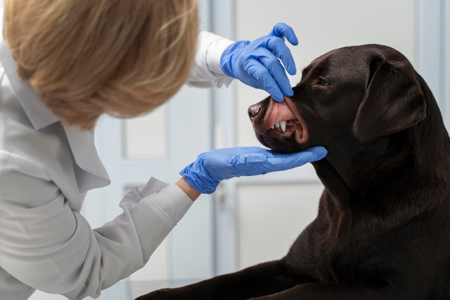
Following the history, a rigorous physical evaluation is performed. During the physical check-up, the veterinarian will meticulously inspect the dog’s external nose, checking for facial symmetry, pain upon palpation over the maxillary sinuses, and the specific character of any discharge. They will thoroughly examine the eyes for secondary inflammation and the oral cavity for severe periodontal disease, fractured teeth, or oronasal fistulas, keeping an eye out for signs of diffuse inflammation, purulent discharge, or traumatic injury – symptoms that can initially appear similar to a common, benign cold in humans. Crucially, the vet will evaluate the dog’s overall systemic well-being, auscultate the lungs to rule out descending pneumonia, and check for any irregular breathing patterns or related symptoms. They will perform an airflow test (often using a chilled glass slide held in front of the nares) to objectively assess for localized nasal blockages or sinus pressure that can make normal physiological breathing exceedingly difficult for the dog.[6]
3. Diagnostic Imaging
Because the complex internal structures of the nasal cavity are hidden within the bony vault of the skull, diagnostic imaging techniques are absolutely mandatory for evaluating chronic nasal disease. Traditional skull X-rays can occasionally provide some basic information regarding severe tooth root abscesses or massive bone destruction, but they are notoriously difficult to interpret due to the superimposition of complex facial bones. Therefore, advanced cross-sectional imaging, specifically Computed Tomography (CT) scans, are now considered the gold standard in veterinary medicine. CT scans provide incredibly detailed, three-dimensional views of the nasal passages and sinuses, unveiling minute structural abnormalities, specific areas of turbinate lysis (classic for fungal disease), soft tissue masses or tumors, and deeper signs of chronic infection. These high-resolution images provide invaluable architectural insights that are definitively required to help diagnose dogs’ destructive sinusitis, map the extent of a tumor before surgery, or identify a deeply lodged foreign object.[7]
4. Rhinoscopy
Once cross-sectional imaging has pinpointed the specific areas of pathology, the clinician needs to visually inspect the tissues. Rhinoscopy is a highly specialized, minimally invasive procedure performed under general anesthesia that enables the veterinarian to directly examine the deep internal mucosa of the nasal passages using either a flexible or rigid fiber-optic endoscope. This powerful diagnostic tool assists the specialist in directly identifying the gross appearance of the mucosal inflammation, spotting characteristic fungal plaques, visualizing specific anatomical blockages, or directly locating hidden foreign bodies, inflammatory polyps, or malignant tumors that could profoundly contribute to the ongoing nasal infection. Rhinoscopy allows the clinician to assess the exact nature of the disease in real-time, thereby decisively helping diagnose specific types of rhinitis.[8]
5. Laboratory Tests
To support the physical and imaging findings, comprehensive laboratory analysis is crucial. Systemic laboratory tests, such as a complete blood count (CBC) and serum biochemistry profile, can help definitively rule out broader systemic illnesses, endocrine disorders, or generalized septic infections that might be indirectly causing the presenting nasal symptoms. Additionally, targeted local sampling is performed. During rhinoscopy, a deep nasal swab or a sterile fluid wash sample can be precisely collected and submitted to an outside laboratory. This fluid is analyzed via aerobic and anaerobic culture and sensitivity testing, and advanced Polymerase Chain Reaction (PCR) panels to screen for the exact presence of specific pathogenic bacteria, destructive fungi, or elusive viral DNA. These advanced assays, alongside cytological examination of the cells, can identify the agents that may be primarily causing the intense inflammation of the delicate mucous membranes, thus accurately pinpointing the specific microbial cause of the stubborn infection.[9]
6. Biopsy
Ultimately, visual inspection and imaging cannot definitively differentiate between severe chronic inflammation and malignant cancer; cellular evidence is required. At times, a mucosal biopsy may be the only method to obtain a definitive, final diagnosis. A deep nasal biopsy entails carefully collecting a small, representative tissue sample directly from the most affected, abnormal area visualized during rhinoscopy or pinpointed on a CT scan. This vital tissue core is then submitted to a veterinary pathologist, who carefully evaluates it under a microscope to definitively identify the exact presence of deeply embedded fungal infection, characterize the precise cellular nature of chronic lymphoplasmacytic inflammation, or grade the aggressive cellular behavior of neoplastic, cancerous cells. Without histopathology, targeted treatment for chronic nasal disease is largely a guessing game.[10]
Given the highly complex and potentially aggressive nature of upper respiratory diseases, it is absolutely crucial to take your dog to a qualified veterinarian if you suspect a persistent nasal infection. Deep-seated sinusitis may also frequently occur in our companion dogs and cats, following pathophysiological pathways highly similar to humans, and these conditions can rapidly cause severe, life-threatening complications—such as extension of the infection into the central nervous system or systemic sepsis—if left undiagnosed and untreated for an extended period.
Treatment for Nasal Infections in Dogs

The successful therapeutic management of nasal infections in dogs is entirely dependent on achieving a highly accurate, definitive diagnosis. Because the etiologies range so widely, a “one size fits all” approach is virtually guaranteed to fail, particularly in chronic cases. The treatment for nasal infections in dogs depends deeply on identifying the primary underlying cause, understanding the extent of structural damage, and assessing the overall severity of the systemic condition. Here are the most standard, evidence-based treatment options frequently utilized for resolving nasal infections in dogs:[11]
1. Medication Management
Pharmacological intervention is the cornerstone of treating uncomplicated nasal disease, and various, highly specific medications might be suggested based on the exact origin of the nasal infection determined via culture or biopsy. For instance, targeted systemic antibiotics are employed strictly for documented bacterial infections, potent systemic or topical antifungal medicines are utilized for deep infections caused by an invasive fungus, and highly specific antiparasitic drugs are required for clearing parasitic mite infections. If diagnostic testing reveals that the persistent infection actually develops secondarily due to severe, underlying environmental allergies, immune-modulating drugs like antihistamines or potent corticosteroids may be utilized to aggressively manage the overactive allergic response and minimize the resulting cascade of inflammation.[12]
- Antibiotics: Antibacterial medications are critical, but they must be used judiciously. Antibiotics tackle active bacterial infections by directly annihilating the invading bacteria (bactericidal) or heavily restraining their cellular growth and replication (bacteriostatic). The attending vet may precisely prescribe systemic antibiotics depending entirely on the specific type of bacteria causing the localized infection, taking into careful consideration the results of advanced laboratory antibiotic susceptibility testing to avoid the growing threat of antibiotic resistance. Some frequently used, highly tissue-penetrating antibiotics specifically chosen for nasal infections in dogs include:
- Amoxicillin-clavulanate (a broad-spectrum penicillin combination highly effective against many oral and respiratory pathogens)
- Enrofloxacin (a potent fluoroquinolone often reserved for severe, deep-tissue infections)
- Doxycycline (a tetracycline antibiotic with excellent respiratory penetration and highly beneficial secondary anti-inflammatory properties)
- Antifungal medications: Treating a fungal infection like Aspergillosis is a major medical undertaking. Systemic antifungal medications aim to eradicate the deeply embedded fungi or severely prevent their further growth, though they often require months of administration and frequent liver enzyme monitoring due to potential hepatotoxicity. In many cases of sinonasal Aspergillosis, topical therapy—where the nasal cavity is flooded with a liquid antifungal under anesthesia—is vastly more effective than oral pills. Common medications prescribed to treat severe fungal nasal infections include:
- Itraconazole (a powerful systemic azole antifungal often used for long-term management)
- Fluconazole (an azole with excellent tissue and central nervous system penetration)
- Terbinafine (an allylamine antifungal frequently used in complex, synergistic combination therapies)
- Antiparasitic medications: If nasal mites are definitively diagnosed, highly targeted antiparasitic medications are employed to quickly annihilate or heavily inhibit the growth and reproduction of the offending parasites. Depending on the specific drug formulation and the exact type of parasite being treated, these potent medications may be administered orally, via injection, or applied topically as a spot-on treatment. Common, highly effective antiparasitic drugs frequently used for clearing nasal mite infestations include:
- Selamectin (a topical avermectin often found in monthly preventative products)
- Ivermectin (a macrocyclic lactone used off-label at specific doses to clear parasitic burdens)
- Milbemycin Oxime (an oral preventative commonly utilized for broad-spectrum parasite control)
- Anti-inflammatory medications: Chronic rhinitis is fundamentally a disease of uncontrolled inflammation. These specific medications are crucial to aggressively reduce severe inflammation and alleviate the profound pain and discomfort associated with nasal infections when the highly sensitive nasal cavity becomes acutely inflamed. Non-Steroidal Anti-Inflammatory Drugs (NSAIDs), like carprofen, meloxicam, or deracoxib, may be judiciously prescribed by a veterinarian for short-term use to safely alleviate painful tissue inflammation. In more severe, immune-mediated, or highly allergic cases, potent corticosteroids like oral prednisone or injectable dexamethasone may also be used to rapidly reduce profound inflammation, but they must always be used with extreme caution and tapered slowly due to their potential for inducing severe systemic side effects and secondary immunosuppression.[13]
- Antihistamines or allergy medications: For dogs diagnosed with atopy or environmental hypersensitivities, these drugs are prescribed to carefully manage allergic rhinitis by competitively blocking massive cellular histamine release and effectively reducing the exaggerated immune response to common environmental allergens. Common, relatively safe over-the-counter antihistamines frequently used off-label in dogs include Diphenhydramine (Benadryl), Cetirizine (Zyrtec), and Hydroxyzine. Sometimes, if the allergies are completely refractory to antihistamines, the vet may prescribe long-term systemic corticosteroids or more advanced immunomodulatory medications, like the calcineurin inhibitor cyclosporine (Atopica) or oclacitinib (Apoquel), to help safely and effectively manage severe, deeply entrenched allergies that drive the rhinitis.
In some highly specialized cases involving severe, chronic lymphoplasmacytic rhinitis that fails to respond to traditional antibiotics and steroids, veterinary internal medicine specialists may also prescribe alternative immunomodulatory medications, specialized hypoallergenic diets, or powerful immunosuppressive drugs to help manage the severe, ongoing mucosal inflammation and grant the dog a comfortable quality of life.
2. Nasal Irrigation
In many complicated instances of chronic rhinosinusitis, particularly when there is a massive accumulation of thick, deeply inspissated nasal discharge or a complete architectural blockage of the delicate turbinates by bacterial biofilms, therapeutic nasal irrigation may be highly recommended. This medical procedure is typically performed under deep sedation or full general anesthesia to ensure the patient’s airway is completely protected via an endotracheal tube. The procedure entails forcefully flushing the deep nasal passages and sinuses with large volumes of a sterile, warmed saline solution, sometimes combined with a specialized, diluted antiseptic or mucolytic nasal spray. This mechanical flushing action serves to physically dislodge and help expel hardened mucus, necrotic tissue debris, and billions of localized infectious agents, effectively breaking down resistant bacterial biofilms, fostering an environment conducive to mucosal healing, and immediately enhancing the dog’s ability to resume normal, unobstructed breathing.[14]
3. Removal of Foreign Bodies or Tumors
When the definitive diagnosis points to a physical obstruction, highly specialized surgical intervention or advanced endoscopic procedures will be absolutely necessary. If the chronic nasal infection is directly due to an inhaled foreign body, such as a barbed grass awn, the object can often be carefully visualized and successfully removed endoscopically using grasping forceps passed through a rigid rhinoscope, sparing the dog invasive surgery. However, if the blockage is caused by a massive, aggressive malignant tumor or a deeply embedded, chronic fungal granuloma that has destroyed the local bone, a highly invasive surgical procedure known as a rhinotomy—where the nasal cavity is surgically opened from the top of the muzzle—might be required to physically debulk the diseased tissue and establish drainage. In many heartbreaking situations involving malignant nasal tumors, surgery alone is insufficient. Instead, advanced veterinary oncology protocols, such as highly targeted stereotactic radiation therapy or specific systemic chemotherapy protocols, might be utilized to aggressively shrink the tumor, alleviate the severe clinical signs, and significantly extend the patient’s overall survival time.[15]
4. Dental Treatment
As previously established, the roots of the upper teeth share a highly intimate bony border with the nasal cavity. Therefore, comprehensive, professional veterinary dental treatment will absolutely be required if a severe, deep-seated dental infection (such as a carnassial tooth root abscess or an advanced oronasal fistula) is definitively identified as the root cause of the unilateral nasal infection. This highly specialized treatment process typically involves a full anesthetic dental procedure with full-mouth intraoral radiographs. The affected, diseased teeth must undergo surgical tooth extraction, or in some specialized instances, advanced endodontic root canal therapy to definitively resolve the deep bony infection. To completely prevent the constant, ongoing recurrence of the nasal infection, the veterinary surgeon must meticulously debride the infected fistula tract and meticulously perform a tension-free gingival mucosal flap to permanently seal the abnormal, open communication between the contaminated oral cavity and the sterile respiratory nasal passages.[16]
5. Supportive Care
Never underestimate the immense value of basic, high-quality nursing in the recovery process. Excellent supportive care is a crucial, non-negotiable component in the overall treatment plan for any upper respiratory patient. Because a dog heavily relies on its nose for thermal regulation and sensing its environment, a blocked nose is highly distressing. Supportive care could include administering subcutaneous or intravenous fluids to ensure the dog stays well-hydrated, which naturally helps to thin and mobilize thick respiratory mucus. Providing a deeply comfortable, stress-free, and immaculately clean recovery environment is essential. Utilizing a room humidifier or placing the dog in a steamy bathroom for short periods can highly effectively moisten the inflamed nasal passages, loosen crusted discharge, and make the dog feel significantly more comfortable. Intensive nutritional support may also be absolutely necessary, especially if the dog has been stubbornly reluctant to eat due to profound facial discomfort, painful mouth lesions, or a complete inability to smell their food. Warming highly palatable, aromatic canned food can often entice a highly anorexic, congested dog to begin eating again, providing the essential caloric energy required to mount a successful immune response against the ongoing infection.[17]
6. Long-Term Management
In the challenging, highly frustrating case of idiopathic chronic rhinitis—which is essentially a chronic, poorly understood, lifelong immune-mediated inflammatory disease characterized by permanent turbinate damage—ongoing, daily management may be absolutely necessary for the remainder of the dog’s life. While a permanent cure is highly unlikely, achieving an excellent quality of life is very possible. This long-term strategy could involve the meticulous, regular administration of targeted medications, such as low-dose oral corticosteroids, daily NSAIDs like piroxicam, or specialized mucolytic drugs. Routine, scheduled veterinary check-ups are required to continuously monitor the stability of the condition, adjust medication dosages, and aggressively treat any acute, secondary bacterial flare-ups as soon as they arise. Additionally, strict lifestyle modifications must be implemented, like actively avoiding known environmental allergens, preventing exposure to secondhand smoke, and utilizing air purifiers, to help effectively manage the dog’s daily symptoms and actively prevent severe, painful inflammatory flare-ups.[18]
Throughout any of these complex therapeutic processes, strict, unwavering adherence to your highly trained veterinarian’s specific recommendations for both active treatment and any necessary, ongoing follow-up care is absolutely vital. Open, regular communication with your dedicated veterinary team throughout the entire, sometimes lengthy treatment process will help ensure the best, most favorable possible outcome for your dog’s full recovery from a debilitating nasal infection. Occasionally, during these follow-up visits, the vet may need to conduct repeat, deep nasal cultures to definitively confirm if a persistent, highly resistant bacterial infection remains, or if another underlying, hidden factor, such as a smoldering viral infection operating silently without presenting initial classic viral symptoms, is actually the primary root cause continuing to trigger the relentless, deeply embedded infection. Remember to consult your veterinarian before making any changes to your pet’s care, particularly regarding long-term medication strategies.[19]
Preventing Nasal Infections in Dogs
While some causes of nasal disease, such as malignant tumors or highly specific immune-mediated conditions, are impossible to predict or entirely prevent, many of the most common infectious, environmental, and parasitic causes of upper respiratory distress are highly preventable. To proactively prevent painful and costly nasal infections in dogs, conscientious owners should strongly consider adopting and consistently practicing the following comprehensive, preventative health care measures:
- Maintain a clean environment: Hygiene is a fundamental pillar of respiratory health. Regularly and thoroughly clean your dog’s primary living area, frequently wash their soft bedding in hot water, and sanitize their chew toys to significantly reduce the overall environmental bacterial load and eliminate potential chances of recurrent infection.
- Avoid allergens: If your dog has a known history of atopy, work to actively identify and strictly minimize their daily exposure to highly irritating allergens, such as seasonal pollen, damp basement mold, or ubiquitous household dust mites, all of which may directly contribute to intense allergic inflammation and secondary nasal infections.
- Regular veterinary check-ups: Proactive medicine is the best medicine. Routine, bi-annual veterinary visits and comprehensive physical exams can help a trained clinician detect and promptly treat any minor underlying health issues, such as early-stage dental disease or mild allergies, long before they have the opportunity to slowly develop into much more severe, deeply entrenched problems, such as chronic, destructive nasal infections.
- Vaccinations: Immunization is your dog’s primary shield against severe viral pathogens. Ensure your dog is strictly kept current on all necessary core and non-core vaccinations, as adhering to modern protocols regarding vaccines can help prevent a multitude of highly contagious, severe respiratory viral infections, including canine distemper, adenovirus, and parainfluenza.
- Parasite control: Do not overlook the threat of microscopic invaders. Consistently administer high-quality, veterinarian-approved regular flea and tick preventatives to reduce the risk of parasite-borne infections and to specifically prevent infestations of irritating nasal mites that can severely affect the delicate mucosal lining of the nasal passages. Dogs are more susceptible to infections due to nasal mites when they are frequently in close, direct contact with unknown, unvetted dogs in public settings like off-leash parks.
- Dental hygiene: Oral health is intrinsically linked to respiratory health. Practice excellent, daily dental hygiene for your dog by brushing their teeth with enzymatic veterinary toothpaste, and schedule regular professional anesthetic dental cleanings, as deeply rooted, progressive dental problems can and frequently do sometimes lead directly to catastrophic, bone-destroying nasal infections.
- Proper grooming: A clean coat supports a healthy immune system. Regular, thorough grooming and bathing can help keep your dog’s thick coat and skin highly clean and physically free of trapped environmental allergens, fungal spores, and chemical irritants that could easily be inhaled into the nasal cavity.
- Monitor outdoor activities: Vigilance during exercise is key. Closely supervise your dog while they are actively playing outdoors, particularly in areas with tall, dry grass, to proactively prevent the accidental, forceful inhalation or ingestion of sharp plant awns, foxtails, or other physical foreign objects that could easily become lodged and cause highly destructive foreign bodies in the nasal passages.
- Avoid exposure to infected animals: Respiratory pathogens are highly contagious. Strictly limit your dog’s direct nose-to-nose contact with other dogs or animals that clearly show active clinical signs of upper respiratory infections, such as coughing, sneezing, or ocular discharge, as these viral and bacterial infections can be rapidly and easily transmitted.
- Strengthen the immune system: A robust host defense mechanism is the ultimate preventative. Provide your dog with a highly balanced, nutritionally complete diet rich in essential fatty acids and antioxidants, ensure they receive regular, appropriate exercise, and provide proper, uninterrupted rest to holistically support a highly functional, healthy immune system and significantly reduce their overall baseline risk of developing opportunistic infections.
By diligently following these highly effective, common-sense preventative measures, proactive dog owners can significantly reduce the statistical likelihood of their beloved dog developing a severe, debilitating nasal infection and help flawlessly maintain its overall, long-term respiratory health. If, despite your best efforts, you suspect your dog is actively experiencing the painful symptoms of a nasal infection, do not delay—consult your trusted veterinarian immediately for a proper, thorough diagnosis and an aggressive, targeted medical treatment plan to ensure a swift return to normal function.
Frequently Asked Questions
What home remedy can I give my dog for nasal infections in dogs?
While definitive diagnosis and targeted medication by a licensed veterinarian are absolutely critical, some supportive home care can help soothe your dog’s symptoms while awaiting a professional appointment. You can temporarily increase ambient humidity by bringing your dog into a steamy bathroom for 15 minutes, which helps liquefy thick nasal secretions. Wiping crusty discharge from the nares with a warm, damp washcloth improves comfort and airflow. Enticing them to eat by slightly warming highly aromatic, wet dog food can also prevent the rapid weight loss often associated with the loss of smell. However, never administer human cold medications, decongestants, or pain relievers without direct veterinary authorization, as many common human drugs, including ibuprofen and certain antihistamine-decongestant combinations, are highly toxic and potentially fatal to canine patients.
Can bad teeth cause sinus problems in dogs?
Yes, advanced dental disease is a highly significant and frequently underdiagnosed cause of chronic sinus and nasal problems in dogs. The roots of the maxillary (upper) canine teeth and the large premolars are separated from the delicate nasal cavity by only a millimeter-thin layer of porous bone. When severe periodontal disease or an endodontic tooth root abscess develops, the aggressive bacterial infection can rapidly dissolve this thin bony barrier. This creates an abnormal, permanent passageway called an oronasal fistula. Once this fistula forms, oral bacteria, food particles, and saliva are constantly pushed up into the nasal passages, resulting in a severe, highly malodorous, and typically unilateral (one-sided) purulent rhinitis that will completely fail to resolve until the diseased tooth is surgically extracted and the fistula is closed by a veterinarian.
How are sinus infections different in dogs and humans?
While the fundamental cellular mechanisms of mucosal inflammation are similar, the structural anatomy, primary underlying causes, and clinical presentation of sinus disease differ significantly between humans and dogs. Humans frequently suffer from primary viral or allergic sinusitis, heavily influenced by our specific sinus drainage anatomy. Dogs, however, have a vastly more complex, highly vascularized labyrinth of nasal turbinates, making them incredibly prone to destructive secondary bacterial infections resulting from inhaled foreign bodies (like grass awns), aggressive fungal pathogens (like Aspergillosis), or severe dental diseases that breach the nasal floor. Furthermore, diagnosing dogs is inherently more complex, as they cannot vocalize their localized facial pain or headaches; instead, they rely on clinical signs like persistent unilateral discharge, severe stertorous breathing, reverse sneezing, or a sudden, profound refusal to eat due to a loss of olfactory function.
References
- Merck Veterinary Manual. Rhinitis and Sinusitis in Dogs and Cats. Merck & Co., Inc., 2023.
- VCA Animal Hospitals. Rhinitis and Sinusitis in Dogs. VCA Hospitals Publishing, 2022.
- PubMed. Canine Sino-nasal Aspergillosis: A Review. Journal of Veterinary Internal Medicine, 2017.
- American Veterinary Medical Association (AVMA). Canine Respiratory Disease Complex. AVMA Publications, 2024.
- ASPCA. Common Dog Diseases: Respiratory Conditions. ASPCA Pet Care, 2021.
- VCA Animal Hospitals. Reverse Sneezing in Dogs. VCA Hospitals Publishing, 2022.
- PubMed. Diagnostics for Canine Chronic Nasal Disease. Veterinary Clinics of North America, 2019.
- Merck Veterinary Manual. Disorders of the Nasal Cavity in Dogs. Merck & Co., Inc., 2023.
- PubMed. Histopathological classification of chronic rhinitis in dogs. Vet Pathol, 2019.
- Veterinary Information Network (VIN). Managing Chronic Upper Respiratory Disease. VIN, 2022.
- PubMed. Bacterial rhinitis and sinusitis in dogs. Compend Contin Educ Vet, 2011.
- American Veterinary Medical Association (AVMA). Antimicrobial Use and Resistance. AVMA, 2024.
- Texas A&M College of Veterinary Medicine. Canine Respiratory Diseases. Vetmed Pet Talk, 2020.
- PubMed. Efficacy of nasal irrigation in chronic rhinitis. J Small Anim Pract, 2018.
- Ohio State University Veterinary Medical Center. Canine Nasal Tumors. OSU Vet Extensions, 2023.
- PubMed. Oronasal fistulas secondary to dental disease in dogs. J Vet Dent, 2021.
- PubMed. Nasal mite infestation (Pneumonyssoides caninum) in dogs. Vet Parasitol, 2013.
- Merck Veterinary Manual. Systemic Pharmacotherapeutics of the Respiratory System. Merck & Co., Inc., 2023.
- Centers for Disease Control and Prevention (CDC). Healthy Pets, Healthy People: Dogs. CDC.gov, 2024.
- VCA Animal Hospitals. Sneezing and Nasal Discharge in Dogs. VCA Hospitals Publishing, 2022.
- PubMed. Foreign bodies in the canine nasal cavity. J Small Anim Pract, 2018.
- Merck Veterinary Manual. Allergies in Dogs. Merck & Co., Inc., 2023.
- PubMed. Allergic rhinitis in the dog: clinical presentation and management. Vet Dermatol, 2016.
- Veterinary Information Network (VIN). Epistaxis (Nosebleeds) in Dogs. VIN, 2021.
- World Health Organization (WHO). One Health: Human-Animal Interface. WHO, 2024.


