What are Corneal Ulcers in Cats?
What is it?
How is it Treated?
Breed Predispositions
Persian cats, Siamese cats, and Himalayan cats
Introduction
When Jenny noticed that her usually playful and energetic cat, Luna, was squinting and keeping one eye closed, she grew increasingly concerned. Luna was never one to shy away from her favorite toy, but she seemed disinterested and uncomfortable. Alarmed, Jenny took Luna to the vet, where she discovered that her precious cat had developed a corneal ulcer.
Corneal ulcers in cats denote the presence of open lesions or wounds on the cornea’s surface, the transparent outer layer of the cat’s eye. These ulcers transpire due to certain processes within the eye that cause corneal tissue damage or disintegration. Much like observing a deep corneal ulcer in a Shih Tzu, understanding the manifestation of corneal ulcers in cats is beneficial in recognizing this condition and seeking appropriate veterinary care.
This knowledge assists in addressing the root causes and fosters the affected eye’s healing process. It’s key to comprehend that a normal cornea should be free of these issues, and the presence of a defect in the cornea’s epithelium is called a corneal ulcer. Therefore, establishing whether there is a corneal ulcer is vital for maintaining your pet’s eye health.
What is the Cornea?
The cornea is the transparent, dome-shaped layer covering the eye’s front part in cats. It is a vital component of the eye’s structure and is crucial in maintaining clear vision. The cornea acts as a protective barrier, shielding the eye’s inner structures from injury and external elements. It also helps to focus light onto the lens and retina, contributing to the cat’s ability to see.
Composed of several layers, the cornea comprises specialized cells called epithelial cells, along with collagen fibers and a thin layer of endothelial cells on the inside. These layers work together to maintain the cornea’s clarity and integrity. The cornea is avascular, meaning it does not have blood vessels, which allows it to remain transparent and transmit light efficiently.
Overall, the cornea is a vital part of the feline visual system, and any damage or condition affecting it, such as corneal ulcers, can significantly impact a cat’s vision and overall eye health.
Causes of Corneal Ulcers in Cats
Corneal ulcers in cats, which may result in corneal scarring, arise from various factors, and grasping these causes is fundamental for successful diagnosis and treatment. Here are some prevalent origins of corneal ulcers in cats:
Injury
Various sources can inflict a traumatic injury on the eye. Incidents involving scratches from sharp objects, such as those experienced during cat fights or contact with prickly plants, can harm the cornea. Moreover, trauma resulting from excessive rubbing or scratching of the eye due to allergies or irritations can also instigate corneal ulcers.
Contaminations
Corneal ulcers can form due to bacterial, viral, or fungal contaminations. A familiar viral source of corneal ulcers in cats is the herpes virus. The Feline Herpesvirus (FHV-1) can trigger severe inflammation and recurring ulcers. Bacterial contaminations, such as Staphylococcus or Streptococcus, can arise as secondary issues to trauma or concurrent conjunctivitis. Although rarer, fungal infections can also lead to corneal ulcers under specific environmental conditions or in certain geographic regions. Hence, corneal ulcers include bacterial infections as a common cause.
Dry Eye (Keratoconjunctivitis Sicca)
A dry eye, a condition marked by insufficient tear production or poor tear quality, can escalate the susceptibility of the cornea to ulcers due to corneal dryness. A dry eye can result from immune-mediated conditions, congenital abnormalities, or medications inhibiting tear production.
Eyelid Irregularities
Eyelid irregularities such as entropion or ectropion can induce corneal ulcers. Entropion, a condition where the eyelid rolls inward, can cause the eyelashes or hair to scrape against the cornea. On the contrary, ectropion, where the eyelid droops outward, exposes the cornea, making it more prone to irritation and damage.
Brachycephalic Syndrome
Brachycephalic cat breeds, like Persians or Himalayans, possess shortened facial structures and are predisposed to corneal ulcers. Their flat faces and shallow eye sockets may lead to insufficient eye protection and poor tear distribution, making the cornea more susceptible to injury and ulcers.
Chemical or Thermal Burns
The cornea is also vulnerable to ulcers following burns from exposure to irritants, chemicals, or high temperatures. This can transpire if a cat interacts with potent cleaning agents, household chemicals, or hot substances.
It’s crucial to note that these causes can sometimes interconnect or occur simultaneously, leading to more severe or complicated corneal ulcers, such as indolent ulcers or superficial corneal ulcers. Foreign bodies in the eye can also prevent the proper healing of these ulcers. Therefore, prompt veterinary care is essential for accurate diagnosis and suitable treatment.
Symptoms of Feline Corneal Ulceration
The clinical signs of corneal ulcers in cats can fluctuate depending on the severity of the condition and its root cause. Here are some typical signs to observe:
- Frequent squinting or keeping the eye closed
- Excessive production of tears
- Redness and inflammation in the eye
- A cloudy or opaque appearance of the eye
- Increased sensitivity to light
- A tendency to rub or paw at the affected eye
- Behavioral changes
It’s crucial to remember that these symptoms can also be linked to other eye diseases, so it is of utmost importance to have your cat evaluated by a veterinarian at an animal hospital for a precise diagnosis. Early detection and treatment of corneal ulcers are critical to preventing complications, such as accumulating dead cells in the eye and preserving the cat’s vision.
Diagnosis of Corneal Ulceration in Cats
Veterinarians employ a variety of diagnostic techniques to diagnose corneal ulcers accurately, also called, also referred to as corneal abrasions, in cats. These techniques include:
Comprehensive Eye Examination
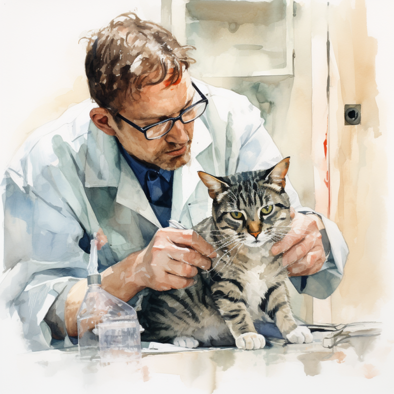
The vet will conduct a detailed eye examination using a slit lamp biomicroscope, facilitating in-depth cornea visualization. This procedure allows them to evaluate the ulcer’s location, size, and depth in the corneal stroma and examine any signs of inflammation or infection associated with the ulcer.
Fluorescein Stain Application
Fluorescein dye is used in the diagnosis of corneal ulcers. This dye is applied to the eye’s surface and adheres to areas of the cornea where the epithelium has been compromised, like ulcers or superficial corneal abrasions. Under blue light, these regions glow bright green, enabling the veterinarian to pinpoint and assess the corneal ulcer’s size, shape, and depth.
Tonometry Test
This test employs a tonometer to measure the intraocular pressure. If the pressure is elevated, it might indicate secondary glaucoma, possibly due to corneal ulceration or inflammation. Regularly checking the pressure helps assess the condition’s severity and directs the course of treatment.
Assessment of Tear Production
The quantity of tear production is assessed using specific tests like the Schirmer tear test. This measures the volume of tears produced by the cat’s eyes over a particular period. Insufficient tear production could lead to corneal ulcers; thus, evaluating tear production assists in recognizing underlying conditions such as dry eye (keratoconjunctivitis sicca).
Corneal Culture and Sensitivity Test
If a suspected infection occurs, a sample from the ulcer might be collected and sent to a lab for culture and sensitivity testing. This helps identify the specific bacteria or fungi involved in the infection and determines the most effective antimicrobial treatment.
Ocular Ultrasound
Ocular ultrasound may be performed in complex cases or when there’s suspicion of deeper involvement in the cornea. This non-invasive imaging technique offers detailed insights into the cornea’s inner layers, allowing the detection of any abnormalities or foreign bodies that might be contributing to the ulceration.
These diagnostic techniques are crucial in understanding the nature and severity of a cat’s corneal ulcer or abrasions. They guide appropriate treatment decisions and ensure optimal care for the cat’s eyes. If a corneal rupture is suspected or a corneal ulcer is diagnosed, prompt veterinary attention should be sought for a thorough examination.
Treatment for Corneal Ulcers in Cats
Diagnosing corneal ulcers in cats involves several steps. Here’s an expanded outline of the process:
Complete Medical History
The veterinarian will start by taking a full medical history of your cat, asking questions about the onset of symptoms, whether the cat has had any recent accidents or injuries, and if there have been any changes in behavior. This can also help the vet identify conditions like corneal sequestrum, a disorder often seen in cats where a dark plaque forms on the cornea.
Physical Examination
The vet will then perform a physical examination, focusing primarily on the eye. They will look for signs of an ulcer or corneal erosion, such as cloudiness, redness, tearing, or pain, such as squinting.
Fluorescein Stain Test
The main diagnostic tool for corneal ulcers is a fluorescein stain test. This is where the vet places a small amount of this orange dye on the eye’s surface. The dye adheres to any damaged areas of the cornea, including ulcers, and glows green under blue light. This allows the vet to see the size, shape, and depth of the ulcer and to assess if corneal perforation is imminent or if there are signs of corneal melting, a serious condition where the cornea starts to dissolve.
Tear Production Test
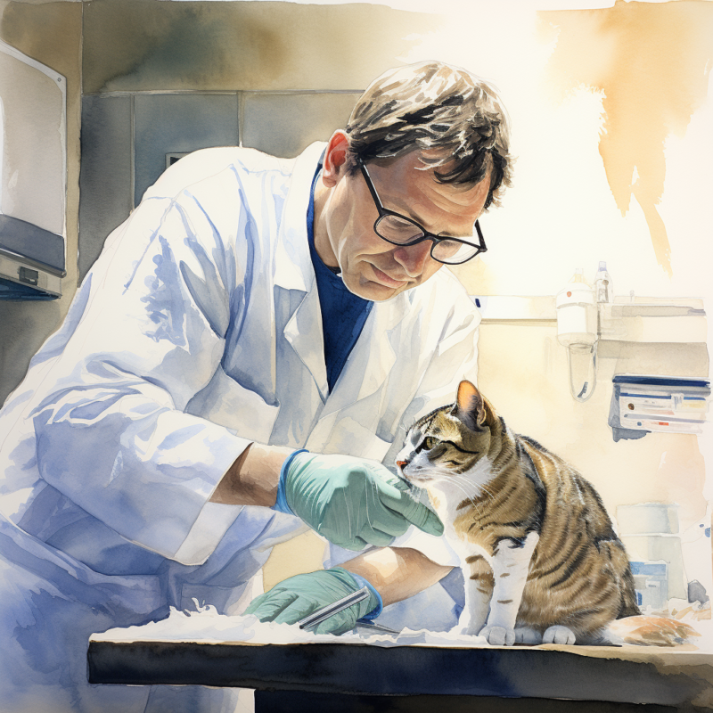
The vet may perform a Schirmer tear test, especially if they suspect the ulcer may have been caused by dry eye, also known as keratoconjunctivitis sicca. This test measures the amount of tear production and can help determine if inadequate tear production could contribute to the problem.
Intraocular Pressure Measurement
To rule out conditions like glaucoma, the vet may measure the intraocular pressure (pressure inside the eye) using a tonometer tool. This is crucial, as high and low intraocular pressure can result in corneal issues.
Additional Diagnostic Tests
In some cases, additional tests may be necessary. These could include bacterial or fungal cultures if an infection is suspected. Should the ulcer not respond to initial treatment, a superficial keratectomy, a procedure where the vet removes the unhealthy layer of the cornea, may be necessary. A veterinary ophthalmologist usually performs this procedure.
In the case of persistent corneal ulcers, or corneal sequestration, the cat may need to wear eye patches between treatments with topical eye medications.
By combining these methods, the veterinarian can not only diagnose the presence of a corneal ulcer but also get insights into possible underlying causes that will guide the days of treatment. If the ulcer is more profound than 50% of the cornea, or if treatment doesn’t show results, seek a board-certified veterinary eye doctor.
Recovery After Treating Corneal Ulcers in Cats
After receiving treatment for corneal ulcers, cats enter the recovery phase, where the healing process begins. The duration of the recovery period can vary depending on the severity of the ulcer and other individual factors. Minor ulcers may heal within a week or two, while more severe or complex ulcers may require several weeks or months to heal fully. Following your veterinarian’s instructions and attending any scheduled follow-up examinations is crucial.
Follow-up examinations are crucial in monitoring the progress of ulcer healing and assessing your cat’s overall eye health. Your veterinarian will carefully evaluate the healing status of the ulcer and make any necessary adjustments to the treatment plan. It is important to continue administering any prescribed medications, such as topical ointments or eye drops, as directed by your veterinarian. These medications help prevent infection, reduce inflammation, and support healing. Additionally, it would be best to take measures to prevent further injury or irritation to the healing cornea, such as protecting your cat’s eyes from potential trauma and avoiding exposure to irritants like dust or chemicals.
Throughout the recovery period, closely monitor your cat’s behavior and progress. Look for any signs of pain, discomfort, or worsening symptoms. If you notice any concerning changes, contact your veterinarian for further guidance. They may provide additional care instructions tailored to your cat’s needs, including continued medication, regular eye rinsing, or dietary modifications. Following these instructions and providing a safe and supportive environment can help facilitate a successful recovery for your cat’s corneal ulcer.
Prevention of Corneal Ulcers in Cats
Pet owners can take several measures to prevent corneal ulcers in their cats. First, it’s important to maintain a safe environment for your cat by removing any potential hazards that could cause eye injuries. This includes sharp objects, cords, and other items that could harm the eyes.
- Good hygiene practices are also crucial. Regularly clean your cat’s face and around the eyes to remove debris and prevent the buildup of irritants. Use a clean, damp cloth or specialized eye wipes recommended by your veterinarian.
- Preventing exposure to irritants is essential. Avoid exposing your cat to chemicals, smoke, dust, or other airborne particles that can irritate the eyes. Keep your cat away from strong cleaning agents or any substances that may be harmful.
- If your cat spends time outdoors, supervise their activities to prevent eye injuries from sharp plants or branches. Consider using a protective collar or harness to discourage rubbing or scratching of the eyes.
- Regular veterinary check-ups are important for monitoring your cat’s eye health. Your veterinarian can identify any underlying conditions or risks for corneal ulcers and provide appropriate preventive measures or treatments.
- Lastly, addressing any underlying health issues, such as dry eye syndrome or feline herpesvirus, is crucial. Work closely with your veterinarian to manage and treat these conditions effectively, as they can increase the risk of corneal ulcers.
Following these preventive measures can help protect your cat’s eyes and reduce the risk of corneal ulcers. Always consult your veterinarian for personalized advice and guidance for your cat’s needs.
Frequently Asked Questions
Disclaimer: The information provided on this veterinary website is intended for general educational purposes only and should not be considered as a substitute for professional veterinary advice, diagnosis, or treatment. Always consult a licensed veterinarian for any concerns or questions regarding the health and well-being of your pet. This website does not claim to cover every possible situation or provide exhaustive knowledge on the subjects presented. The owners and contributors of this website are not responsible for any harm or loss that may result from the use or misuse of the information provided herein.

