


 February 10, 2023
 Phil Good, DVM
Introduction
Meet Max, an energetic and lovable golden retriever who loves to play fetch with his owner, Emily. One day, while chasing his favorite ball, Max suddenly limps to a halt, whimpering in pain. A trip to the vet reveals that Max is suffering from cranial cruciate disease, a common but serious orthopedic condition in dogs.
At Beyond Pets Animal Hospital, we know your pets are family, and we understand how difficult it can be when your pet suffers from cranial cruciate disease. As the vet who knows your name, we are here to provide you with gentle, compassionate care and all the information you need. Our experienced veterinarians will work closely with you to determine the best course of action for your beloved companion.
The cranial cruciate ligament (CCL) is an essential knee-joint component found in the hind limb of pets. It runs between the intercondylar fossa of the femur and the middle of the tibial plateau within the joint. In humans, it’s similar to the ACL in front of the knee. Unfortunately, many pets tear their CCL during regular activity, so pet owners need to be aware of this common problem. A CCL injury can cause limping, pain, and eventually chronic changes such as osteoarthritis if left untreated.
Incidence and Prevalence of Cruciate Ligament Injury in Dogs
The incidence and prevalence of cruciate disease (CCLD) in dogs is a growing concern among pet owners. This condition affects dogs of all sizes and ages, though it is rarely seen in cats. Certain breeds are more prone to CCLD than others, with Newfoundlands particularly susceptible to genetic inheritance. Female and neutered dogs are also at greater risk for developing CCLD, although the exact reason for this is unknown.
Early diagnosis and treatment of CCLD can help reduce symptoms’ severity and improve the affected animals’ quality of life. Pet owners should be aware of the signs and symptoms associated with CCLD, such as limping or difficulty rising from a resting position, so they can seek veterinary care as soon as possible if their pet shows any signs of the condition. Regular checkups with a veterinarian can also help identify any potential issues before they become serious problems.
What Causes and Symptoms of a Cranial Cruciate Ligament Injury in Dogs?
Trauma is the most common cause of ACL injury in humans but is rare in dogs. Instead, a mix of aging, obesity, physical fitness, conformation, and breed is more often responsible for CCLD (Cranial Cruciate Ligament Disease) in dogs. In dogs, tearing of the CCL is also common, and if left untreated, it may progress to a complete tear.
Common symptoms of CCL damage include difficulty rising, trouble jumping into the car, decreased activity level, and muscle atrophy. Other signs may include a popping noise, swelling inside the shin bone, and shifting weight away from the damaged leg when standing. If a dog’s ligament is partly torn or the meniscus is injured, it might become unable to bear any weight, hopping with three legs.
The most common sign of CCL damage is a limp in the affected limb. Lameness may begin as mild on-and-off lameness but can progress to non-weight bearing if left untreated.

Without prompt intervention, lameness will not resolve and cause progressive arthritis and pain. Therefore, you must seek veterinary care immediately if you suspect your dog has suffered from CCL damage. This way, appropriate treatment can be administered to reduce pain and improve mobility.
How is an ACL Tear Diagnosed?
A canine ACL (anterior cruciate ligament) tear is usually diagnosed through a combination of physical examination and imaging studies. The most common physical examination test used to diagnose an ACL tear in dogs is the “cranial drawer test.” This test involves gently pulling the tibia forward while stabilizing the femur and observing for abnormal movement of the tibia relative to the femur. If the tibia moves abnormally, it is indicative of the ligament rupture of the ACL.
Imaging studies, such as X-rays and MRIs, can also diagnose an ACL tear, and MRIs determine the extent of the injury. In addition, these studies provide a more detailed view of the knee joint and can help to rule out other conditions that may be causing similar symptoms.
Please note that a veterinarian should be experienced with orthopedic examinations to make a definitive diagnosis of an ACL tear.
What are the Risk Factors?
Canine cruciate ligament disease (CCLD) is more prevalent in dogs with a poor physical condition and a large weight gain. Pet owners can help to reduce these risks by providing regular exercise and avoiding intermittent, excessive activity. Regular exercise will help to prevent obesity and maintain a good fitness level, which is essential for the health of the dog’s joints. In addition, torn cranial cruciate ligament surgery carries a 15% complication rate, with potential complications including stretching or breakage of the suture and infection of the surgery site.
Surgical site infection, patellar tendonitis/luxation, surgical implant loosening or breakage, and tibial/fibula fractures are all possible side effects of TPLO and TTA surgeries. Medications and physical rehabilitation therapy can often fix most complications, although second surgery may be required to replace faulty implants or eliminate severe infections in certain circumstances. Pet owners need to understand these risks before deciding on any pet surgery.
When Should You Ask for Veterinary Advice for Repair Surgery?
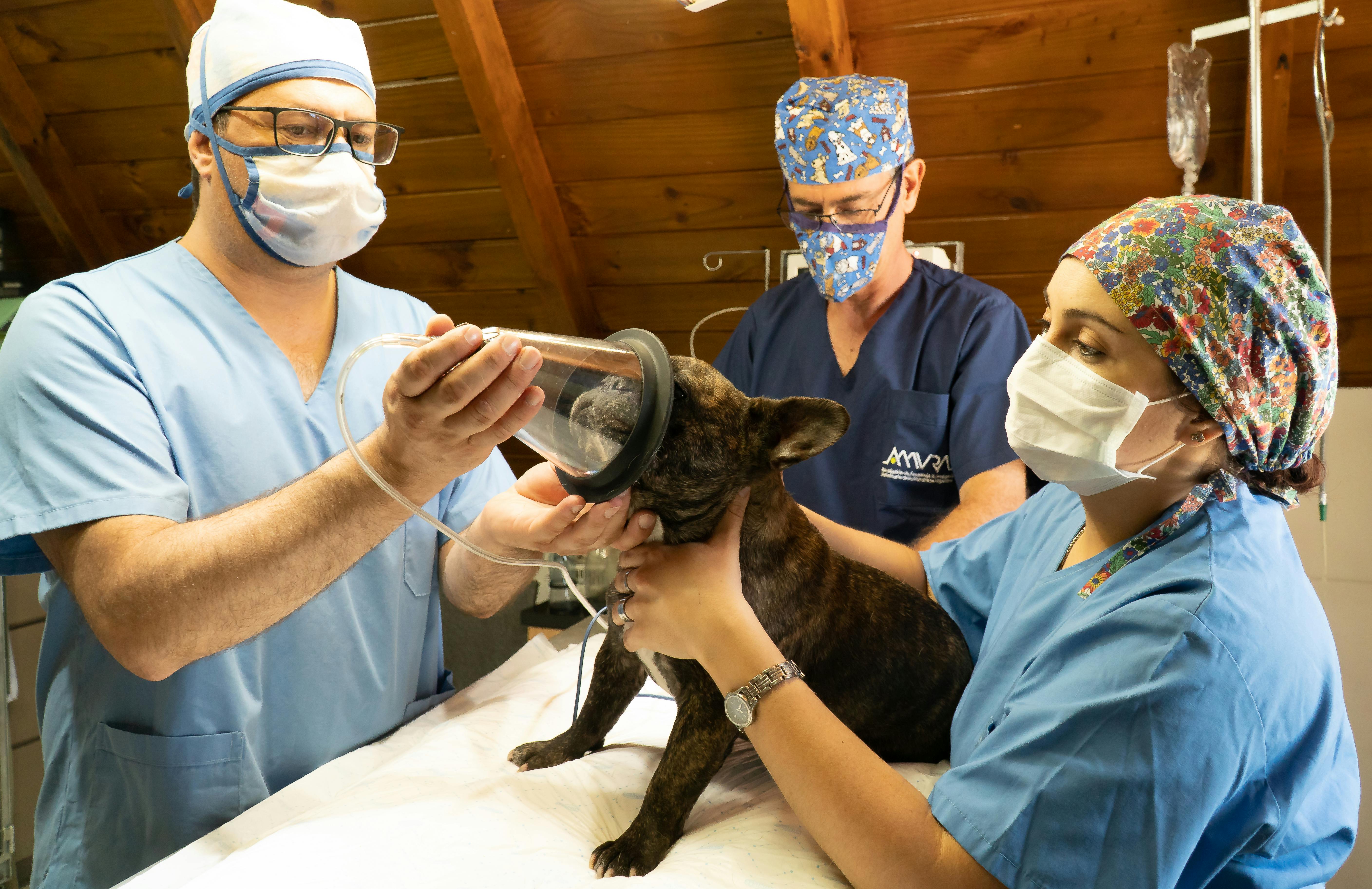
When it comes to your pet’s health, it is essential to know when to seek veterinary surgical advice. You should immediately contact your veterinarian if your dog exhibits obvious lameness that lasts more than a day or is in obvious discomfort. If the lameness is mild and there is no significant discomfort, it’s also important to rest your dog for a few days (no off-leash activity). Human pain medications and other over-the-counter drugs are commonly poisonous to your cat, so do not give them to him.
Before surgery, x-rays must be taken, and special views may be needed depending on the surgery. Routine blood work will also be performed before anesthesia to ensure that your pet can safely undergo the procedure. Your veterinarian will discuss all of these steps with you before scheduling any surgical procedures so that you know what needs to be done and why it needs to be done.
Surgical Treatment Options
Surgery is the only way to permanently control the instability in the stifle joint and evaluate the structures within the joint. Different surgical techniques can address stifle instability, including those based on other concepts. Possible complications associated with surgical treatment include anesthesia-related issues, wound problems, infection, patellar luxation, and failure to return to normal function. In addition, delayed bone healing, non-healing of the bone, bone healing in an incorrect position, and screw/plate failure/breakage are all complications specific to osteotomy techniques.
When considering surgical treatment for stifle instability, it is crucial to be aware of potential complications. An experienced surgeon must perform any procedure, and that exercise restriction guidelines must be followed closely. However, these complications are uncommon and may need multiple revision operations if they need to be appropriately managed. Therefore, patients must associate surgery with their doctor before deciding to proceed.
Osteotomy Techniques
Osteotomy techniques change how the quadriceps muscles act on the tibial plateau to stabilize the stifle joint. At Colorado State University, Tibial Plateau Leveling Osteotomy (TPLO) is generally preferred for large, active dogs. This technique involves making a circular cut in the tibial plateau and rotating it until it attains a relatively level orientation. The benefit of this technique is that it provides superior outcomes compared to traditional suture techniques, especially in young, large-breed dogs. However, there is also a significant drawback: performing an osteotomy may require multiple revision surgeries if complications arise.
The TTA technique also provides stability without replacing the CCL by advancing the attachment of the muscle. This method is successful in some cases but not as reliable as TPLO for larger breeds. Therefore, discussing with your veterinarian which technique would be best for your pet’s case is essential.
Tibial Plateau Leveling Osteotomy (TPLO)
Tibial plateau leveling osteotomy (TPLO) is a surgical technique to treat dogs’ cranial cruciate ligament (CCL) injuries. This procedure involves making a circular cut in the tibial plateau and rotating the contact surface of this bone until it attains a relatively level orientation. The most significant benefit of TPLO is the perceived superior outcome compared to traditional suture techniques, especially in young, large-breed dogs.
In addition, this technique can provide excellent stability to the stifle joint by either advancing the attachment of the muscle (TTA) or rotating the plateau (TPLO). At Colorado State University, TPLO is generally preferred for large, active dogs due to its higher success rate. More than 90% of dogs will resume their normal activities after undergoing TPLO or TTA surgery.
The major drawback of TPLO is that it requires an osteotomy, which may require multiple revision surgeries if complications arise. To modify the function of the quadriceps muscles on the top of the shin bone, osteotomies involve making a bone cut.
This can be a difficult and complex procedure, so it is essential to discuss all risks associated with surgery with your veterinarian before deciding on whether or not to proceed.
Tibial Tuberosity Advancement (TTA)
Tibial Tuberosity Advancement (TTA) is a surgical procedure used to treat small animals suffering from patellar luxation with retro patellar chondromalacia. It involves making a linear cut along the front of the shin bone and advancing the tibial tuberosity forward to orient the attachment of the quadriceps approximately 90 degrees to the tibial plateau, rendering the knee stable and independent of the CCL.
A biomechanical model of joint forces in the canine stifle was developed, which showed that moving the patellar ligament at 90° to the tibial plateau at full extension would annihilate cranial shear and cranial tibial thrust.
Measurements of the angle between the patellar ligament and tibial plateau during motion showed that TTA could shift loading from the cranial cruciate ligament to the caudal cruciate ligament at 90° flexion. In addition, an in vitro study demonstrated that TTA could reduce the cranial tibial thrust force by up to 75%.
TTA is a less invasive procedure than TPLO and is sometimes successful. However, it is less reliable than TPLO for larger breeds and may require additional surgery if complications arise. So, you’ll need to talk with your veterinarian about the best technique for your pet’s specific condition.
Suture Techniques
Suture techniques are a standard method of stabilizing the knee joint in dogs. Intra-articular replacement of the ACL has been studied extensively but found to be unsuccessful due to anatomical and disease process differences. Therefore, extracapsular suture stabilization is currently the most commonly used technique, utilizing strong suture material placed outside the knee joint to stabilize it.
This technique replaces the function of a defective CCL on the outside of the joint and has several benefits, such as lower cost and no bone cut, meaning fewer associated complications. However, some potential complications are associated with this technique, such as suture failure or progressive development of arthritis, with more significant active dogs being more prone to suture failure.
Overall, suture techniques can effectively stabilize a dog’s knee joint when other methods are not available or appropriate. Veterinarians must assess each case carefully before deciding which approach is best for their patients. With proper design and postoperative care, suture techniques can achieve a successful outcome for many dogs.
Extra-Capsular Suture Stabilization
Extracapsular suture stabilization is a technique used to replace the function of a defective CCL on the outside of the joint. This procedure involves placing strong suture material just outside the knee joint to stabilize it and is currently performed in an extra-articular fashion in dogs. Results are largely successful, with success rates of at least 85% and infection rates between 1% and 4%.
It has several benefits over the intra-articular replacement of the ACL, including lower cost and lack of a bone cut, reducing associated complications. However, some potential complications are associated with this technique, such as suture failure and progressive development of arthritis, with more significant active dogs being more prone to suture failure.
Overall, extracapsular suture stabilization is a viable option for replacing the function of a defective CCL in dogs. While there are some potential risks involved with this procedure, its benefits make it an attractive option for many pet owners looking for an effective way to treat their pet’s injury without breaking the bank. In addition, with proper care and monitoring after surgery, most pets can return to normal activities with no long-term complications.
Potential Complications Following Cruciate Ligament Repair

Surgical treatment can be a beneficial and effective way to treat certain medical conditions, but it is essential to be aware of the potential complications that may arise. Complications associated with surgical treatment can range from anesthesia-related issues, wound problems, infection, patellar luxation, and failure to return to normal function.
Osteotomy procedures risk delayed bone healing, non-healing of the bone, misalignment during the healing process, bone breakage, and screws or plates becoming damaged. These complications can be severe and require multiple revision surgeries; however, they are rare when performed by an experienced surgeon and exercise restriction guidelines are followed.
Facts about Canine and Feline Cranial Cruciate Disease
Cranial cruciate disease (CCD) is a common orthopedic condition in dogs and cats. It is caused by the rupture of the cranial cruciate ligament, which stabilizes the knee joint. CCD can cause pain, lameness, and instability in affected pets, leading to decreased quality of life and mobility. Treatment options vary depending on the severity of the condition.
Still, they can include non-surgical management, such as rest and physical therapy, or surgical intervention, such as extracapsular suture stabilization or tibial plateau leveling osteotomy. Regardless of the treatment chosen, following up with regular checkups and exercise/activity restrictions is essential to ensure a successful outcome.
#1: Hindlimb lameness in pets is often caused by a torn cruciate ligament.
Tearing a cruciate ligament is one of the most frequent reasons pets become lame in their hindlimb. Cranial cruciate disease is a condition that affects large and giant breeds more than smaller breeds. While cats can suffer from this injury, it is rare.
The cause of cruciate ligament injury in dogs is largely unknown, but genetic factors are likely to be the most important. Certain breeds are more predisposed to this injury, including Labradors, Rottweilers, Boxers, West Highland White Terriers, and Newfoundlands.
Other factors such as obesity, joint conformation, hormonal imbalance, and certain inflammatory disorders may also raise the danger of injury.
Pet owners need to be aware of the signs and symptoms of a ruptured cruciate ligament so they can seek prompt medical attention if their pet shows any signs of lameness or discomfort in their hindlimbs. In addition, early diagnosis and treatment can help to reduce the risk of long-term complications such as osteoarthritis.
Approximately 40-60 percent of dogs with a ruptured CCL will also rupture the other.
Cranial cruciate disease in pets is a common condition that can cause significant pain and lameness. Unfortunately, dogs are more likely to injure the opposite leg after suffering one CCL tear, suggesting that pets are not damaging normal, healthy ligaments but rather ones undergoing slow, chronic degeneration. In addition, partial tears often progress into complete tears, making it an unfortunate reality for pet owners when treated for the cranial cruciate disease.
Surgery is the most efficient way to stabilize a ruptured CCL in pets. Different surgical approaches are available, including suture-based techniques and TPLO procedures. You must consult a board-certified veterinary surgeon to determine which method suits your pet’s needs.
Recovery time varies depending on the type of surgery performed and the severity of the injury; however, up to 40% to 60% of dogs that rupture one CCL will eventually rupture the other. Therefore, following up with regular checkups and exercise/activity restrictions is essential to ensure a successful outcome.
Surgery is usually the preferred method of treatment for ruptured CCL in animals.
Several surgical techniques, such as suturing and TPLO procedures, are available. You should consult a board-certified veterinary surgeon to determine the most appropriate treatment method for your animal. Following the surgery, your pet must have lots of rest and therapy to avoid any postoperative issues.
This can comprise using a sling, active range of motion exercises, gentle lead strolls, and delivering pain control. In addition, our veterinary staff can offer precise directions and recommendations to certified rehabilitation specialists if necessary.
Owners must follow all postoperative care instructions provided by their veterinarian to ensure their pet’s recovery goes as smoothly as possible. Proper care after surgery can help reduce pain and swelling while promoting healing and preventing further injury or complications. With good care and attention, pets can fully recover from a ruptured CCL with minimal long-term effects.
Postoperative care is essential for animals with CCL disease.
Following surgery, it’s essential to monitor pets with CCL disease closely. After surgery, it is vital to provide adequate rest and rehabilitation for the pet to ensure a successful recovery. This may include using a sling, passive range of motion exercises, light leash walks, and pain control. Certified rehabilitation professionals can provide additional support during this time.
Veterinarians typically advise surgery as a solution for a CCL rupture in animals. Different surgical approaches are available, including suture-based techniques and TPLO procedures. It is essential to consult with a board-certified veterinary surgeon before making any decisions about treatment options. With proper care and attention during the postoperative period, pets with CCL disease can fully recover and return to normal activities.
Obese pets are at a higher risk of developing CCL disease.
Pets who are overweight are more likely to suffer from cranial cruciate ligament disease. This condition can cause orthopedic injuries and internal dysfunction, such as diabetes mellitus. To prevent this from happening, it is essential to ensure that your pet maintains a healthy weight through regular exercise and a balanced diet with little treats. Scripps Ranch Veterinary Hospital offers the gold standard experience for pets suffering from CCL disease.
Following orthopedic surgery for CCL sickness, animals necessitate therapeutic activity and rehabilitation to avoid potential difficulties. For instance, postoperative care can include using a sling, performing passive movement exercises, taking short-leashed strolls, and checking pain levels. To reach desired recuperation targets, it is suggested that you seek advice from a qualified rehab professional. With the proper care and consideration, your pet can completely heal from CCL illness and lead a life free of anguish or distress.


