


 October 23, 2022
 Phil Good, DVM
Canine Hip Dysplasia
This content was prepared with AI assistance and reviewed by a licensed professional for accuracy.
Introduction
Canine hip dysplasia is one of the most frequently diagnosed orthopedic conditions in veterinary medicine, representing a significant cause of chronic pain, lameness, and diminished quality of life in dogs. At its core, canine hip dysplasia is a developmental disorder characterized by an abnormal formation of the coxofemoral joint, which is the anatomical term for the hip joint. To understand this condition fully, it is essential to first understand the anatomy of a healthy canine hip. The hip is a classic ball-and-socket joint. The “ball” is the head of the femur (thigh bone), and the “socket” is the acetabulum, a concave structure located on the pelvis. In a structurally sound and healthy dog, the femoral head rests deeply and snugly within the acetabulum, providing a massive degree of stability while allowing for a smooth, frictionless range of motion during walking, running, and jumping.[1]
This seamless mechanical function is facilitated by several crucial anatomical components. The surfaces of both the femoral head and the acetabulum are coated with a layer of articular cartilage. This hyaline cartilage is incredibly smooth and acts as a shock absorber, distributing concussive forces across the joint. Furthermore, the entire joint is encased in a fibrous joint capsule that secretes synovial fluid, a viscous biological lubricant that further reduces friction and nourishes the avascular cartilage. A strong ligament called the round ligament (ligamentum teres) connects the tip of the femoral head directly to the deepest part of the acetabulum, anchoring it securely in place. The surrounding pelvic and thigh muscles provide dynamic stability, ensuring the joint remains congruous during intense physical activity.[2]
In a dog afflicted with canine hip dysplasia, this flawless biomechanical system breaks down. The condition begins during the puppy’s rapid growth phase, where a disparity occurs between the development of the skeletal structures and the supporting soft tissues. Specifically, the joint capsule and surrounding ligaments fail to hold the femoral head tightly within the acetabulum. This looseness is clinically referred to as joint laxity. Because the joint is loose, the ball partially slips out of the socket during weight-bearing activities—a phenomenon known as subluxation. This abnormal movement creates highly concentrated areas of stress on the cartilage and bone, setting off a cascade of destructive events that inevitably lead to joint degeneration and osteoarthritis. While it is widely known that large and giant breed dogs—such as German Shepherds, Labrador Retrievers, Golden Retrievers, Rottweilers, and Mastiffs—are disproportionately affected, it is a clinical misconception that small dogs are immune. Breeds like the French Bulldog, Pug, and even mixed-breed dogs of all sizes frequently suffer from this debilitating developmental condition.[3]
Degenerative Hip Dysplasia
Degenerative hip dysplasia refers to the chronic, progressive form of the disease that manifests as the dog ages, typically becoming most clinically apparent in middle-aged to senior dogs. This condition is intrinsically linked to secondary osteoarthritis (also known as degenerative joint disease), which develops as a direct consequence of long-term joint laxity and abnormal biomechanical wear. Over months and years, the constant subluxation and pounding of the loose femoral head against the rim of the acetabulum cause the protective layer of articular cartilage to degrade. This cartilage wear is not merely a mechanical scraping; it triggers a profound biological response. The injured cartilage cells (chondrocytes) release inflammatory mediators, including prostaglandins and destructive enzymes called matrix metalloproteinases, which further break down the cartilage matrix.[4]
As the cartilage layer thins, the underlying subchondral bone is exposed to unnatural friction and pressure. In an attempt to stabilize the unstable joint and repair the damage, the body begins to remodel the bone. The bone beneath the cartilage hardens and becomes incredibly dense, a process known as subchondral sclerosis. Simultaneously, the body produces new, abnormal bone growths around the margins of the joint, called osteophytes or bone spurs. These osteophytes physically alter the shape of the joint, changing it from a smooth, round ball-and-socket into a flattened, irregular, and highly dysfunctional structure. The joint capsule itself becomes chronically inflamed (synovitis) and thickens significantly, a process known as capsular fibrosis.[5]
The resulting degenerative hip dysplasia is intensely painful for the mature dog. Because the disease process is cyclical and progressive, the pain and loss of mobility tend to worsen over time. The once-fluid synovial fluid becomes thin and loses its lubricating properties, turning joint movement into a painful, grinding ordeal. While the genetic predisposition for the condition was present at birth, the degenerative changes seen in these older dogs are the culmination of a lifetime of micro-traumas to the joint structure. Managing this advanced stage of the disease requires comprehensive, multimodal veterinary intervention to control pain, reduce inflammation, and maintain a functional quality of life for the senior pet.[6]
Juvenile Hip Dysplasia
Unlike the chronic degenerative changes seen in older dogs, juvenile hip dysplasia presents entirely different clinical challenges and mechanisms of pain. This variant of the condition affects young, rapidly growing puppies, typically manifesting clinical signs between four and twelve months of age. The primary hallmark of juvenile hip dysplasia is extreme joint laxity. During this critical developmental window, the puppy’s skeletal frame is expanding at a remarkable rate, but in affected dogs, the supporting musculature, ligaments, and joint capsule do not develop synchronously to keep the hip joint tight. This profound looseness allows the femoral head to repeatedly subluxate—sliding in and out of the shallow acetabulum with every step the puppy takes.[7]
The pain experienced by puppies with juvenile hip dysplasia is acute and often severe, but it is not initially caused by arthritis. Instead, the pain stems from the overstretching and tearing of the joint capsule and the round ligament. The hip joint capsule is packed with highly sensitive nerve endings. When the femoral head subluxates, it places immense, unnatural traction on this capsule, resulting in sharp, tearing pain. Additionally, because the ball does not sit deeply in the socket, the entire weight of the dog’s hind end is concentrated on a very small area of the dorsal acetabular rim (the top edge of the socket). This point-loading forces the soft, pliable, growing bone to bear excessive weight, leading to painful microfractures along the rim of the socket.[8]
Interestingly, the clinical presentation of juvenile hip dysplasia often appears to spontaneously improve as the dog reaches 12 to 18 months of age. This period of apparent recovery is frequently misunderstood by pet owners as a “cure.” In reality, the body’s response to the continuous stretching and micro-trauma is to lay down thick, fibrotic scar tissue within the joint capsule. This fibrotic response ultimately thickens the capsule, reducing the severe laxity and stabilizing the joint just enough to alleviate the acute tearing pain. While the young dog may seem to move better and exhibit less overt pain, the underlying structural damage remains. The joint surfaces have already been compromised, the anatomy is malformed, and the foundation for lifelong, progressive osteoarthritis has been permanently set.[9]
Causes of Canine Hip Dysplasia

Canine hip dysplasia is universally recognized by the veterinary community as a multifactorial disease. This means that its onset and severity are not dictated by a single, isolated factor, but rather by a complex interplay of genetic predispositions and environmental influences. The abnormal formation of the hip joint is the fundamental defect, and understanding the myriad of factors that contribute to this developmental flaw is crucial for both management and prevention. As dogs age, the primary laxity driven by these causes invariably develops into degenerative joint disease, leading to the discomfort and mobility challenges that characterize the condition. A comprehensive understanding of the root causes is the first step in protecting vulnerable dogs.[10]
Genetic Predisposition: Genetics plays the most significant and foundational role in the development of hip dysplasia. The condition is considered polygenic, meaning that the genetic risk is determined by multiple genes interacting with one another, rather than a single dominant or recessive gene. Because of this polygenic nature, the inheritance pattern is highly complex. A dog can be a carrier of the genes that cause hip dysplasia without ever showing clinical signs themselves, making it incredibly difficult to eradicate from breeding lines. Certain large and giant breeds are genetically predisposed, including the Labrador Retriever, Golden Retriever, German Shepherd, Great Dane, Saint Bernard, and Rottweiler. However, smaller breeds with specific conformational traits, such as Bulldogs and Pugs, also carry a high genetic load for the disease. If a dog lacks the genetic code for hip dysplasia, they will not develop the condition, regardless of environmental factors. Conversely, if the genetic blueprint is present, environmental factors will heavily influence how severely the disease manifests.[11]
Rapid Growth: The speed at which a puppy grows during its first year of life is a critical environmental trigger for hip dysplasia. Puppies that experience exceptionally swift growth spurts are at a markedly elevated risk. When the skeletal system (the bones) lengthens and expands faster than the surrounding soft tissues (the muscles, ligaments, and tendons) can strengthen and adapt, the joints lose their necessary support structure. This asynchronous growth results in joint laxity. High-calorie diets that promote maximum, speedy growth or cause excessive weight gain in young puppies place extreme mechanical pressure on the soft, developing cartilaginous joints. This excessive force distorts the growing bones, leading to the flattened femoral heads and shallow acetabula that typify the abnormal development known as hip dysplasia.[12]
Nutrition: Unbalanced nutrition, particularly during the critical developmental window of a large-breed puppy (between two and eight months of age), is a potent catalyst for hip dysplasia. Diets that are too rich in calories drive the rapid growth mentioned above. Furthermore, the specific mineral balance of the diet—especially the ratio of calcium to phosphorus—is of paramount importance. Unlike adult dogs, young puppies lack the physiological ability to regulate how much calcium they absorb from their intestines. If they are fed a diet excessively high in calcium, they will absorb it all, leading to disruptions in endochondral ossification, the process by which cartilage turns into bone. This atypical bone development weakens the joint structure and encourages the subluxation characteristic of dysplastic hips.[13]
Obesity: Carrying excessive body fat is incredibly detrimental to canine joint health. Overweight dogs are more likely to develop hip dysplasia due to the immense mechanical stress the excess weight bears down on the vulnerable hip joints. Every extra pound exponentially increases the concussive forces transmitted through the coxofemoral joint during walking or running. This chronic mechanical overload accelerates the breakdown of the articular cartilage, leading to abnormal wear and tear and, ultimately, severe degenerative joint disease. Furthermore, adipose (fat) tissue is not inert; it acts as an active endocrine organ, secreting pro-inflammatory hormones called adipokines. These inflammatory mediators circulate throughout the body, directly contributing to the degradation of joint tissues and amplifying the pain associated with dysplasia.[14]
Exercise: The type, duration, and intensity of exercise a puppy receives profoundly impact the development of their hip joints. While moderate, low-impact exercise is essential for building the muscle mass required to support the hips and contributes positively to a dog’s overall health, strenuous or high-impact exercise during a puppy’s growth phase can severely enhance the risk of clinical hip dysplasia. Engaging a young, growing dog in activities like repetitive running on hard concrete surfaces, aggressive Frisbee jumping, or leaping from significant heights can impose excessive concussive stress on the soft, immature joints. These repetitive micro-traumas can stretch the joint capsule and damage the developing cartilage, resulting in permanent joint abnormalities.[15]
Environmental and Endocrine Factors: Beyond diet and exercise, other environmental and physiological factors can exacerbate the condition. Conditions such as cold and damp environments can intensify joint pain and stiffness in dogs already suffering from osteoarthritis secondary to hip dysplasia, potentially aggravating their clinical symptoms. Additionally, recent veterinary research suggests that endocrine factors, specifically the timing of spaying or neutering, may influence joint development. Early removal of sex hormones (estrogen and testosterone) delays the closure of the long bone growth plates. This allows the long bones of the legs to grow longer than they naturally would, altering the biomechanical angles of the hips and knees, which may place additional stress on dysplastic joints.[16]
In summary, while canine hip dysplasia is fundamentally an abnormal developmental condition strongly linked to genetic factors, the clinical expression of the disease is highly malleable. Responsible breeding practices form the first line of defense, but proper nutrition, strict weight management, and age-appropriate exercise can assist tremendously in minimizing the risk. By controlling the environmental variables, pet owners can significantly alter the trajectory of the disease, potentially preventing genetically susceptible young and mature dogs from developing the most severe, debilitating forms of this condition.
Signs of Hip Dysplasia in Dogs
The clinical manifestations of hip dysplasia in dogs are diverse and depend heavily on the age of the patient, the degree of joint laxity, and the amount of secondary osteoarthritis present. Because the underlying pathology changes as the dog ages—shifting from acute laxity and soft tissue pain to chronic structural degeneration and bone pain—veterinarians categorize the symptoms into two distinct phases: the juvenile form and the mature form. While these two forms share some overlapping similarities and can appear fluidly as a dog transitions through different life stages, they present unique characteristics that require careful observation by pet owners.
Juvenile Form
The juvenile form of hip dysplasia typically affects puppies between the ages of four and twelve months. During this rapid growth phase, the primary issue is profound joint laxity, meaning the ball is constantly slipping out of the socket. This subluxation causes intense, sharp pain due to the stretching of the highly innervated joint capsule and the creation of microfractures along the acetabular rim. Owners of affected puppies often notice a specific set of clinical signs related to this acute pain and mechanical instability.[17]
- A ‘bunny hopping’ gait: When running or climbing stairs, the puppy will move both hind legs forward together simultaneously, much like a rabbit. This mechanical compensation allows the dog to propel themselves forward while avoiding the painful extension and independent range of motion required by a normal trotting gait.
- Lameness in the rear leg or legs: The puppy may limp, favor one side, or exhibit a generalized weakness in the hindquarters, particularly after a bout of play.
- Challenges in rising from a resting position: Puppies with acute capsular pain will often struggle, slip, or exhibit reluctance when trying to stand up after a nap.
- A noticeable clicking or popping sound: When the dog is walking, standing, or simply shifting weight, a palpable or audible “clunk” may occur as the subluxated femoral head violently slips back into the acetabular socket.
- A shift of weight to the front limbs: To relieve the painful pressure on their dysplastic hips, young dogs will purposefully shift their center of gravity forward, heavily relying on their front legs and shoulders.
- A limited ability to exercise for extended periods: Affected puppies will often sit down repeatedly during a walk or refuse to continue playing, displaying a profound exercise intolerance compared to their healthy littermates.
- Narrow stance: The puppy may stand with their hind feet placed unusually close together, attempting to mechanically lock the unstable hip joints in a more secure position.
It is essential to note that young dogs have a remarkable ability to adapt, and some may rarely show overt, crying signs of hip dysfunction despite severe joint pathology. Furthermore, as the puppy reaches 12 to 18 months of age, many can appear to recover from mild hip dysplasia. This apparent improvement occurs because the body produces thick fibrotic scar tissue around the joint capsule to stabilize the excessively loose joints. While the dog may seem pain-free, the underlying structural deformity remains. Research indicates that while approximately 30 percent of these dogs might require advanced surgical treatment later in life due to severe osteoarthritis progression, many dogs can manage the middle years of their life without further aggressive intervention, provided their weight is kept under strict control.[18]
Mature Form
The mature form of hip dysplasia represents the chronic, end-stage progression of the disease, typically seen in middle-aged to senior dogs. At this stage, the acute pain of joint laxity has been replaced by the deep, aching, chronic pain of severe osteoarthritis. The protective articular cartilage has been worn away, leading to bone-on-bone friction, dense subchondral sclerosis, and the formation of extensive osteophytes (bone spurs) that physically restrict the joint’s movement. The symptoms in mature dogs reflect this profound structural deterioration.[19]
- Progressive lameness in the rear limbs: The limping becomes a daily, chronic issue, worsening over months and years rather than appearing acutely.
- A ‘bunny hopping’ gait: The dog continues to use this compensatory movement to minimize the painful grinding of their arthritic joints during locomotion.
- Lameness following exercise: While the dog may “warm out” of their stiffness during a walk, they will often exhibit severe lameness, stiffness, and profound discomfort a few hours after exercise or the following morning.
- Loss of muscle mass in one or both rear limbs: Due to chronic disuse and reluctance to bear weight, the gluteal and thigh muscles (quadriceps and hamstrings) undergo severe atrophy, giving the dog’s hind end a sunken, bony appearance.
- Compensatory forelimb hypertrophy: Because the dog has spent years shifting their weight forward to avoid hip pain, the muscles of the chest, shoulders, and forelimbs often become heavily muscled and disproportionately large.
- Challenges in performing routine activities: The dog will display marked hesitation or outright refusal when asked to perform activities that require hindlimb power and hip extension, such as jumping into the car, climbing stairs, or getting onto furniture.
- Behavioral changes: The chronic, grinding pain of osteoarthritis takes a significant psychological toll. Dogs may become irritable, display uncharacteristic aggression when their hindquarters are touched, or become withdrawn and depressed.
These clinical signs vary depending on the severity of the original degree of hip laxity and the current state of joint degeneration. However, they uniformly indicate that the dog is experiencing a painful, debilitating condition, often secondary to severe hip dysplasia. Therefore, recognizing these insidious symptoms of hip dysplasia is absolutely essential for initiating timely veterinary intervention. Early detection and comprehensive management are the only ways to provide meaningful relief from the chronic pain from hip dysplasia that affected dogs must endure daily.
Diagnosis of Hip Dysplasia in Dogs

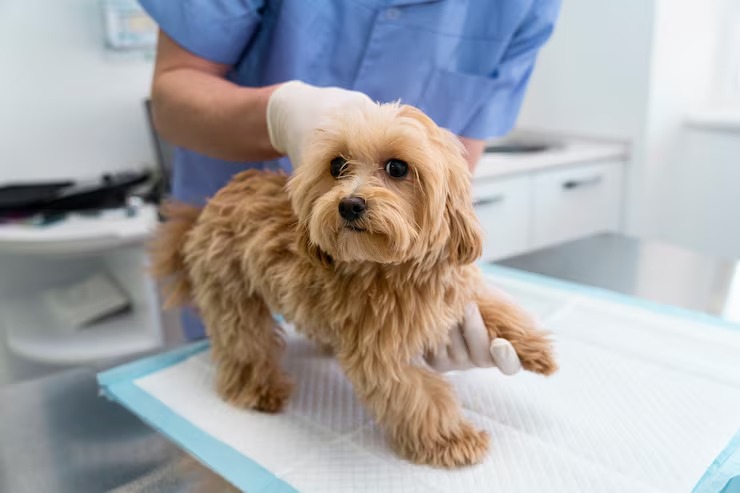
Identifying and accurately diagnosing hip dysplasia in dogs entails a highly structured, comprehensive approach. A definitive diagnosis cannot be made by visual observation alone; it requires combining thorough medical history analysis, detailed orthopedic physical assessment, and precise imaging techniques to determine both the presence of the disease and the specific severity of the condition. The typical diagnostic protocol employed by veterinary professionals involves several distinct but complementary stages:[20]
Medical History
The diagnostic process always begins with a detailed medical history. The veterinarian will gather precise information about the dog’s breed, current age, exact dietary history (including puppy growth formulas), and any observed symptoms like discomfort, exercise intolerance, or mobility difficulties. The clinician will ask targeted questions about when the signs first appeared, whether the lameness worsens after rest or after exercise, and if the dog exhibits specific compensatory behaviors like bunny hopping. This subjective data enables the veterinarian to establish if the dog fits the typical profile of a patient vulnerable to hip dysplasia and helps gauge the severity of the potential condition before laying hands on the animal.[21]
Physical Examination
Following the history, the veterinarian conducts a thorough, hands-on physical and orthopedic examination. The doctor will perform a visual gait analysis, watching the dog walk and trot to identify subtle weight shifts, lameness, or neurological deficits. During the hands-on assessment, the veterinarian checks for indications of pain, joint effusion (swelling), crepitus (a crunching feel within the joint), or a diminished range of motion in the dog’s hips—particularly focusing on pain elicited during the extension of the rear legs. They assess the symmetry and mass of the gluteal and thigh muscles to quantify any atrophy. Crucially, they carry out specific manipulative evaluations designed to mechanically test the integrity of the joint capsule. Tests such as the Ortolani sign or the Bardens test are designed specifically to check for joint laxity and subluxation. The Ortolani test, often performed under light sedation for accuracy, involves applying pressure to the femur to subluxate the hip, then slowly abducting the leg until a palpable “clunk” is felt as the femoral head drops back into the acetabulum, definitively confirming laxity.[22]
Radiographs (X-rays)
Employing advanced imaging techniques is strictly critical in diagnosing hip dysplasia since it provides the only way to visually examine the bony architecture of the hip joint and assess the extent of the pathology. For diagnostically accurate results, orthopedic X-rays must be taken while the dog is heavily sedated or fully anesthetized. This chemical restraint is mandatory to ensure complete muscle relaxation and perfectly symmetrical, straight positioning of the pelvis; even a slight pelvic tilt can artificially mimic or mask the signs of dysplasia. The standard view is the ventrodorsal (VD) hip-extended projection. These high-resolution images assist the veterinarian in evaluating the depth of the acetabulum, determining the degree of joint laxity (by checking if less than 50% of the femoral head is covered by the socket), identifying the presence of early osteoarthritis (such as the Morgan line, an early enthesophyte formation on the femoral neck), and noting any profound alterations to the overall bone structure of the hip joint.[23]
OFA (Orthopedic Foundation for Animals) Evaluation
In some instances, mainly when it concerns screening dogs for future breeding programs, the dog’s standard VD extended X-rays may be formally forwarded to the Orthopedic Foundation for Animals (OFA) for a professional, independent assessment. At the OFA, three board-certified veterinary radiologists evaluate the radiographs and provide a consensus grading of the dog’s hip conformation. The OFA employs a standardized, seven-point grading system (Excellent, Good, Fair, Borderline, Mild, Moderate, Severe) to ascertain the severity of hip dysplasia. The foundation provides official certification and a registry number only for dogs with normal hip function (Fair, Good, or Excellent). It is important to note that a dog must be at least 24 months of age to receive a permanent OFA certification, as skeletal maturity must be complete to ensure the final joint conformation is accurately represented.[24]
PennHIP Evaluation
An advanced, alternative, and highly predictive method to assess hip dysplasia is the PennHIP (University of Pennsylvania Hip Improvement Program) evaluation. Unlike the OFA, which largely assesses the presence of arthritis in mature dogs, PennHIP measures passive joint laxity far more accurately than traditional hip-extended X-rays and can be performed on puppies as young as 16 weeks of age. This specialized technique requires specific training and uses three different radiographic views: a standard VD extended view, a compression view to seat the femoral heads deeply, and a unique distraction view. The distraction view utilizes a specialized, custom-designed device called a distractor, which safely acts as a fulcrum to physically measure the maximum degree of joint laxity. The resulting radiographic measurements are mathematically expressed as a distraction index (DI), a number ranging from 0 to over 1. A lower DI (e.g., < 0.3) indicates a exceptionally tighter, healthier hip joint with virtually no risk of developing osteoarthritis, while a higher DI (e.g., > 0.7) signifies severe laxity and a near certainty of future dysplastic degeneration.[25]
Early diagnosis and aggressive, proactive intervention are absolutely crucial in managing hip dysplasia in dogs. Whether diagnosed via clinical suspicion, OFA screening, or early PennHIP testing, timely and appropriate medical or surgical treatment can help to significantly slow down the progression of the degenerative condition, preserve joint biomechanics, and drastically improve the dog’s long-term comfort and quality of life.
Treatment Options for Hip Dysplasia in Dogs

Managing canine hip dysplasia requires a highly tailored, patient-specific approach. Because the disease presents on a wide spectrum—from mildly loose hips in an asymptomatic puppy to severe, bone-on-bone arthritis in a geriatric dog—there is no single “cure-all.” Different therapeutic strategies are rigorously employed to manage the condition, with the optimal path mainly depending on the disease’s overall severity, the patient’s age, body weight, intended activity level, and comprehensive overall health status. In most clinical cases, veterinarians may propose a multimodal blend of medical treatment, strict weight regulation, targeted physical therapy, and, when indicated, surgical intervention. The overarching goal of any treatment plan is to alleviate clinical symptoms, halt or slow the progression of osteoarthritis, and enhance the dog’s daily quality of life. Below are the standard, evidence-based treatment methods for hip dysplasia in dogs:
Surgical Intervention
Surgical intervention may be strongly recommended in more severe cases of hip dysplasia, in very young dogs where we aim to alter the developmental trajectory of the joint, or when conservative, medical management is insufficient to control the animal’s chronic pain. Veterinary orthopedic surgery has advanced tremendously, offering procedures tailored to specific stages of the disease. If successful, early interventions can be highly curative; only 30% of young dogs treated with early, proactive surgical methods will need advanced, salvage therapies later in life. There are several primary surgical options for hip dysplasia in dogs, including:
- Juvenile Pubic Symphysiodesis (JPS): This highly time-sensitive, minimally invasive procedure is typically performed on very young puppies, strictly between the ages of 12 and 20 weeks (up to 5 months old). The goal of JPS is to permanently alter the angle of the growing pelvis to provide better coverage over the femoral head. The veterinary surgeon uses electrocautery to prematurely fuse the symphysis (the cartilage uniting the two halves of the pelvis on the bottom). As the puppy continues to grow dorsally but is restricted ventrally, the developing hip sockets are forced to rotate outward and downward into a more horizontal, aligned position over the ball. This dramatically increases joint stability, allows their hips to develop more normally, and can completely circumvent severe osteoarthritic complications later in life.
- Triple Pelvic Osteotomy (TPO) / Double Pelvic Osteotomy (DPO): This major surgical option greatly benefits young dogs (typically under 10-12 months of age) that have significant joint laxity but minimal to no arthritic joint damage. In a TPO or DPO, the veterinary surgeon physically cuts the pelvic bones in two or three specific locations. They then physically rotate the acetabulum outward to provide vastly better bony coverage for the loose femoral head, securing the newly aligned bone segments with specialized orthopedic bone plates and screws. By altering the biomechanics to eliminate subluxation, this surgery halts the destructive process before cartilage damage occurs.
- Femoral Head Osteotomy (FHO): This salvage procedure, formally known as an FHO surgery or femoral head and neck excision, is used when the joint is damaged beyond repair or when advanced joint replacements are financially unfeasible. It involves the complete surgical removal of the femoral head and neck—the ball-like part of the femur that connects to the hip joint. By removing the bone, the painful bone-on-bone grinding of severe arthritis is instantly eliminated. The body consequently heals by forming a “pseudo-joint” composed of thick, dense fibrous scar tissue, supported by the strong gluteal muscles. While it alters the dog’s biomechanical gait, it can reliably alleviate pain and improve functional mobility, especially in dogs weighing under 50 pounds.
- Total Hip Replacement (THR): Considered the absolute gold standard in veterinary orthopedics, this hip dysplasia surgery completely replaces the diseased, damaged biological hip joint with a highly advanced, bio-compatible artificial one, much like total hip replacement surgery in humans. The surgeon implants a titanium or cobalt-chrome stem into the femur and high-density polyethylene cup into the pelvis, which can be affixed using specialized bone cement or utilizing a cementless, bone-ingrowth technology. This surgical option provides the best, most normal long-term functional outcome for dogs with severe, crippling hip dysplasia, fully restoring normal range of motion and eliminating pain, though it is undeniably the most invasive, technically demanding, and expensive option available.
Prompt, precise intervention and a holistic, thoughtfully constructed treatment plan are vital for successfully managing dog hip dysplasia and preserving their long-term quality of life. Because the window for certain surgeries (like JPS and TPO) closes very early in a puppy’s life, working closely and proactively with a veterinarian or board-certified veterinary surgeon is crucial to determine the most suitable, stage-appropriate treatment options for each individual dog.
Conservative Treatment
When surgery is not an option due to financial constraints, the dog’s advanced age, concurrent medical conditions, or because the clinical signs are mild, conservative medical management becomes the cornerstone of care. This approach utilizes a multimodal strategy to reduce joint inflammation, block pain pathways, and support the remaining healthy cartilage. Conservative management is a lifelong commitment.
- Physical therapy: Formal veterinary rehabilitation comprises a highly structured set of strengthening exercises designed to bolster weak gluteal and thigh muscles, providing superior dynamic support to the unstable joint and improving the overall range of motion. Modalities such as underwater treadmill hydrotherapy are exceptional, as the water provides buoyancy to reduce weight-bearing stress while the resistance builds profound muscle mass. Additionally, this form of controlled exercise, combined with passive range of motion stretches, aids in averting future mobility complications associated with severe hip dysplasia.
- Acupuncture and Laser Therapy: Acupuncture is a traditional Chinese medical practice involving the precise insertion of tiny needles to stimulate nerve pathways, which has been used to treat various conditions for centuries. Recently, it has been gaining significant, evidence-based popularity as an adjunctive treatment for canine hip dysplasia. Some scientific evidence supports using acupuncture for treating osteoarthritis pain; for example, a study published in the Journal of Veterinary Medicine found that acupuncture effectively reduced pain scores and improved measurable mobility in dogs with hip dysplasia. Similarly, Class IV cold laser therapy (photobiomodulation) reduces tissue inflammation and increases local blood flow. If you are considering acupuncture to treat your dog’s hip dysplasia, always consult a qualified veterinarian or a certified veterinary acupuncturist.
- Medication: Pharmaceuticals are critical and can effectively alleviate clinical symptoms, particularly when the dog suffers extreme, chronic pain. Medications range from standard pain relievers to highly specific targeted therapies. The foundation of medical therapy relies on prescription nonsteroidal anti-inflammatory drugs (NSAIDs) to reduce joint swelling. Veterinarians often add prescription nerve-pain medication or other targeted therapies to block chronic pain wind-up in the spinal cord. Newer biological therapies, such as prescription monoclonal antibodies, target nerve growth factor to safely provide profound relief. Prescription anti-inflammatory steroids or advanced prescription pain medications may be used in acute crises. However, owners must be aware that chronic use of some systemic oral drugs can cause side effects like vomiting, diarrhea, gastrointestinal ulceration, liver enzyme elevation, and drowsiness.
- Supplemental feeding and Therapeutic Diets: Dietary intervention is another powerful option for dogs suffering from hip dysplasia. Supplemental feeding aims to drastically reduce excessive weight gain while supporting cartilage health and slowing bone density loss. When optimizing nutrition to manage weight and inflammation, some dogs require specialized diets. Many clinical diets are available for dogs with hip dysplasia, including grain-free, gluten-free, and hypoallergenic diets, which can be essential for systemic health. Grain-free diets contain fewer carbohydrates than regular diets, making them better choices for dogs who experience gastrointestinal issues, as maintaining optimal gut health is critical when administering chronic oral pain medications to prevent compounding systemic inflammation. Gluten-free diets are typically formulated from rice, corn, potato starch, soy protein, and tapioca. Hypoallergenic diets utilize novel proteins or hydrolyzed proteins and exclude common triggers like wheat, barley, rye, oats, milk, eggs, fish, shellfish, peanuts, tree nuts, and sesame seeds to reduce overall bodily inflammation. Supplemental feeding strategies should only be considered and implemented after consulting with a veterinarian.
- Joint supplements and Chondroprotectants: Nutraceuticals are widely utilized and highly beneficial as they supply essential biological building blocks and nutrients (typically glucosamine, chondroitin sulfate, avocado/soybean unsaponifiables, and Green Lipped Mussel extracts) that support healthy joint capsule function, enhance synovial fluid viscosity, and alleviate the underlying inflammation and pain associated with hip dysplasia. Furthermore, high doses of Omega-3 fatty acids (specifically EPA and DHA from marine sources) competitively inhibit the inflammatory pathways in the body. Prescription joint-protectant injections administered by your veterinarian are also highly effective at inhibiting destructive cartilage enzymes. Joint supplements present a remarkably safe and effective, long-term way to manage canine hip dysplasia without the risk of organ toxicity.
Prevention of Hip Dysplasia in Dogs
Prevention of hip dysplasia in dogs is an incredibly complex, multifaceted process encompassing meticulous genetic screening, precise nutritional management, and careful lifestyle moderation. Because the underlying genetic mechanics are polygenic, completely eradicating the disease from the canine population is extraordinarily difficult. While not every instance of hip dysplasia can be definitively forestalled, proactive dog owners and dedicated breeders can undertake specific, evidence-based steps to drastically mitigate the risk and foster the healthy, stable development of the hip joint, which relies heavily on its structurally sound ball and socket architecture. Here is how to potentially prevent or minimize the severity of hip dysplasia, which is notably common and devastating in large-breed dogs:
Prudent Breeding Practices
Undeniably, one of the most impactful methods to prevent hip dysplasia is the rigorous, scientific breeding of dogs with proven, robust hip health. Breeders must take responsibility and should conduct comprehensive radiographic hip evaluations like those offered by the Orthopedic Foundation for Animals (OFA) at two years of age, or ideally, the highly predictive PennHIP tests as early as 16 weeks. These objective metrics ensure only dogs with exceptional hip conformation and exceptionally low distraction indices are chosen for breeding programs. Advanced breeders also utilize Estimated Breeding Values (EBVs) to track genetic health across entire pedigrees. Prospective puppy buyers must be vigilant; always steer clear of buying puppies from breeders who fail to provide certified, verifiable proof of official hip evaluations for both parental dogs and ideally the grandparents.
Appropriate Nutrition
A strictly balanced, age-suitable, and precisely formulated diet is absolutely vital for a young dog’s healthy hip development. Feeding a standard adult food or an all-life-stages diet to a genetically prone large-breed puppy is a critical error. Overfeeding energy-dense foods can result in dangerous, rapid skeletal growth and puppyhood obesity, exponentially escalating the risk of the soft tissues failing to support the developing hip dysplasia. Large-breed specific puppy formulas are designed with slightly restricted calories and, most importantly, rigorously controlled calcium and phosphorus ratios to ensure slow, steady endochondral ossification. It’s highly recommended to consult your veterinarian to determine the absolute best commercial options for your dog’s specific nutritional diet and strictly measure portion sizes based on breed, age, and activity level rather than free-feeding.
Weight Control
Ensuring your dog maintains a healthy weight throughout their entire life is quite possibly the single most crucial non-surgical intervention to prevent undue mechanical stress on the vulnerable hip joints. Canine obesity is an epidemic that can catastrophically worsen a mild case of hip dysplasia and lead to a cascade of other crippling joint issues like cranial cruciate ligament tears. Excess fat mechanically overloads the joints and chemically drives inflammation via circulating adipokines. Consequently, adhering to strict Body Condition Scoring (aiming for a lean 4/9 or 5/9), committing to regular exercise, and enforcing strict portion control become absolutely critical, daily factors in managing your dog’s weight and protecting their orthopedic health.
Appropriate Exercise
Regular, low-impact, thoughtfully conditioned exercise can immensely assist in strengthening your dog’s gluteal and thigh muscles, which are necessary to provide dynamic stability to the pelvis and preserve long-term joint health. Swimming is exceptional, as are controlled leash walking and gentle leash play, which offer excellent cardiovascular and muscular exercise without imposing dangerous, excessive concussive stress on the developing hip joints. Conversely, high-impact, bone-jarring activities such as aggressive jumping for a Frisbee, forced jogging on hard concrete surfaces, or repetitive leaping from heights should be strictly avoided, especially for young, growing dogs whose growth plates remain open and whose joints are soft and highly susceptible to micro-trauma.
Environmental Adjustments
Simple modifications to the dog’s living environment can drastically reduce daily micro-trauma. Providing a comfortable, highly supportive sleeping area for your dog can significantly lessen the resting stress on their joints. Thick, high-density memory foam orthopedic dog beds or specifically cushioned surfaces can help relieve the intense pressure on the hips and prevent pressure sores. Additionally, slippery hardwood or tile floors can cause a dog with weak hips to constantly splay, stretching the joint capsule; laying down yoga mats or area rugs provides essential traction. Furthermore, strictly reducing the use of stairs for puppies, and providing supportive ramps or padded steps for entry and exit from vehicles, can effectively minimize sudden joint strain and prevent acute capsular injuries in dogs predisposed to hip dysplasia.
While entirely preventing hip dysplasia in dogs with a heavy genetic load is not always scientifically feasible, strictly adhering to these comprehensive lifestyle guidelines can unequivocally help lower the environmental risk and foster the most healthy hip development biologically possible. Prompt detection and immediate, multimodal intervention are absolutely essential for managing clinical hip dysplasia, so it’s vital to regularly consult your veterinarian, request physical orthopedic exams, and closely monitor your dog’s hip health throughout their entire life. As always, please consult your veterinarian before making any changes to your pet’s care, particularly when considering surgical interventions, altering a prescription pain management protocol, or changing their nutritional plan. It’s also essential to educate yourself comprehensively about hip dysplasia and its subtle, early symptoms to identify it in affected dogs as early as possible, helping keep your dog remarkably less prone to the devastating progression of this painful hip condition. Through early detection, weight control, joint support, and, when indicated, advanced surgical care and pain medication, it is entirely possible for some dogs to manage the pain and lead incredibly happy, functional, and fulfilling lives.
Frequently Asked Questions
How long can dogs live with hip dysplasia?
The lifespan of a dog diagnosed with canine hip dysplasia is typically normal, as the condition itself is not fatal and does not directly affect the vital internal organs. However, the disease profoundly impacts the dog’s quality of life. The dog’s functional lifespan—the period during which they can move comfortably and enjoy daily activities—depends heavily on the severity of the initial joint laxity, the speed at which secondary osteoarthritis develops, and how aggressively the condition is medically or surgically managed. Dogs with mild dysplasia, maintained at an exceptionally lean body weight and supported with multimodal pain management, often live their full, average lifespan with excellent mobility. Conversely, dogs with severe, unmanaged hip dysplasia may face a reduced lifespan, not because the disease is lethal, but because the intractable chronic pain, complete loss of hindlimb mobility, and resulting poor quality of life often necessitate the heartbreaking decision to pursue humane euthanasia. Proactive veterinary care is the key to maximizing their comfortable years.
How much does it cost to fix hip dysplasia in dogs?
The financial cost to “fix” or manage hip dysplasia in a dog varies astronomically depending on the chosen treatment path, the dog’s physical size, and the geographic location of the veterinary surgical specialty center. Early interceptive surgeries for puppies under five months, like Juvenile Pubic Symphysiodesis (JPS), are relatively inexpensive, often ranging from $500 to $1,000. A Femoral Head Osteotomy (FHO), considered a salvage procedure for smaller or lighter dogs, typically costs between $1,500 and $3,000 per hip. However, the gold standard treatment for severe adult dysplasia—a Total Hip Replacement (THR) performed by a board-certified veterinary surgeon—is a major financial investment. A THR generally costs between $5,000 and $8,000 per hip, covering the advanced titanium implants, surgical time, specialized anesthesia, and mandatory post-operative hospitalization. Beyond surgery, owners must also budget for lifelong conservative management, including the ongoing costs of prescription NSAIDs, prescription monoclonal antibody injections, high-quality joint supplements, prescription diets, and physical rehabilitation, which can easily total thousands of dollars over the dog’s lifetime.
Does dog hip dysplasia happen suddenly?
No, canine hip dysplasia is fundamentally a chronic, developmental condition, meaning the structural abnormalities do not occur suddenly overnight. The genetic blueprint for the malformed joint is present from the moment of conception, and the physical manifestation—the laxity and abnormal growth of the ball and socket—develops gradually as the puppy rapidly grows during its first several months of life. However, while the structural disease is developmental, the clinical symptoms can sometimes appear to arise very suddenly to the pet owner. For example, a young dog with underlying joint laxity may jump off a couch, acutely overstretching the diseased joint capsule or causing a microfracture on the edge of the socket, resulting in a sudden, crying lameness. Similarly, an older dog with chronic, underlying arthritis may compensate well for years until a minor slip or a sudden drop in barometric pressure causes an acute flare-up of joint inflammation, resulting in severe, sudden-onset stiffness. In both cases, the disease was present long before the sudden appearance of symptoms.
References
- American Veterinary Medical Association. Canine Hip Dysplasia Overview. AVMA Guidelines, 2023.
- Merck Veterinary Manual. Orthopedic Diseases of Dogs. Merck & Co., 2022.
- VCA Animal Hospitals. Hip Dysplasia in Dogs. VCA Clinical Articles, 2023.
- Smith, G. Pathophysiology of Canine Osteoarthritis. Journal of Veterinary Internal Medicine, 2021.
- American College of Veterinary Surgeons. Degenerative Joint Disease and Hip Dysplasia. ACVS, 2022.
- Johnson, J. et al. Multimodal Pain Management in Senior Canines. Veterinary Clinics of North America, 2020.
- Fries, C. Juvenile Canine Hip Dysplasia: Mechanisms of Pain. Journal of Small Animal Practice, 2019.
- Merck Veterinary Manual. Developmental Orthopedic Disease. Merck & Co., 2021.
- Kapatkin, A. Natural History of Canine Hip Dysplasia. Veterinary Surgery, 2018.
- Todhunter, R. Multifactorial Inheritance of Canine Hip Dysplasia. Genetics, 2020.
- Orthopedic Foundation for Animals. The Genetics of Hip Dysplasia. OFA Research, 2022.
- Kealy, R. Effects of Diet Restriction on Life Span and Age-Related Changes in Dogs. JAVMA, 2002.
- Hazewinkel, H. Nutritional Influences on Skeletal Development. Veterinary Clinics of North America, 2019.
- American Veterinary Medical Association. The Link Between Canine Obesity and Osteoarthritis. AVMA, 2021.
- Krontveit, R. Housing- and Exercise-Related Risk Factors Associated with the Development of Hip Dysplasia. AJVR, 2012.
- Torres de la Riva, G. Neutering Dogs: Effects on Joint Disorders and Cancers in Golden Retrievers. PLOS One, 2013.
- VCA Animal Hospitals. Clinical Signs of Juvenile Hip Dysplasia. VCA Health, 2022.
- Ginja, M. Diagnosis and Management of Canine Hip Dysplasia. Veterinary Journal, 2010.
- American College of Veterinary Surgeons. End-Stage Osteoarthritis in the Canine Hip. ACVS, 2021.
- Witte, P. Diagnosis of Canine Hip Dysplasia. In Practice, 2019.
- Merck Veterinary Manual. Orthopedic Examination Protocol. Merck & Co., 2020.
- Chalman, J. The Ortolani Test and its Clinical Significance. Veterinary Radiology, 2018.
- Adams, W. Radiographic Signs of Canine Hip Dysplasia. Veterinary Clinics of North America, 2017.
- Orthopedic Foundation for Animals. Standardized Grading Protocol for Hip Dysplasia. OFA, 2023.
- PennHIP. Distraction Index as a Predictor of Canine Osteoarthritis. University of Pennsylvania, 2022.
Schedule a Veterinary Appointment
If you suspect your dog may be suffering from hip dysplasia or experiencing any signs of joint pain, early detection and intervention are crucial. Contact your local licensed veterinarian today to schedule a comprehensive orthopedic evaluation and discuss a tailored treatment plan for your pet’s long-term comfort and mobility.


