


 March 4, 2023
 Phil Good, DVM
What is Hyperadrenocorticism (Cushing’s Disease) in Dogs?
This content was prepared with AI assistance and reviewed by a licensed professional for accuracy.
Introduction
When diagnosing a pet with an endocrine disorder, veterinarians frequently encounter Hyperadrenocorticism, which is more commonly known by pet owners as Cushing’s disease. This complex, multi-systemic condition is one of the most frequently diagnosed endocrine pathologies in canine internal medicine. To truly understand the profound impact that this condition has on a dog’s body, we must first explore the fascinating intricacies of the canine endocrine system. The endocrine system is a highly coordinated network of glands that secrete hormones directly into the circulatory system, serving as the body’s primary chemical communication network. These hormones regulate virtually every essential physiological process, from metabolic rate and growth to mood, reproductive cycles, and the body’s life-saving response to stress and danger. [1]
At the very center of the Cushing’s disease puzzle are the adrenal glands. These are two small, bean-shaped organs positioned precisely just in front of the kidneys within the dog’s abdominal cavity. Despite their diminutive size, the adrenal glands are absolute powerhouses of hormonal production. Each adrenal gland is divided into two main sections: the inner adrenal medulla, which is responsible for producing catecholamines like epinephrine (adrenaline) and norepinephrine, and the outer adrenal cortex. The adrenal cortex itself is further subdivided into three distinct microscopic zones. The outermost zone, the zona glomerulosa, produces mineralocorticoids like aldosterone, which regulate sodium and potassium balance. The innermost zone, the zona reticularis, produces sex hormones. However, it is the middle layer, known as the zona fasciculata, that is the primary focus of Hyperadrenocorticism. [2]
The zona fasciculata is responsible for the synthesis and secretion of glucocorticoids, the most prominent of which is cortisol. Cortisol is often colloquially referred to as the “stress hormone,” but this moniker vastly oversimplifies its indispensable role in canine physiology. In a healthy dog, cortisol is produced in a precise, rhythmic manner and is absolutely vital for sustaining life. It plays a critical role in mobilizing glucose for energy, thereby ensuring the brain and muscles have the fuel they need during times of exertion or starvation. Cortisol also modulates the metabolism of fats, proteins, and carbohydrates. Furthermore, it acts as a potent natural anti-inflammatory agent, carefully regulating the immune system to prevent it from overreacting to minor stimuli. [3]
The production of cortisol is not a rogue operation; it is tightly controlled by a masterful physiological feedback loop known as the hypothalamic-pituitary-adrenal (HPA) axis. When a dog experiences physical or emotional stress—whether that is a sudden loud noise, a systemic illness, or simply the daily fluctuations of waking up and being active—the hypothalamus in the brain releases Corticotropin-Releasing Hormone (CRH). This hormone travels a microscopic distance to the pituitary gland, a pea-sized master gland located at the base of the brain. The pituitary gland responds by releasing Adrenocorticotropic Hormone (ACTH) into the systemic bloodstream. When ACTH reaches the adrenal glands, it acts like a key in a lock, stimulating the zona fasciculata to synthesize and release cortisol. Once cortisol levels in the blood reach an appropriate threshold, they send a “stop” signal back to the hypothalamus and pituitary gland, halting further production. This is known as a negative feedback loop. [4]
In a dog suffering from Cushing’s disease, this beautiful, elegant feedback loop is catastrophically broken. Hyperadrenocorticism literally translates to an overactive (“hyper”) adrenal cortex (“adrenocortico”) condition (“ism”). For reasons we will explore in detail, the adrenal glands are essentially locked into an “always on” state, pumping out massive, toxic levels of cortisol into the dog’s bloodstream continuously. While normal amounts of cortisol are life-saving, chronic exposure to excessive cortisol is highly toxic to the body. It essentially bathes the dog’s internal organs in an overwhelming hormonal storm, leading to widespread cellular dysfunction, immune suppression, and a slow but progressive breakdown of normal physiological functions. [5]
The disease was first characterized in human medicine by the pioneering neurosurgeon Dr. Harvey Cushing in the early 20th century, which is why it bears his name today. In veterinary medicine, it is most commonly diagnosed in middle-aged to senior dogs, typically between the ages of seven and twelve years old. While any breed can theoretically develop the condition, decades of epidemiological data have revealed strong breed predispositions. Poodles (especially Miniature Poodles), Dachshunds, Boston Terriers, Boxers, Beagles, Yorkshire Terriers, and Staffordshire Bull Terriers are heavily overrepresented in clinical case studies. The slow, insidious onset of the disease often means that pet owners mistake the early clinical signs for normal aging, which unfortunately delays definitive diagnosis and therapeutic intervention. [6]
The Three Types of Cushing’s Disease in Dogs
When a veterinarian diagnoses a dog with Hyperadrenocorticism, the diagnostic journey is far from over. The disease is not a single, monolithic entity; rather, it is an umbrella term that encompasses three distinct etiological types. Understanding exactly which type of Cushing’s disease a dog has is not merely an academic exercise—it is an absolute clinical necessity that dictates the entire therapeutic approach, the dog’s long-term prognosis, and the specific monitoring protocols the veterinary team will employ. The three recognized types of the disease are Pituitary-Dependent Hyperadrenocorticism (PDH), Adrenal-Dependent Hyperadrenocorticism (ADH), and Iatrogenic Hyperadrenocorticism. [7]
The most statistically prevalent form of the disease is Pituitary-Dependent Hyperadrenocorticism, commonly abbreviated as PDH. According to extensive veterinary literature and internal medicine specialty boards, PDH accounts for approximately 80 to 85 percent of all naturally occurring (spontaneous) cases of Cushing’s disease in dogs. This means that if ten dogs walk into a clinic with the disease, eight or nine of them will have the pituitary-dependent form. In PDH, the primary defect lies not within the adrenal glands themselves, but rather within the pituitary gland situated at the base of the dog’s brain. A tiny, usually benign tumor forms within the pituitary tissue. This tumor disrupts the normal regulatory mechanisms of the HPA axis, causing the pituitary gland to continuously secrete massive amounts of Adrenocorticotropic Hormone (ACTH), regardless of the amount of cortisol already circulating in the bloodstream. [8]
The adrenal glands, for their part, are perfectly healthy in a dog with early-stage PDH. They are simply following the chemical orders they are receiving from the brain. However, because they are constantly bombarded with excessive ACTH stimulation, both adrenal glands will undergo bilateral hyperplasia—meaning they both physically grow larger and become hyperactive to meet the demanded output. The negative feedback loop is completely ignored by the autonomous pituitary tumor. Interestingly, PDH is seen predominantly in small-breed dogs, specifically those weighing less than 20 kilograms (44 pounds), although large breed dogs can occasionally be affected. [9]
The second type of the disease is Adrenal-Dependent Hyperadrenocorticism, or ADH. This form accounts for the remaining 15 to 20 percent of spontaneous cases. In dogs with ADH, the pituitary gland is completely normal and functioning exactly as it should. The primary pathology lies within one (and very rarely, both) of the adrenal glands. A tumor develops within the adrenal cortex—specifically the zona fasciculata—and begins to secrete cortisol autonomously. This adrenal tumor operates completely independently of the brain’s control mechanisms. It ignores ACTH stimulation and continuously pumps out excessive cortisol. [10]
Because the body is flooded with cortisol from the adrenal tumor, the normal pituitary gland attempts to fix the problem by shutting down its production of ACTH entirely, hoping to starve the adrenal glands of stimulation. The tumor, however, does not care; it continues to produce cortisol anyway. As a fascinating result, the non-tumorous, healthy adrenal gland on the opposite side of the dog’s body will actually atrophy (shrink) because it is no longer receiving any ACTH stimulation from the pituitary gland. ADH is diagnosed more frequently in larger breed dogs, particularly those weighing over 20 kilograms, such as Labrador Retrievers and German Shepherds. Furthermore, while pituitary tumors are almost always benign, adrenal tumors carry a roughly 50 percent chance of being malignant carcinomas that can metastasize to the liver or lungs. [11]
The third and final classification is Iatrogenic Cushing’s disease. The word “iatrogenic” is derived from Greek and translates to “caused by the healer” or physician-induced. This form of the disease is not caused by a naturally occurring tumor in the brain or the abdomen. Instead, it is the direct clinical consequence of a dog receiving prolonged, high-dose therapy with exogenous glucocorticoid medications. These medications, such as anti-inflammatory steroid, anti-inflammatory steroid, or anti-inflammatory steroid, are structurally and functionally similar to the dog’s naturally produced cortisol. They are incredibly potent and highly useful drugs utilized in veterinary medicine to manage severe allergies, immune-mediated hemolytic anemia, inflammatory bowel disease, and asthma. However, when these powerful synthetic steroids are administered at high doses over a long period, they enter the bloodstream and exactly mimic the physiological effects of massive endogenous cortisol overproduction. [12]
The dog’s body cannot distinguish between the synthetic steroid pill given in a piece of cheese and the natural cortisol produced by its own adrenal glands. Consequently, the HPA axis detects massive amounts of circulating steroid, the pituitary completely shuts down ACTH production, and both adrenal glands atrophy into tiny, non-functional slivers of tissue. Yet, because the dog is continually receiving the exogenous medication, it develops all the classic, severe physical symptoms of Cushing’s disease. Recognizing iatrogenic hyperadrenocorticism requires a meticulous review of the patient’s medical history by the attending veterinarian, as the clinical presentation is visually indistinguishable from the naturally occurring forms of the disorder. [13]
Causes of Cushing’s Disease in Dogs
 in Dogs? 1")
To effectively manage this complex endocrine disorder, we must delve deeply into the precise anatomical and pathological mechanisms that drive it. The overproduction of cortisol is merely the end result; the root cause lies in cellular mutations, neoplastic growths, or human medical interventions. Understanding these three distinct etiological pathways empowers pet owners to make informed decisions alongside their veterinary team. [14]
Pituitary Gland Tumor: The Culprit Behind Pituitary Dependent Cushing’s
The most frequent driver of Cushing’s disease in dogs is a neoplastic growth within the pituitary gland, resulting in Pituitary-Dependent Hyperadrenocorticism (PDH). The pituitary gland is anatomically situated in a small, bony depression at the base of the skull known as the sella turcica. It is physically connected to the hypothalamus via the pituitary stalk. In the vast majority of PDH cases, the underlying cause is an adenoma—a benign, non-cancerous tumor—originating within the pars distalis or, less commonly, the pars intermedia of the pituitary gland. These tumors consist of hyperplastic corticotroph cells, the specific cells responsible for synthesizing and secreting ACTH. [15]
In a healthy animal, corticotroph cells are highly sensitive to the negative feedback provided by circulating cortisol. When blood cortisol levels rise, these cells immediately cease ACTH production. However, in a dog with a pituitary adenoma, these neoplastic cells have lost their sensitivity to this feedback mechanism. They become autonomous, continuously manufacturing and dumping massive quantities of ACTH into the systemic circulation. This relentless barrage of ACTH forces both adrenal glands to work overtime, leading to bilateral adrenal hyperplasia and a continuous, unabated outpouring of cortisol. [16]
Veterinary endocrinologists further classify these pituitary tumors based on their physical size. A microadenoma is defined as a tumor measuring less than 10 millimeters in diameter. These small tumors do not exert significant physical pressure on the surrounding brain tissue, meaning the dog will only exhibit the classic metabolic and physical signs of cortisol excess. Conversely, a macroadenoma is a tumor that exceeds 10 millimeters in diameter. Because the pituitary gland sits in a confined bony space, a large, expanding macroadenoma has nowhere to go but upward, directly compressing the overlying diencephalon and hypothalamus. [17]
Dogs suffering from a pituitary macroadenoma may eventually develop severe neurological complications, collectively referred to as “macroadenoma syndrome.” These neurological signs can include aimless pacing, profound lethargy, pressing the head against walls or furniture, behavioral changes, stupor, and even blindness as the expanding mass compresses the optic chiasm. The systemic immunosuppression caused by the excessively high cortisol levels also drastically diminishes the dog’s ability to fight off opportunistic pathogens, leading to an increased risk of urinary tract infections. These infections are often “silent,” meaning the dog does not show the typical signs of straining or crying during urination, because the excess cortisol suppresses the local inflammatory response in the bladder wall. [18]
Adrenal Gland Tumor: The Underlying Cause of Adrenal Dependent Cushing’s
The second primary cause of spontaneous Cushing’s disease is an autonomous tumor originating directly within the adrenal cortex, leading to Adrenal-Dependent Hyperadrenocorticism (ADH). The adrenal glands are retroperitoneal organs, meaning they are located just outside the main abdominal cavity, tucked neatly next to the cranial poles of the kidneys. When a tumor develops in the zona fasciculata of one of these glands, the cells undergo rapid, uncontrolled division and begin secreting cortisol in a completely unregulated fashion. This hormone production occurs independently of ACTH stimulation from the pituitary gland. [19]
Adrenal tumors in dogs present a unique clinical challenge because of their biological behavior. Statistically, approximately 50 percent of cortisol-secreting adrenal tumors are benign adenomas. These adenomas grow slowly, remain encapsulated, and do not spread to other organs. Their primary threat to the dog’s health is purely hormonal—the systemic damage caused by the massive amounts of cortisol they produce. The other 50 percent, unfortunately, are malignant adrenocortical carcinomas. These are aggressive cancers that not only secrete lethal amounts of cortisol but also exhibit a high propensity for local invasion and distant metastasis. [20]
Malignant adrenal carcinomas have a notorious tendency to invade local vascular structures, particularly the phrenicoabdominal vein, and can even grow into a massive tumor thrombus that extends directly into the caudal vena cava, the largest vein in the animal’s body returning blood to the heart. If the tumor thrombus breaks free, it can cause a sudden, fatal pulmonary thromboembolism. Furthermore, adrenal carcinomas frequently metastasize to the liver, lungs, and regional lymph nodes. Because of the sheer volume of cortisol produced by either benign or malignant adrenal tumors, the dog experiences profound systemic changes, including severe protein catabolism and elevated liver enzymes. Over time, the relentless filtration of concentrated cortisol metabolites, combined with systemic hypertension induced by the disease, can cause significant damage to the renal glomeruli. This structural damage impairs the kidneys’ filtration ability, making kidney disease a frequent and serious secondary complication of untreated adrenal-dependent Cushing’s. [21]
Prolonged Use of Corticosteroid Medications: The Iatrogenic Cause
The final recognized cause of Hyperadrenocorticism is iatrogenic, meaning it is a direct consequence of medical treatment. Glucocorticoids, such as anti-inflammatory steroid, anti-inflammatory steroid, anti-inflammatory steroid, and anti-inflammatory steroid, are among the most frequently recommended and highly effective medications in all of veterinary practice. They are indispensable tools for managing acute allergic reactions, intense pruritus (itching) from atopic dermatitis, immune-mediated diseases where the body attacks its own red blood cells or platelets, and severe gastrointestinal inflammation. They are also sometimes utilized as a component of treatment protocols for conditions like chronic inflammatory liver disease. [22]
However, the potency of these drugs is a double-edged sword. When a dog receives exogenous glucocorticoids, the medication binds to the exact same cellular receptors as the dog’s naturally produced cortisol. The dog’s brain, constantly monitoring blood steroid levels via the HPA axis, detects this massive influx of glucocorticoid activity. Operating precisely as evolution designed it to, the hypothalamus halts the release of CRH, and the pituitary gland stops releasing ACTH. This effectively places the dog’s own adrenal glands in a dormant state. Without the daily stimulation of ACTH, the zona fasciculata of both adrenal glands begins to atrophy, shrinking down to a fraction of their normal size and entirely losing their ability to produce natural cortisol. [23]
If the exogenous steroid medications are administered at high doses or for extended periods (usually weeks to months), the dog will inevitably develop all the classic physical and biochemical signs of Cushing’s disease, including profound muscle wasting, a pendulous abdomen, extreme thirst, and severe hair loss. This is iatrogenic Cushing’s disease. It is crucial to understand that this condition can be caused not only by oral pills but also by long-acting steroid injections, chronic use of potent steroid-containing eye drops, and even heavy application of topical steroid creams absorbed through the skin. [24]
The medical management of iatrogenic Cushing’s disease requires immense clinical finesse. A pet owner must never abruptly stop administering the steroid medication when they notice signs of Cushing’s. Because the dog’s own adrenal glands have atrophied, suddenly removing the synthetic steroid will plunge the dog into an acute Addisonian crisis—a life-threatening state of severe shock, electrolyte collapse, and cardiovascular failure due to a sudden total lack of any glucocorticoid in the body. Instead, the veterinarian must design a very slow, highly calculated tapering schedule. This gradual reduction of the medication over weeks or months slowly coaxes the dormant pituitary gland back into action, allowing the adrenal glands time to regenerate, rebuild their tissue, and resume their vital natural production of cortisol. [25]
Symptoms of Hyperadrenocorticism in Dogs
 in Dogs? 2")
The clinical presentation of Cushing’s disease is extraordinarily diverse, as cortisol receptors are located on virtually every cell in the canine body. Because the disease typically develops slowly and insidiously over many months or even years, the early warning signs are frequently overlooked or mistakenly attributed to the natural aging process. However, as the toxic levels of cortisol continue to wreak havoc on the dog’s systemic physiology, a highly characteristic cluster of symptoms begins to emerge. Recognizing these clinical signs is the first vital step toward achieving a definitive diagnosis and restoring the dog’s quality of life. [26]
One of the earliest, most prominent, and most frustrating symptoms reported by pet owners is profound polydipsia (excessive thirst) accompanied by polyuria (excessive urination). A dog that previously slept through the night may suddenly need to be let outside several times before dawn, or a well-house-trained pet may begin having large urinary accidents indoors. This occurs because elevated cortisol directly interferes with the action of Anti-Diuretic Hormone (ADH) at the level of the renal tubules. Normally, ADH instructs the kidneys to conserve water and concentrate the urine. Cortisol blocks this signal, causing the kidneys to excrete massive volumes of dilute, watery urine. To prevent severe dehydration, the dog is forced to drink enormous quantities of water to compensate for the continuous fluid loss. [27]
Another hallmark symptom is polyphagia, an insatiable, ravenous appetite. Dogs with Cushing’s disease often act as if they are constantly starving. They may begin stealing food off counters, guarding their food bowls aggressively, begging relentlessly, or getting into the household trash—behaviors they may have never exhibited before. This extreme hunger is driven directly by the glucocorticoid effect on the brain’s appetite centers, combined with altered glucose metabolism that leaves the body feeling chronically under-fueled despite adequate caloric intake. [28]
Excessive panting, even when the dog is resting in a cool, air-conditioned room, is another frequent clinical sign. Cortisol induces weakness in the respiratory muscles, particularly the diaphragm, and alters the central thermoregulatory centers in the brain, causing the dog to pant heavily to blow off heat and maintain oxygenation. This panting is often accompanied by profound, generalized muscle weakness and persistent lethargy. Owners often report that their previously active dog is now reluctant to go for walks, struggles to climb a flight of stairs, or cannot jump onto the couch. This is due to cortisol’s potent catabolic effect on protein. It literally breaks down the protein in the dog’s skeletal muscles to convert it into glucose, leading to severe muscle wasting, particularly in the hind limbs and over the spine. [29]
The physical appearance of a dog with advanced Cushing’s disease is striking and distinct. They develop a classic “pot-bellied” or pendulous abdomen. This striking abdominal distension is not simply weight gain; it is the result of a “perfect storm” of three distinct physiological failures caused by cortisol. First, the protein breakdown causes severe atrophy and weakness of the abdominal wall muscles, allowing the internal organs to sag outward. Second, cortisol causes dramatic hepatomegaly (enlargement of the liver) due to massive glycogen accumulation within the liver cells, taking up significant space in the abdomen. Finally, cortisol alters lipid metabolism, causing fat to be redistributed from the dog’s limbs directly into the abdominal cavity, further contributing to the bloated appearance. [30]
Dermatological changes are incredibly common and often serve as the visual trigger that prompts a veterinary visit. Dogs with Cushing’s frequently develop profound, bilateral symmetrical alopecia (hair loss) that typically spares the head and lower legs but severely affects the trunk, flanks, and tail. Cortisol essentially forces the hair follicles into a permanent resting state (telogen phase), preventing new hair growth. Furthermore, the skin itself becomes paper-thin, fragile, and inelastic due to the breakdown of dermal collagen. In severe cases, dogs may develop a unique and painful condition called calcinosis cutis, where calcium salts precipitate and deposit directly within the skin, creating hard, gritty, raised plaques that are highly prone to secondary bacterial infections. [31]
Because cortisol is a potent immunosuppressant, dogs with Hyperadrenocorticism are highly susceptible to recurrent infections, particularly in the skin, ears, and urinary tract. It is critical to note that excess cortisol also increases the blood’s tendency to clot by increasing the production of clotting factors in the liver. This hypercoagulable state places the dog at a severe risk of developing a sudden, catastrophic pulmonary thromboembolism (a blood clot in the lungs), which presents as acute, life-threatening respiratory distress and requires immediate emergency veterinary intervention. [32]
Diagnosis of Cushing’s Disease in Dogs
 in Dogs? 3")
Arriving at a definitive diagnosis of Hyperadrenocorticism is widely considered one of the most intellectually demanding challenges in veterinary internal medicine. There is no single, simple “Cushing’s test” that can provide a simple yes or no answer. Instead, the diagnostic process is a meticulous, multi-tiered investigation. It requires the veterinarian to synthesize data from a detailed patient history, a comprehensive physical examination, baseline laboratory screening, highly specific endocrine function tests, and advanced diagnostic imaging. [33]
Detailed Physical Examination and Medical History
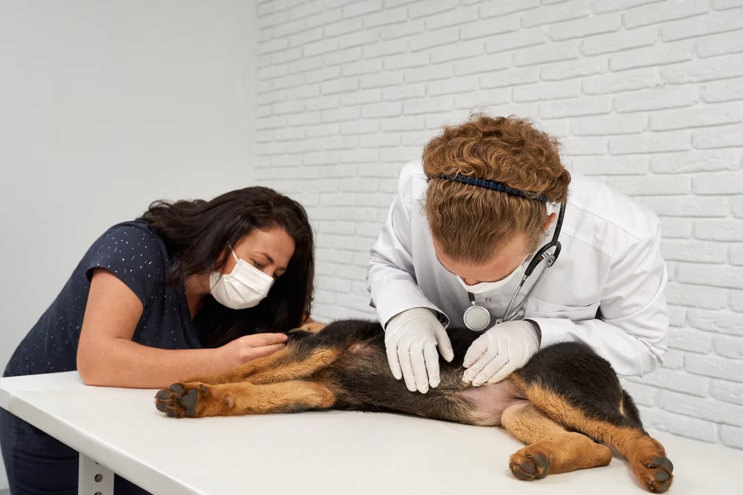
The diagnostic cascade always begins with an exhaustive medical history and a thorough hands-on physical examination. The veterinarian will ask the owner highly detailed questions about the dog’s daily habits, focusing specifically on water consumption, urination frequency, changes in appetite, exercise tolerance, and any recent behavioral shifts. During the physical exam, the clinician will palpate the abdomen to evaluate for hepatomegaly (an enlarged liver) and assess the tone of the abdominal musculature. The dog’s skin and coat will be examined under bright lighting for signs of bilateral alopecia, epidermal thinning, comedones (blackheads), and the gritty dermal plaques indicative of calcinosis cutis. Crucially, the veterinarian will also measure the dog’s blood pressure, as systemic hypertension is a very common complication of cortisol excess that can cause silent damage to the eyes, kidneys, and heart. [34]
Laboratory Tests: Complete Blood Panel and Urinalysis
If the history and physical exam raise a clinical suspicion of Cushing’s disease, the next step is to run a comprehensive baseline laboratory profile, including a Complete Blood Count (CBC), a serum biochemistry panel, and a complete urinalysis. The CBC may reveal a classic “stress leukogram,” characterized by an increase in neutrophils and monocytes, accompanied by a decrease in lymphocytes and eosinophils—a direct cellular response to high circulating cortisol. The serum biochemistry panel is often highly revealing. The most consistent finding is a dramatic elevation in Alkaline Phosphatase (ALP). In dogs, a specific isoenzyme of ALP is produced directly in response to glucocorticoids. The veterinarian will also look for elevated cholesterol, triglycerides, and fasting blood glucose, as well as elevated liver enzymes that may indicate gallbladder disease or secondary hepatic damage due to glycogen storage. [35]
A complete urinalysis is absolutely vital in the Cushing’s workup. Because cortisol inhibits ADH, the dog’s urine specific gravity will almost always be very low (typically less than 1.015), indicating highly dilute, watery urine. The urinalysis will also check for proteinuria (excess protein leaking through damaged renal filters) and glycosuria (sugar in the urine, indicating concurrent diabetes). Most importantly, the veterinarian will perform a urine culture to check for a “silent” urinary tract infection, as the immunosuppressive effects of cortisol can allow bacteria to thrive in the bladder without causing any visible white blood cells or outward signs of discomfort. [36]
Hormone Tests: ACTH Stimulation Test
Once the baseline tests support the suspicion of Hyperadrenocorticism, the veterinarian must perform specialized endocrine function tests to definitively confirm the diagnosis. One of the primary screening tools is the ACTH Stimulation Test. This test evaluates the adrenal glands’ reserve capacity to produce cortisol. The protocol involves drawing a baseline blood sample to measure the resting cortisol level. Next, the dog is injected with a precise dose of synthetic ACTH (cosyntropin). Exactly one hour later, a second blood sample is drawn to measure the stimulated cortisol level. [37]
In a healthy dog, the post-stimulation cortisol level will rise moderately into an expected normal range. However, a dog with Cushing’s disease (either pituitary or adrenal dependent) has hyperplastic or tumorous adrenal glands that are primed and hyper-responsive. When hit with the synthetic ACTH, these abnormal glands will produce a massive, exaggerated spike in cortisol, confirming the diagnosis. Conversely, if the veterinarian suspects Iatrogenic Cushing’s disease, this test is the gold standard for confirmation. A dog with iatrogenic Cushing’s will have a flat-line response—both the baseline and the stimulated cortisol levels will be extremely low, proving that the dog’s adrenal glands have atrophied due to chronic exogenous steroid administration. [38]
Imaging Studies
Diagnostic imaging plays a pivotal role not only in confirming the diagnosis but also in differentiating between pituitary-dependent and adrenal-dependent forms of the disease. Abdominal radiographs (X-rays) are often taken first. They can reveal the classic enlarged liver (hepatomegaly) and may occasionally show mineralization (calcification) in the area of the adrenal glands, which is a strong indicator of an adrenal tumor. However, the most valuable imaging modality in a Cushing’s workup is a comprehensive abdominal ultrasound performed by a board-certified veterinary radiologist or a highly experienced ultrasonographer. [39]
An abdominal ultrasound allows the veterinarian to visually inspect the exact size, shape, and internal architecture of both adrenal glands. If the ultrasound reveals two symmetrically enlarged, plump adrenal glands, it strongly points to Pituitary-Dependent Hyperadrenocorticism, as both glands are being overstimulated by brain ACTH. On the other hand, if the ultrasound reveals one massively enlarged, misshapen adrenal gland containing a tumor, while the contralateral gland is severely shrunken and atrophied, it confirms Adrenal-Dependent Hyperadrenocorticism. The ultrasound is also critical for evaluating whether an adrenal tumor has invaded the local blood vessels, such as the vena cava, and checking the liver for signs of metastatic cancer. [40]
Additional Diagnostic Tests: High Dose anti-inflammatory steroid Suppression Test
While the ACTH stimulation test is excellent for screening, the Low-Dose anti-inflammatory steroid Suppression Test (LDDST) is widely considered the most sensitive diagnostic tool for naturally occurring Cushing’s disease. The LDDST involves taking a baseline blood sample, injecting a very small dose of anti-inflammatory steroid, and then drawing blood again at 4 hours and 8 hours post-injection. In a normal dog, the brain detects the anti-inflammatory steroid and suppresses cortisol production for the entire 8 hours. In a dog with Cushing’s disease, this normal suppression fails, and cortisol levels remain elevated at the 8-hour mark. [41]
Once a diagnosis is confirmed, the veterinarian must determine the specific type (pituitary vs. adrenal) if the ultrasound was inconclusive. The High-Dose anti-inflammatory steroid Suppression Test (HDDST) is specifically designed for this differentiation. By administering a massive dose of anti-inflammatory steroid, the veterinarian attempts to overcome the resistance of a pituitary tumor. About 75 percent of pituitary tumors will show a temporary suppression of cortisol at the 4-hour or 8-hour mark during the HDDST. In contrast, an adrenal tumor is completely autonomous and will never suppress, even when hit with a massive dose of anti-inflammatory steroid. Other advanced diagnostics may include the Urine Cortisol:Creatinine Ratio (UCCR) as a highly sensitive initial screening tool, or measuring endogenous plasma ACTH concentrations. In cases where a pituitary macroadenoma is suspected due to neurological signs, advanced neuroimaging such as a Magnetic Resonance Imaging (MRI) or Computed Tomography (CT) scan of the brain is strongly recommended to evaluate the size of the tumor and plan potential treatments. [42]
Treatment for Canine Cushing’s Disease
 in Dogs? 4")
The primary objective of treating canine Hyperadrenocorticism is not necessarily to achieve a complete cure, but rather to medically control the severe clinical symptoms, thereby dramatically improving the dog’s quality of life and preventing life-threatening secondary complications such as pulmonary thromboembolism or acute diabetes mellitus. The therapeutic strategy chosen by the veterinarian is highly individualized and depends entirely on the specific etiology of the disease (pituitary vs. adrenal), the size and invasiveness of any tumors present, the presence of concurrent medical conditions, and the pet owner’s financial and logistical constraints. Treatment pathways generally fall into three main categories: lifelong medical management, complex surgical intervention, or advanced radiation therapy. [43]
Medication
For the vast majority of dogs diagnosed with Pituitary-Dependent Hyperadrenocorticism, as well as for dogs with adrenal tumors whose owners decline surgery, pharmacological medical management is the absolute cornerstone of therapy. The goal of medication is to artificially suppress the adrenal glands’ ability to manufacture cortisol, bringing circulating blood levels back down to a normal, safe range. Today, the most widely recommended and safest medication approved for this purpose is Trilostane (marketed under the trade name Vetoryl). [44]
Trilostane is a highly effective, reversible enzyme inhibitor. Once administered orally, it specifically blocks the action of 3-beta-hydroxysteroid dehydrogenase, a critical enzyme in the adrenal cortex responsible for the synthesis of cortisol. By blocking this enzyme, Trilostane rapidly drops the circulating levels of cortisol, providing fast relief from the clinical signs of excessive thirst, hunger, and panting. Because Trilostane’s effects are dose-dependent and reversible, it is generally considered safer than older medications. However, finding the exact correct dose requires a delicate balancing act. If the dose is too low, the dog continues to suffer from Cushing’s symptoms. If the dose is too high, the drug can suppress the adrenal glands too much, causing a sudden and potentially fatal Addisonian crisis. Therefore, dogs on Trilostane require intense, lifelong monitoring, with ACTH stimulation tests and blood electrolyte panels performed 10 to 14 days after starting the drug, and then every 3 to 6 months for the rest of the dog’s life. [45]
An older, but still utilized, medication is Mitotane (Lysodren). Unlike Trilostane, which merely blocks an enzyme, Mitotane is a potent adrenocorticolytic agent—a derivative of the insecticide DDT. It works by actively destroying and causing necrosis of the cortisol-producing cells in the zona fasciculata and zona reticularis of the adrenal gland. Mitotane therapy involves a rigorous “induction phase” where high doses are given daily until the dog’s appetite or water intake drops, followed by a lifelong “maintenance phase” given once or twice a week. While highly effective, Mitotane carries a significantly higher risk of causing permanent, irreversible damage to the adrenal glands, resulting in a lifelong need for treatment of Addison’s disease. Other less commonly used medications include Selegiline (Anipryl), which alters brain dopamine levels to reduce ACTH, and Ketoconazole, an antifungal drug that inhibits steroid synthesis as a side effect, though both are generally considered less efficacious than Trilostane. [46]
Surgery
Surgical intervention is considered the treatment of choice for a very specific subset of Cushing’s patients: those diagnosed with Adrenal-Dependent Hyperadrenocorticism caused by a unilateral adrenal tumor that has not yet metastasized. The surgical procedure, known as an adrenalectomy, involves opening the abdomen and completely excising the diseased adrenal gland. If the tumor is a benign adenoma, an adrenalectomy can be entirely curative, completely resolving the Cushing’s disease and allowing the dog to live a normal lifespan without the need for lifelong daily medication. [47]
However, it is critical to emphasize that an adrenalectomy is a highly complex, technically demanding, and high-risk surgical procedure. The adrenal glands are located in the deepest part of the abdominal cavity, intimately attached to the caudal vena cava and the phrenicoabdominal vein. Adrenal tumors, especially carcinomas, are highly vascular and prone to massive hemorrhage during dissection. Furthermore, because these dogs have been flooded with cortisol for months, they suffer from delayed wound healing, severe muscle weakness, and a dramatically increased risk of developing pulmonary thromboembolisms during the perioperative period. Due to these extreme risks, an adrenalectomy should only be performed by a board-certified veterinary surgeon in a specialized 24-hour facility equipped to handle severe anesthetic complications and provide intensive post-operative care. In highly specialized veterinary teaching hospitals, hypophysectomy (the surgical removal of the pituitary gland via the roof of the mouth) is occasionally performed for pituitary tumors, but this is incredibly rare and highly specialized. [48]
Radiation Therapy
For dogs suffering from Pituitary-Dependent Hyperadrenocorticism caused by a large, expanding pituitary macroadenoma, medical therapy with Trilostane will control the cortisol overproduction, but it will do absolutely nothing to stop the physical growth of the brain tumor. As the macroadenoma continues to expand, it will eventually cause severe, life-limiting neurological signs. In these specific cases, radiation therapy is the treatment of choice. [49]
Advanced veterinary oncology centers utilize highly targeted, high-energy radiation beams to irradiate the pituitary mass located at the base of the brain. The goal of radiation therapy is to damage the DNA of the tumor cells, thereby halting their division, shrinking the physical size of the tumor, and relieving the mechanical pressure on the surrounding delicate brain tissue. Modern techniques, such as Stereotactic Radiation Therapy (SRT), allow oncologists to deliver massive doses of radiation with pinpoint sub-millimeter accuracy in just one to three sessions, sparing the healthy surrounding brain tissue and minimizing side effects, which may occasionally include temporary issues with the eyes in dogs or surrounding skin. While radiation therapy is highly effective at resolving neurological symptoms and significantly extending the dog’s lifespan, it does not always completely cure the hormonal overproduction, meaning the dog may still require a low daily dose of Trilostane to manage the Cushing’s symptoms. [50]
Prevention of Cushing’s Disease in Dogs
Because the vast majority of Cushing’s disease cases (80 to 85 percent) are caused by spontaneous, naturally occurring microscopic tumors in the pituitary gland or the adrenal glands, there is unfortunately no known lifestyle change, diet, or preventative medication that can completely stop these tumors from forming. The genetic and cellular mutations that trigger Pituitary-Dependent and Adrenal-Dependent Hyperadrenocorticism are largely out of a pet owner’s control. However, there are proactive strategies that a diligent pet owner can employ to minimize risks, ensure early detection, and completely prevent the iatrogenic form of the disease. [51]
The most absolute and direct preventative measure applies solely to Iatrogenic Cushing’s disease. Because this form is caused by the chronic administration of exogenous steroids, pet owners must be hyper-vigilant when their dogs are recommended medications like anti-inflammatory steroid, anti-inflammatory steroid, or strong topical cortisone creams. Pet owners must adhere strictly to the veterinarian’s dosing instructions and never arbitrarily increase the dose or the frequency of the medication. Furthermore, if a dog has been on steroids for an extended period, the medication must never be stopped abruptly; it must be slowly and carefully tapered under strict veterinary supervision to allow the dog’s suppressed adrenal glands time to wake up and resume function. [52]
For the naturally occurring forms of the disease, early detection is the absolute best form of defense. Cushing’s disease is easiest to manage medically before the excess cortisol has caused irreversible secondary damage, such as severe hypertension, torn cruciate ligaments due to muscle weakness, or permanent structural damage to the kidneys. Pet owners with breeds known to be predisposed to the disease—such as Miniature Poodles, Dachshunds, Boxers, and Boston Terriers—should be particularly observant as their dogs enter their senior years (typically over the age of seven). [53]
Maintaining a baseline of exceptional overall health is crucial. Providing a high-quality, biologically appropriate diet and ensuring regular, moderate daily exercise helps maintain a lean body condition score. While obesity does not cause Cushing’s disease, an overweight dog will suffer far more severe complications—such as diabetes and joint failure—if they do develop the condition. Finally, routine bi-annual veterinary wellness exams, complete with comprehensive senior blood panels and urinalysis, are non-negotiable. These routine screenings can detect the subtle early warning signs of disease, such as a slightly elevated ALP enzyme or mildly dilute urine, long before the dog develops the classic pot-bellied appearance or severe hair loss, allowing for prompt, life-saving intervention. As always, please consult your veterinarian before making any changes to your pet’s care. [54]
Frequently Asked Questions
What is the life expectancy of a dog diagnosed with Cushing’s disease?
The life expectancy of a dog with Cushing’s disease depends heavily on the type of the disease, the age at diagnosis, and how well the dog responds to medical or surgical treatment. For dogs with Pituitary-Dependent Hyperadrenocorticism that receive proper medical management with drugs like Trilostane, the average survival time is roughly two to three years post-diagnosis, though many dogs live much longer. Because the disease primarily strikes senior dogs, many ultimately pass away from unrelated age-related conditions rather than the Cushing’s disease itself. If the dog has a malignant adrenal tumor, the prognosis is unfortunately much shorter, whereas a surgically removed benign adrenal tumor can result in a complete cure and a normal lifespan.
What should I feed a dog that has been diagnosed with Cushing’s disease?
Dietary management is a critical supportive component of treating Cushing’s disease. Because dogs with this condition are highly prone to muscle wasting, hyperlipidemia (high fat in the blood), and an increased risk of developing pancreatitis, veterinarians generally recommend a highly digestible, low-fat, and moderate-protein diet. Protein should be of exceptionally high biological value to help combat the severe muscle atrophy caused by excess cortisol. Additionally, keeping dietary sodium restricted can help manage the systemic hypertension (high blood pressure) that frequently accompanies the disease. You should always work directly with your veterinarian or a board-certified veterinary nutritionist to select a medication diet tailored to your dog’s specific bloodwork and metabolic needs.
Can Cushing’s disease cause behavioral changes or aggression in my dog?
Yes, Cushing’s disease can definitely cause noticeable behavioral changes in dogs, though overt aggression is relatively rare. The massive, continuous influx of cortisol essentially keeps the dog’s brain in a constant state of physiological “fight or flight” stress. This can cause dogs to become restless, anxious, irritable, and prone to aimless pacing, particularly at night. Furthermore, the extreme polyphagia (ravenous hunger) associated with the disease can lead to resource-guarding behaviors, where a dog becomes highly protective of their food bowl or stolen items. If a dog has a growing pituitary macroadenoma pressing on their brain, they may exhibit more severe neurological behaviors, such as staring blankly at walls or acting disoriented. Proper medical treatment usually resolves these behavioral shifts as cortisol levels normalize.
References
- Merck Veterinary Manual. Hyperadrenocorticism (Cushing Syndrome) in Dogs. Merck & Co., Inc., 2023.
- Behrend, E. N., et al. “Diagnosis of spontaneous canine hyperadrenocorticism: 2012 ACVIM consensus statement (small animal).” Journal of Veterinary Internal Medicine, 2013.
- American Veterinary Medical Association (AVMA). Endocrine Disorders in Dogs. AVMA, 2022.
- Feldman, E. C., Nelson, R. W. “Canine and Feline Endocrinology and Reproduction.” Elsevier, 2015.
- VCA Animal Hospitals. Cushing’s Disease in Dogs. VCA Hospitals, 2021.
- Gilor, C., et al. “Trilostane therapy for canine hyperadrenocorticism.” Veterinary Clinics of North America, 2011.
- Washington State University. Pet Health Topics: Cushing’s Disease. WSU College of Veterinary Medicine, 2020.
- Sanders, K., et al. “Treatment of canine pituitary-dependent hyperadrenocorticism.” Veterinary Clinics of North America: Small Animal Practice, 2018.
- Veterinary Information Network (VIN). Pituitary-Dependent Hyperadrenocorticism. VIN, 2019.
- Reusch, C. E. “Hyperadrenocorticism.” Textbook of Veterinary Internal Medicine, 2010.
- Merck Veterinary Manual. Adrenal Tumors in Dogs. Merck & Co., Inc., 2023.
- Kaplan, A. J., et al. “Iatrogenic hyperadrenocorticism.” Compendium on Continuing Education for the Practicing Veterinarian, 1995.
- VCA Animal Hospitals. Iatrogenic Cushing’s Disease in Dogs. VCA Hospitals, 2021.
- Bennaim, M., et al. “Diagnostic pathways for spontaneous hyperadrenocorticism in dogs.” Veterinary Journal, 2014.
- Meij, B. P. “Hypophysectomy as a treatment for canine and feline pituitary-dependent hyperadrenocorticism.” Veterinary Clinics of North America, 2001.
- JAVMA News. New consensus statement on canine Cushing’s. American Veterinary Medical Association, 2012.
- Pollard, R. E., et al. “Cross-sectional imaging of the pituitary gland in dogs.” Veterinary Radiology & Ultrasound, 2005.
- Veterinary Information Network (VIN). Macroadenoma Syndrome in Dogs. VIN, 2018.
- Schwartz, P., et al. “Adrenal-dependent hyperadrenocorticism in dogs.” Journal of Veterinary Internal Medicine, 2007.
- Massari, F., et al. “Adrenalectomy in dogs with adrenal gland tumors.” Journal of the American Veterinary Medical Association, 2011.
- Merck Veterinary Manual. Adrenocortical Carcinoma. Merck & Co., Inc., 2023.
- Lowe, A. D., et al. “Clinical approach to glucocorticoid therapy in dogs.” Journal of Veterinary Pharmacology and Therapeutics, 2013.
- VCA Animal Hospitals. Steroid Treatment Long-term Effects in Dogs. VCA Hospitals, 2022.
- Frank, L. A. “Dermatologic manifestations of canine hyperadrenocorticism.” Veterinary Clinics of North America, 2015.
- AVMA. Glucocorticoid Therapy in Dogs. American Veterinary Medical Association, 2020.
- Herrtage, M. E., Ramsey, I. K. “Canine hyperadrenocorticism.” BSAVA Manual of Canine and Feline Endocrinology, 2012.
- Nichols, R. “Complications of hyperadrenocorticism.” Veterinary Clinics of North America: Small Animal Practice, 2001.
- Veterinary Information Network (VIN). Polyphagia in the Endocrine Patient. VIN, 2017.
- De Marco, V., et al. “Myopathy in dogs with hyperadrenocorticism.” Journal of Veterinary Internal Medicine, 2005.
- Pessina, P., et al. “Abdominal fat distribution in hyperadrenocorticism.” Veterinary Radiology & Ultrasound, 2009.
- Outerbridge, C. A. “Cutaneous manifestations of internal diseases.” Veterinary Clinics of North America, 2001.
- Bosje, J. T., et al. “Thromboembolism in dogs with hyperadrenocorticism.” Journal of Veterinary Internal Medicine, 2006.
- Behrend, E. N. “Canine hyperadrenocorticism: diagnosis.” Compendium on Continuing Education, 2013.
- Veterinary Information Network (VIN). Blood Pressure Monitoring in Endocrine Disease. VIN, 2016.
- Sepesy, L. M., et al. “Alkaline phosphatase isoenzymes in dogs.” Journal of Veterinary Internal Medicine, 2008.
- Smets, P. M., et al. “Urinary tract infections in dogs with Cushing’s.” Journal of Veterinary Internal Medicine, 2002.
- Lemetayer, J., et al. “ACTH stimulation test protocols.” Canadian Veterinary Journal, 2011.
- Martin, V., et al. “Assessment of ACTH testing in iatrogenic Cushing’s.” Veterinary Record, 2009.
- Besso, J. G., et al. “Ultrasonography of the adrenal glands in dogs.” Veterinary Radiology & Ultrasound, 2000.
- American College of Veterinary Radiology (ACVR). Diagnostic Imaging in Canine Hyperadrenocorticism. ACVR, 2021.
- Reusch, C. E., et al. “Low-dose anti-inflammatory steroid suppression testing.” Journal of Veterinary Internal Medicine, 2004.
- Feldman, E. C. “High-dose anti-inflammatory steroid suppression test.” Compendium on Continuing Education, 2008.
- Merck Veterinary Manual. Treatment of Hyperadrenocorticism. Merck & Co., Inc., 2023.
- Neiger, R., et al. “Trilostane treatment of canine hyperadrenocorticism.” Veterinary Record, 2006.
- Macfarlane, L., et al. “Monitoring dogs receiving trilostane.” Journal of Small Animal Practice, 2015.
- Kintzer, P. P., Peterson, M. E. “Mitotane treatment for hyperadrenocorticism.” Veterinary Clinics of North America, 2002.
- Schwartz, P. “Surgical management of adrenal tumors.” Veterinary Clinics of North America, 2011.
- American College of Veterinary Surgeons (ACVS). Adrenal Tumors. ACVS, 2021.
- Bley, C. R., et al. “Radiation therapy of pituitary macroadenomas.” Journal of Veterinary Internal Medicine, 2005.
- UC Davis Veterinary Medicine. Radiation Therapy for Cushing’s Disease. University of California, 2022.
- O’Neill, D. G., et al. “Epidemiology of canine hyperadrenocorticism.” Journal of Small Animal Practice, 2014.
- American Veterinary Medical Association (AVMA). Safe Use of Steroids in Dogs. AVMA, 2021.
- Ling, G. V., et al. “Breed predispositions to hyperadrenocorticism.” Journal of the American Veterinary Medical Association, 2005.
- VCA Animal Hospitals. Wellness Examination in Senior Dogs. VCA Hospitals, 2022.


