


 March 4, 2023
 Phil Good, DVM
What is Anal Gland Disease in Dogs?
This content was prepared with AI assistance and reviewed by a licensed professional for accuracy.
Introduction
Understanding anal gland disease in dogs is crucial for every pet owner, as these hidden anatomical structures can cause immense discomfort when they fail to function correctly. Imagine taking your beloved companion for a walk, only to notice them constantly stopping to drag their hindquarters across the grass or living room carpet. This peculiar and often comical behavior, commonly known as scooting, is rarely a laughing matter for the dog experiencing it. In most clinical scenarios, this scooting indicates that a dog is suffering from a condition impacting their perianal region. When we talk about anal gland disease, we are referring to a broad veterinary term that encompasses a variety of painful, irritating, and sometimes life-threatening conditions affecting the small secretory sacs located near a dog’s rectum.[1] Whether the issue is a simple mechanical blockage, a severe bacterial infection, or even a neoplastic growth, early identification and prompt veterinary intervention are essential for preserving the animal’s quality of life.
For many dog owners, the mere mention of anal glands conjures up unpleasant thoughts of foul odors and messy veterinary visits. However, these glands play a fascinating evolutionary role in canine communication and territorial marking. In a healthy dog, the anal sacs empty their contents naturally during routine bowel movements, coating the feces with a unique scent signature that conveys biological information to other animals.[2] Unfortunately, modern canine lifestyles, dietary factors, and genetic predispositions often disrupt this natural emptying process. When the normal physiological mechanisms fail, the secretory fluid becomes trapped within the narrow confines of the sacs. This trapped fluid provides an ideal breeding ground for opportunistic bacteria that naturally reside in the gastrointestinal tract and surrounding perianal skin.
As the condition progresses, what begins as a mild irritation can rapidly escalate into a significant medical emergency. The spectrum of anal gland disease ranges from simple impactions, where the fluid thickens into a dense paste, to severe sacculitis (inflammation of the sacs), painful abscesses, and even malignant tumors known as apocrine gland anal sac adenocarcinomas (AGASACA).[3] Each of these distinct pathologies requires a specific diagnostic approach and a tailored treatment plan formulated by a licensed veterinarian. Recognizing the subtle behavioral changes and physical symptoms associated with these disorders empowers pet owners to seek timely medical care, thereby preventing the escalation of minor discomfort into excruciating pain or systemic illness. In the comprehensive guide below, we will explore the anatomy, causes, symptoms, diagnostic procedures, and advanced treatments associated with canine anal gland problems.
What is Anal Gland in Dogs?
To fully grasp the complexities of the conditions that affect them, it is necessary to answer the fundamental question: what is anal gland in dogs? Anatomically referred to as anal sacs, these structures are two small, oval-shaped pouches located beneath the skin on either side of a dog’s anus. If you were to visualize the canine anus as the face of a clock, the anal glands are typically situated at approximately the 4 o’clock and 8 o’clock positions.[4] They reside within the delicate connective tissue sandwiched between the internal and external anal sphincter muscles. Each sac is connected to the outside world by a tiny, narrow duct that opens just inside the mucocutaneous junction of the anal opening.
Microscopically, the inner lining of the anal sacs is highly specialized. It is composed of a combination of sebaceous (oil-producing) glands and apocrine (sweat-producing) glands. Together, these microscopic glands continuously secrete a liquid substance that collects within the reservoir of the sac. In healthy dogs, this anal gland fluid varies in consistency from a thin, watery liquid to a slightly thicker, serous discharge. The color can range from pale yellow to a murky brown. The most notable characteristic of this secretion, however, is its incredibly potent, musky, and often overwhelmingly foul odor, which many pet owners liken to the smell of rotting fish or old pennies.[5]
Biologically, the anal glands are the canine equivalent of the scent glands found in skunks, although dogs have lost the ability to voluntarily spray their secretions as a primary defense mechanism. Instead, the fluid serves as an intricate chemical communication system. Whenever a dog passes a bowel movement, the expansion of the rectum and the contraction of the anal sphincter muscles exert natural physical pressure on the anal sacs. This pressure forces a small amount of the pungent fluid out through the narrow ducts and onto the surface of the feces. To a human nose, this scent is undetectable under normal circumstances, but to the highly sensitive olfactory system of another dog, it provides a wealth of information.[1] The scent signature conveys details about the dog’s age, sex, reproductive status, health, and diet. This biological mechanism explains why dogs are so intently focused on sniffing the feces of other animals and why they greet each other by sniffing rear ends.
In addition to intentional scent marking during defecation, the anal glands can also empty spontaneously in response to sudden fear, extreme stress, or intense excitement. During a “fight or flight” response, involuntary muscle contractions can cause the sacs to express their contents suddenly, leaving a lingering odor on the dog’s fur or the surrounding environment. While this is a normal physiological reaction, it highlights the dynamic nature of these glands. However, because the ducts leading from the sacs to the exterior are incredibly narrow—often no wider than a pinhead—they are highly susceptible to becoming blocked. When the delicate balance of fluid production and natural expression is disrupted, the foundation for anal gland disease is laid, setting off a cascade of uncomfortable and potentially dangerous health issues for the dog.[2]
Common Anal Gland Problems
Anal gland issues are among the most frequently diagnosed problems in small animal veterinary practice. They rarely resolve on their own once the normal physiological mechanisms fail. The overarching term “anal sac disease” is generally divided into four distinct clinical categories, ranging from easily managed mechanical blockages to aggressive, life-threatening malignancies. Understanding these common anal gland problems is essential for recognizing the severity of a dog’s condition and anticipating the required level of veterinary care.[3]
1. Anal Gland Impaction: This is by far the most common manifestation of anal gland disease in dogs. Impaction occurs when the anal gland secretions fail to empty normally during defecation. Because the glandular cells continuously produce fluid, the inability to express the fluid causes it to pool within the sac. Over time, the watery components of the fluid are reabsorbed by the body, leaving behind a thick, dehydrated, and pasty substance that resembles dark clay or peanut butter. This thickened material cannot pass through the narrow excretory ducts. As the glands become engorged and distended, they place significant pressure on the surrounding sensitive tissues of the perineum. This stretching and pressure cause a dull, constant ache, leading to the classic scooting behavior as the dog attempts to manually relieve the physical pressure. While impaction itself is not an infection, it completely compromises the normal environment of the sac, setting the stage for more severe complications if left unaddressed.[4]
2. Anal Gland Infection (Sacculitis): When an impaction is not resolved promptly, the stagnant, nutrient-rich fluid trapped inside the anal sac becomes an ideal incubator for bacteria. The perianal region is naturally populated with billions of bacteria from the gastrointestinal tract, including species like Escherichia coli, Enterococcus faecalis, Staphylococcus, and Streptococcus. When these opportunistic microbes migrate up the blocked duct and enter the stagnant fluid of the sac, they rapidly multiply, leading to active inflammation and infection known as anal sacculitis. The dog’s immune system responds to the bacterial invasion by sending white blood cells to the area, which results in the production of pus. The character of the anal gland fluid changes dramatically, shifting from a thick brown paste to a yellowish, greenish, or blood-tinged purulent discharge. The sacs become intensely painful, hot to the touch, and highly inflamed, causing the dog severe distress even during simple movements like sitting or walking.[2]
3. Anal Gland Abscess and Rupture: If an infection is allowed to progress without antibiotic therapy and physical drainage, it will inevitably culminate in the formation of an anal gland abscess. An abscess is a walled-off pocket of severe infection and necrotic (dead) tissue. Because the natural excretory duct remains blocked, the sheer volume of pus accumulating within the enclosed space causes the sac to swell massively. The pressure inside the abscess builds to an excruciating level. The intense inflammation weakens the wall of the anal sac and the overlying perianal skin. Eventually, the pressure exceeds the structural integrity of the tissue, and the abscess violently ruptures. This rupture creates a fistulous tract—a new, abnormal opening in the skin alongside the anus—through which foul-smelling, bloody pus drains freely. While the rupture often provides the dog with immediate relief from the extreme pressure, it leaves an open, contaminated wound that requires intensive surgical and medical management to heal properly and prevent systemic sepsis.[5]
4. Apocrine Gland Anal Sac Adenocarcinoma (AGASACA): Although benign conditions like impactions and infections are the most common, the anal glands can also be the site of aggressive neoplastic disease. AGASACA is a malignant tumor arising from the apocrine sweat glands lining the anal sac. This specific type of cancer accounts for approximately 17% of all perianal tumors in canines. It is an insidious disease because the tumors are often small and difficult to detect without a thorough digital rectal exam, yet they are highly invasive to local tissues. Furthermore, AGASACA has a notoriously high rate of early metastasis, frequently spreading to the sublumbar (iliosacral) lymph nodes located deep within the pelvic canal, as well as to distant organs like the liver, spleen, and lungs.[6] Additionally, up to 50% of dogs with AGASACA develop a paraneoplastic syndrome called hypercalcemia of malignancy. The tumor cells secrete a hormone-like protein that causes the dog’s blood calcium levels to spike dangerously high, which can lead to catastrophic kidney failure, severe neurological signs, and life-threatening cardiac arrhythmias.[7]
Causes of Anal Gland Disease in Dogs

The exact etiology of anal gland disease in dogs is often multifactorial, meaning that a combination of several different physiological, environmental, and anatomical factors contributes to the breakdown of normal anal sac function. Identifying the underlying root causes is a critical component of veterinary care, as simply treating the immediate impaction or infection without addressing the precipitating factors will almost certainly lead to a recurrence of the problem. A comprehensive understanding of these causes helps veterinarians and pet owners develop effective, long-term management strategies.[4]
Gastrointestinal and Dietary Factors: The most significant driver of anal gland impaction is the quality and consistency of a dog’s stool. The normal physiological emptying of the anal sacs relies entirely on the mechanical pressure exerted by a firm, well-formed column of feces passing through the rectum and stretching the anal sphincter. If a dog experiences chronic gastrointestinal upset, diarrhea, or persistently soft and mushy stools, the feces will pass through the anal canal without applying adequate pressure to the sacs. Without this physical stimulation, the fluid is never squeezed out, leading directly to stagnation and impaction. Diets that are highly processed or deficient in adequate dietary fiber are often the primary culprits behind poor stool quality. Fiber is essential for absorbing excess water in the colon and providing the necessary bulk to create a firm stool capable of emptying the glands naturally.[3]
Anatomical Irregularities: The structural anatomy of the perineal region varies widely among individual dogs, and certain anatomical quirks can predispose a dog to chronic anal gland disease. In some animals, the anal sacs are situated unusually deep within the muscular layers of the pelvic floor, making it impossible for even a firm stool to exert enough pressure against them. In other cases, the tiny excretory ducts that connect the sac to the anal opening may be congenitally narrow, tortuous, or malformed, creating a physical bottleneck that prevents the thick fluid from escaping. Additionally, dogs that have suffered from previous bouts of severe sacculitis or ruptured abscesses may develop extensive scar tissue in and around the ducts. This fibrosis permanently alters the local anatomy, strictly limiting the flow of secretions and ensuring that the dog will suffer from recurrent, lifelong impactions that require manual veterinary intervention.[5]
Obesity and Body Condition: A dog’s overall physical condition plays a surprisingly significant role in the health of their anal glands. Overweight or obese dogs are at a substantially higher risk of developing impactions and infections. When a dog carries excess body weight, thick layers of adipose (fat) tissue accumulate around the tail base, perineum, and rectum. This excess fat physically cushions the anal sacs, effectively absorbing the mechanical pressure generated during defecation. As a result, the sacs are insulated from the forces needed to express them. Furthermore, obese dogs often lead sedentary lifestyles, resulting in poor generalized muscle tone, including a weakened anal sphincter, which further compromises the natural emptying process.[2]
Allergies and Dermatological Disease: The skin lining the perianal region is continuous with the rest of the body’s integumentary system, meaning that generalized skin conditions directly impact the anal sacs. Atopic dermatitis (environmental allergies) and adverse food reactions frequently manifest as severe pruritus (itching) and inflammation around the tail base and anus. In fact, dogs may have food allergies where the only presenting clinical sign is chronic, recurrent anal gland inflammation. The allergic response causes the skin surrounding the anal ducts to become swollen, red, and edematous. This localized tissue swelling physically compresses the delicate excretory ducts, trapping the fluid inside the sacs and rapidly leading to impaction and secondary bacterial infection.[1]
Age and Breed Predispositions: While anal gland issues can afflict any dog of any age or size, clinical data clearly demonstrates that genetics play a vital role. Certain dog breeds, especially small or toy breeds, may be more vulnerable to anal gland disease. Breeds such as Chihuahuas, Miniature Poodles, Toy Poodles, Cocker Spaniels, Basset Hounds, and Beagles are vastly overrepresented in veterinary clinics for benign anal gland impactions and infections. The exact reason for this breed susceptibility is likely a combination of genetic anatomical variations in duct size and the higher prevalence of food allergies in these specific breeds. Conversely, when it comes to malignant anal gland disease, older, large-breed dogs, particularly English Cocker Spaniels, German Shepherds, Alaskan Malamutes, and Golden Retrievers, show a higher predisposition for developing apocrine gland anal sac adenocarcinomas (AGASACA).[7]
Symptoms of Anal Gland Disease in Dogs

The clinical manifestations of anal gland disease in dogs can range from mild, intermittent behavioral changes to severe, acute systemic illness, depending entirely on whether the animal is dealing with a simple impaction, a raging abscess, or a malignant tumor. Because dogs cannot verbally communicate their pain, they rely on specific, often intense behavioral cues to indicate that something is wrong in their perianal region. Pet owners must remain vigilant and observe their dogs closely, as missing the early warning signs of an impaction allows the condition to rapidly deteriorate into a surgical emergency.[3]
The most iconic and frequently observed symptom of anal gland disease is “scooting.” Scooting involves the dog sitting down on its hindquarters, lifting its rear legs slightly off the ground, and using its front legs to drag its anus across the floor, grass, or carpet. This action is a desperate attempt to scratch an itch they cannot reach, soothe inflamed tissue, and apply mechanical pressure to the swollen sacs in hopes of expressing the trapped fluid. While occasionally scooting after a messy bowel movement can be normal, persistent, repetitive scooting is a definitive red flag that requires veterinary attention.[4]
In addition to scooting, dogs with anal gland discomfort will exhibit intense, obsessive grooming behaviors. They will frequently whip their heads around to aggressively lick or chew at the base of their tail, their flanks, or directly at the anal region. The area may become completely denuded of hair and red from the constant trauma of the dog’s tongue and teeth. When the glands are severely impacted or infected, they become exquisitely painful. A normally docile and loving dog may suddenly display signs of aggression, growling or snapping if an owner attempts to touch their tail, brush their hindquarters, or lift them up. Their posture may change; they often walk with a clamped-down tail tucked tightly between their legs to protect the sensitive area, or they may appear hesitant and reluctant to sit down on hard surfaces.[1]
Physical signs are equally telling. Owners may notice a profoundly unpleasant, fishy, or metallic odor wafting from the dog’s rear end, often leaving a foul-smelling residue on the dog’s bedding or the owner’s furniture. During defecation, the dog may exhibit tenesmus (straining without producing much stool) or dyschezia (vocalizing or crying out in pain while passing feces) because the expansion of the rectum presses against the painfully swollen, infected sacs. Visual inspection of the perineum may reveal obvious, asymmetrical swelling on one or both sides of the anus, accompanied by intense redness (erythema). If an abscess has ruptured, owners will be confronted with the alarming sight of a bloody, purulent, yellowish-green discharge draining from an open wound right next to the anal sphincter.[5]
When dealing with apocrine gland anal sac adenocarcinoma (AGASACA), the symptoms can be far more insidious and systemic. While some dogs with tumors may show the classic signs of scooting and swelling, many exhibit symptoms related to the paraneoplastic hypercalcemia caused by the cancer. Extreme elevations in blood calcium lead to polyuria (excessive, voluminous urination) and compensatory polydipsia (excessive thirst and drinking). The hypercalcemia acts as a systemic toxin, causing profound lethargy, generalized muscle weakness, severe loss of appetite (anorexia), vomiting, and constipation. In advanced cases of AGASACA, the massive enlargement of the sublumbar lymph nodes due to metastasis can physically compress the colon from the outside, making it physically impossible for the dog to pass stool, leading to obstipation and severe abdominal pain.[8]
Diagnosis of Anal Gland Issues in Dogs

Accurate diagnosis of anal gland disease requires a systematic, thorough, and highly clinical approach by a licensed veterinarian. Because the symptoms of a simple benign impaction can overlap heavily with those of an aggressively malignant tumor, veterinarians cannot rely on visual inspection alone. The diagnostic protocol involves a combination of detailed historical data collection, hands-on physical manipulation, microscopic analysis, and advanced diagnostic imaging techniques to accurately stage the severity of the disease and formulate an appropriate treatment plan.[2]
Health Background and Physical Check-up
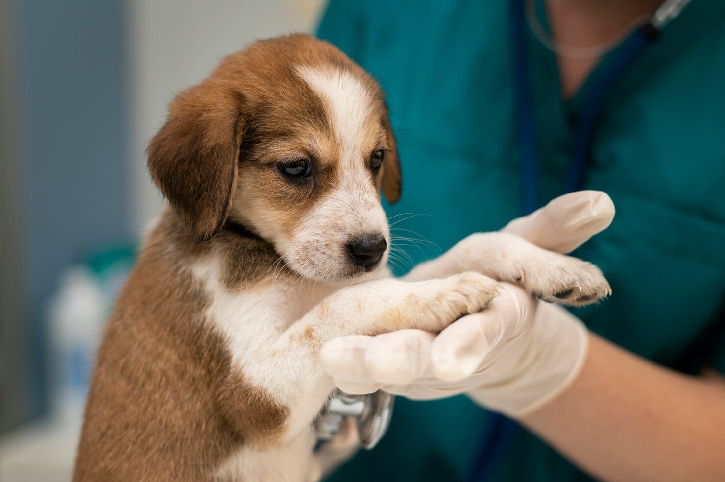
The diagnostic process invariably begins in the examination room with a comprehensive gathering of the dog’s health background. The veterinarian will ask probing questions regarding the dog’s diet, the frequency and consistency of their bowel movements, any history of food or environmental allergies, and the exact timeline of the current clinical signs. Following the history, a complete nose-to-tail physical check-up is conducted to ensure the dog is stable and to rule out other concurrent medical issues. The critical component of the examination is the evaluation of the perineum. The veterinarian will visually inspect the external perianal area for signs of swelling, redness, fistulous tracts, or matting of the hair with purulent discharge.
Visual inspection is immediately followed by the gold standard diagnostic tool for anal gland issues: the digital rectal examination (DRE). Wearing a lubricated exam glove, the veterinarian gently inserts a finger into the dog’s rectum. This allows the doctor to meticulously palpate (feel) both anal sacs simultaneously by squeezing the tissue between the index finger inside the rectum and the thumb on the external perianal skin. The veterinarian assesses the size, symmetry, temperature, and consistency of the glands. They feel for the doughy firmness of an impaction, the fluctuant heat of an abscess, or the hard, immovable, nodular feel characteristic of a neoplastic tumor. Additionally, the vet will attempt to sweep their finger dorsally within the pelvic canal to feel for any enlargement of the sublumbar lymph nodes, which is a critical indicator of metastatic cancer spread.[1]
Anal Gland Expression
During the digital rectal exam, if the glands are enlarged but not ruptured, the veterinarian will attempt to perform a diagnostic and therapeutic anal gland expression. By applying steady, controlled pressure from the deepest portion of the sac moving outward toward the duct, the vet attempts to milk the contents out through the natural opening. This internal expression technique is far superior and much safer than the external squeezing often attempted by groomers, as it ensures complete emptying without bruising the delicate surrounding tissues. The veterinarian pays close attention to the character of the expressed material. A thick, dry, clay-like, brown substance confirms a severe impaction. A thin, foul-smelling liquid with streaks of blood, or a frankly purulent yellow-green discharge, confirms active bacterial sacculitis. If the gland feels rock-hard and no fluid can be expressed despite appropriate pressure, it significantly raises the index of suspicion for a solid tumor mass.[4]
Microscopic Examination
When the character of the expressed fluid is abnormal, or if a tumor is suspected, the next diagnostic step is a microscopic examination, formally known as cytology. The veterinarian will collect a sample of the anal gland exudate or perform a fine-needle aspirate (FNA) of a solid mass using a small syringe. This sample is smeared onto a glass slide, stained with specific biological dyes, and examined under a high-powered microscope. Cytology allows the veterinarian to peer into the cellular environment of the gland. The presence of massive numbers of degenerative neutrophils (white blood cells) actively engulfing intracellular bacteria (such as cocci or rods) provides a definitive diagnosis of septic sacculitis. If the cytology reveals clusters of atypical, highly nucleated epithelial cells demonstrating criteria of malignancy (such as variable cell size, prominent nucleoli, and frequent mitotic figures), it strongly points toward a diagnosis of apocrine gland anal sac adenocarcinoma. In cases of chronic, non-responsive infection, a sample may also be sent to an outside laboratory for aerobic and anaerobic bacterial culture and antibiotic sensitivity testing to determine exactly which pathogens are present and which specific antibiotics will effectively kill them.[2]
Ultrasound Scanning
In cases that are complex, chronic, or highly suspicious for cancer, advanced diagnostic imaging is required. Ultrasound scanning of the abdomen and the perineal region is a powerful, non-invasive tool. A specialized ultrasound probe applied to the perianal area can differentiate between a fluid-filled abscess pocket and a solid, vascularized neoplastic tumor mass. More importantly, a complete abdominal ultrasound is the primary modality used for clinical staging if an AGASACA tumor is confirmed. The ultrasonographer will meticulously evaluate the internal abdominal organs, specifically searching for the medial iliac, hypogastric, and sacral lymph nodes deep within the sublumbar region. Because anal gland tumors metastasize to these specific nodes early in the disease process, detecting enlarged, abnormal lymph nodes on an ultrasound scan drastically alters the prognosis and the surgical approach. Ultrasound can also identify metastatic lesions scattered throughout the liver or the spleen.[7]
Biopsy
While cytology provides a rapid provisional diagnosis, the absolute gold standard for diagnosing cancer in the anal glands is a surgical biopsy combined with formal histopathology. If an ultrasound confirms a solid mass, the veterinarian will recommend removing a wedge of the tissue (incisional biopsy) or surgically excising the entire anal sac (excisional biopsy). This solid tissue sample is preserved in formalin and sent to a board-certified veterinary pathologist. The pathologist examines the architecture of the tissue to determine the exact tumor type, grade its aggressiveness, and evaluate the surgical margins to ensure that no microscopic cancer cells were left behind in the surrounding muscle bed. A biopsy is the only way to definitively differentiate an AGASACA from a benign adenoma or a severe, chronic, granulomatous inflammatory reaction. Alongside these specific tests, a comprehensive blood panel (Complete Blood Count and Serum Biochemistry) will be run to evaluate the patient’s overall organ function and specifically check for the presence of hypercalcemia, which is a hallmark paraneoplastic indicator of anal gland malignancy.[8]
Treatment for Anal Gland Disease in Dogs

The medical and surgical management of anal sac issues in dogs requires a highly tailored, phase-specific approach. Treatment protocols are dictated entirely by the precise diagnosis—whether the clinician is dealing with a simple mechanical impaction, a necrotizing bacterial abscess, or an aggressive, metastatic carcinoma. An accurate diagnosis ensures that the prescribed interventions are not only effective in providing immediate pain relief but also appropriate for achieving long-term resolution and preventing chronic recurrence.[3]
Anal Sac Expression and Lavage: For benign, uncomplicated impactions, the primary therapy is mechanical relief. A veterinarian or highly trained veterinary technician will perform an internal manual expression, applying careful, steady pressure to evacuate the thickened, clay-like secretions from the sacs. If the material is too inspissated (dried out and dense) to pass through the narrow duct, the veterinarian may need to perform a therapeutic flush, also known as an anal sac lavage. Often requiring mild sedation or light anesthesia due to the inherent discomfort, a small, flexible catheter is gently threaded directly into the anal duct. Sterile saline is flushed into the sac under mild pressure to break down, hydrate, and flush out the hardened plug of material, restoring the patency of the duct and cleaning the reservoir.[4]
Topical and Systemic Antibiotics: When bacterial sacculitis is present, simply emptying the gland is insufficient; the active infection must be eradicated. Following a thorough lavage to remove the infected pus and debris, the veterinarian will often infuse a specialized topical medication directly into the empty sac via the catheter. These veterinary-specific ointments typically contain a potent combination of a broad-spectrum antibiotic to kill the remaining bacteria, an antifungal agent to address secondary yeast overgrowth, and a corticosteroid (like dexamethasone or triamcinolone) to rapidly reduce the intense localized inflammation and swelling of the duct. In cases where the infection is severe, or if an abscess has ruptured, systemic (oral) antibiotics are mandatory. Medications such as amoxicillin-clavulanate (Clavamox), cephalexin, or clindamycin are frequently prescribed for a duration of 10 to 14 days. These systemic antibiotics penetrate the inflamed perianal tissues from the bloodstream, fighting the infection from the inside out.[2]
Management of Abscesses and Pain Alleviation: The treatment of a ruptured anal gland abscess is akin to managing severe localized trauma. The intense pressure has blown a hole through the dog’s perianal skin, creating a painful, open, oozing wound. The veterinarian will clip the fur widely around the area to prevent hair from matting into the wound and acting as a nidus for further infection. The abscess cavity is aggressively flushed with dilute antiseptic solutions like chlorhexidine or povidone-iodine. Owners are often instructed to apply warm, moist compresses to the area several times a day at home to encourage continuous drainage and improve localized blood flow to speed healing. Because these conditions are excruciatingly painful, systemic pain relief is a vital component of the treatment plan. Non-steroidal anti-inflammatory drugs (NSAIDs) such as carprofen, meloxicam, or grapiprant are prescribed to drastically reduce pain and systemic inflammation. Furthermore, the dog must wear an Elizabethan collar (the “cone of shame”) at all times to prevent them from licking or chewing the healing wound, which would rapidly introduce new oral bacteria and destroy the delicate repairing tissues.[5]
Surgical Intervention (Anal Sacculectomy): When medical management fails, surgery becomes necessary. If a dog suffers from chronic, intractable impactions requiring manual expression every few weeks, or if they endure recurrent, painful abscesses despite appropriate antibiotic therapy, the veterinarian will strongly recommend an anal sacculectomy. This surgical procedure involves the complete, permanent removal of one or both anal sacs and their associated ducts. The surgery is delicate and requires advanced surgical skill. The surgeon must meticulously dissect the sac away from the highly vascular and heavily innervated internal and external anal sphincter muscles. If the dissection is too aggressive or if the local nerves are damaged, the dog may suffer from permanent fecal incontinence—the inability to control their bowel movements. However, in the hands of an experienced veterinary surgeon, anal sacculectomy boasts a very high success rate and provides a permanent, lifelong cure for dogs plagued by chronic anal sacculitis, drastically improving their overall quality of life.[4]
Oncological Treatment for AGASACA: The treatment paradigm shifts dramatically when a biopsy confirms apocrine gland anal sac adenocarcinoma. The management of AGASACA requires a multidisciplinary oncological approach. The cornerstone of treatment is aggressive surgical excision of the primary perianal tumor, often combined with the surgical removal of any metastatic sublumbar lymph nodes in the abdomen. Because AGASACA is highly locally invasive, achieving clean, cancer-free surgical margins around the rectum is incredibly challenging. Therefore, surgery is frequently followed by a course of localized radiation therapy to eradicate microscopic tumor cells left behind in the sphincter muscles. Finally, systemic chemotherapy, using drugs like carboplatin or mitoxantrone, is administered to delay or prevent distant metastasis to the lungs and internal organs. Additionally, dogs presenting with paraneoplastic hypercalcemia require immediate intravenous fluid diuresis, bisphosphonates, and steroids to rapidly lower their blood calcium levels, protecting their kidneys from irreversible failure before surgery can even be attempted.[11]
How to Prevent Anal Gland Problems in Dogs
While certain anatomical defects and genetic predispositions cannot be altered, the vast majority of benign anal gland issues, such as chronic impactions and recurrent sacculitis, are highly preventable through diligent, proactive home care and mindful lifestyle management. By understanding the biomechanical requirements necessary for normal anal sac expression, pet owners can implement specific, targeted strategies that vastly reduce the risk of their dog ever experiencing the pain and trauma of anal gland disease. Preventative care relies heavily on optimizing gastrointestinal health, maintaining ideal body condition, and establishing routine veterinary monitoring.[3]
1. The single most effective preventative measure is providing a high-quality, biologically appropriate, fiber-rich diet. Because the anal sacs rely exclusively on the mechanical pressure of firm feces to empty, bulking up the stool is paramount. Incorporating an optimal blend of soluble and insoluble fiber into the dog’s daily meals significantly improves stool consistency. Commercial therapeutic veterinary diets formulated specifically for gastrointestinal health are excellent options. Alternatively, under veterinary guidance, owners can supplement their dog’s regular food with natural fiber sources such as plain, unsweetened canned pumpkin puree, unflavored psyllium husk powder, or specially formulated over-the-counter anal gland supplements designed to promote firm, bulky bowel movements that naturally squeeze the glands clean.[9]
2. Maintaining a lean, athletic body condition is critical for perianal health. Regular physical activity helps maintain a healthy weight, which directly prevents the accumulation of dense adipose (fat) tissue around the tail base and rectum. Excess perineal fat cushions the anal sacs, preventing the stool from applying the necessary pressure required for natural expression. Daily, vigorous exercise regimens tailored to the dog’s age and breed—such as brisk walking, running, swimming, or active fetch sessions—not only burn calories to keep the dog slim but also stimulate normal gastrointestinal motility and strengthen the pelvic floor and anal sphincter muscles, facilitating more efficient gland emptying.[2]
3. Consistent, rigorous hygiene and attentive grooming practices offer a front line defense against developing severe complications. Regularly scheduled grooming sessions allow you to closely inspect your dog’s skin and coat and spot early signs of problems before they escalate into emergencies. Keeping the fur around the perianal area meticulously clipped, clean, and free of dried fecal matter prevents external bacteria from migrating up the anal ducts. Furthermore, managing underlying dermatological issues, such as strict adherence to flea prevention and rigorous control of food or environmental allergies, prevents the perineal inflammation and tissue swelling that so frequently leads to acute duct blockage.[1]
4. Proactive, routine veterinary care is indispensable. Scheduling comprehensive wellness exams every six to twelve months allows your veterinarian to perform a thorough digital rectal examination. During these visits, the vet can palpate the size and consistency of the anal sacs, identifying early, silent impactions or small, asymptomatic neoplastic masses long before the dog begins scooting or showing signs of clinical distress. Always consult your veterinarian before making any changes to your pet’s care, particularly before adding high doses of fiber supplements or radically changing their primary diet, to ensure the new regimen is safe for your dog’s specific metabolic needs.
5. Vigilance regarding behavioral changes is essential for early intervention. Pet owners must learn to recognize the subtle signs of anal discomfort—such as infrequent scooting, sudden obsessive licking of the flanks or tail base, an abnormal reluctance to sit on hard surfaces, or the sudden appearance of a pungent, metallic odor. Immediate presentation to a veterinary clinic at the first sign of these behaviors allows the veterinarian to resolve a simple impaction quickly, entirely bypassing the painful, costly, and traumatic cascade of infection, abscessation, and surgical rupture.[10]
6. Finally, it is vital to address the topic of unnecessary expression. Unless explicitly directed by a veterinarian, pet owners and groomers should never routinely squeeze or express a dog’s anal glands if the dog is asymptomatic. Unnecessary, forceful external squeezing can severely traumatize the delicate tissues, create micro-abrasions within the ducts, cause severe inflammation, and actually induce the very impactions and infections you are trying to prevent. The goal of prevention is to foster an environment where the body naturally performs the function, intervening manually only when a medical necessity is verified by a professional.[4]
Frequently Asked Questions
What foods cause anal gland issues in dogs?
There is rarely one specific food ingredient that directly causes anal gland disease; rather, it is the overall nutritional profile and its effect on digestion that leads to problems. Diets that are excessively high in fat, heavily processed, or woefully lacking in adequate dietary fiber often result in chronically soft, mushy, or small-volume stools. Because the anal sacs require the mechanical pressure of a large, firm stool to empty properly, these poor-quality stools pass through the rectum without squeezing the glands, leading directly to fluid retention and impaction. Additionally, adverse food reactions and allergies play a massive role. If a dog is allergic to a common protein source—such as beef, chicken, dairy, wheat, or soy—the resulting allergic response causes severe, systemic skin inflammation. This inflammation heavily affects the perianal skin, causing the tissue around the anal ducts to swell shut, trapping the fluid inside and predisposing the dog to recurrent sacculitis. Switching to a high-fiber, easily digestible, or novel-protein veterinary diet is often required to resolve these diet-induced anal gland issues.[3]
How often do dogs need their anal glands expressed?
The ideal frequency for anal gland expression is entirely dependent on the individual dog, and for the vast majority of healthy dogs, the answer is “never.” Dogs are anatomically designed to express their anal sacs naturally and autonomously every time they pass a healthy, firm bowel movement. If a dog is not showing any signs of discomfort—such as scooting, licking, or emitting a foul odor—their glands should be left completely alone. Routine, unnecessary manual expression by groomers or owners can actually cause localized tissue trauma, leading to inflammation and subsequent impaction where no problem previously existed. However, for dogs with anatomical abnormalities, chronic allergies, or a history of severe sacculitis, their natural mechanisms are permanently compromised. These specific patients may require professional manual expression by a veterinarian every 3 to 6 weeks to prevent the fluid from thickening and turning into a painful abscess. Your veterinarian will help you determine the appropriate, customized schedule based on your dog’s specific clinical history.[4]
Can anal gland disease be completely cured?
The ability to completely cure anal gland disease depends entirely on the root cause and the severity of the specific condition. For dogs experiencing an isolated impaction or a single, acute episode of bacterial sacculitis due to a brief bout of dietary indiscretion and diarrhea, the condition can be entirely resolved with manual expression and a short course of targeted antibiotics, often never returning. However, for dogs suffering from chronic, recurrent impactions caused by deep-seated anatomical defects, severe obesity, or intractable severe allergies, the disease is managed rather than cured. These dogs will require lifelong dietary management, weight control, and periodic manual expressions to keep them comfortable. If medical management fails and the dog suffers from continuous abscesses, a permanent surgical cure can be achieved via an anal sacculectomy, which entirely removes the problematic glands. In the case of malignant apocrine gland anal sac adenocarcinoma (AGASACA), a “cure” is challenging due to the high rate of early metastasis, but aggressive surgical excision, radiation, and chemotherapy can result in extended, high-quality remission times.[8]
References
- VCA Animal Hospitals. Anal Sac Disease in Dogs. VCA Hospitals, 2024.
- Merck Veterinary Manual. Anal Sac Disease in Dogs and Cats. Merck & Co., Inc., 2023.
- PetMD. Dog Anal Glands: Common Problems, Treatment, and Prevention. PetMD Editorial, 2026.
- Vetster. Anal Gland Impaction, Infection, and Abscesses in Dogs. Vetster Inc., 2022.
- Cornell University College of Veterinary Medicine. Anal sac diseases. Cornell University, 2024.
- National Library of Medicine. Is There Anything New in Canine AGASACA? PMC, 2023.
- Cornell University College of Veterinary Medicine. Anal sac adenocarcinoma. Cornell University, 2024.
- MDPI. A Retrospective Study of Clinical and Histopathological Features of Canine Apocrine Gland Adenocarcinoma of the Anal Sac. Animals, 2021.
- The Honest Kitchen. Impacted Anal Glands in Dogs: Causes, Signs & Relief. THK, 2025.
- The Kennel Club. Anal gland impaction. The Kennel Club UK, 2024.
- Veterinary Specialty Center. Radiation Therapy – Apocrine Gland Anal Sac Adenocarcinoma. VSC, 2023.


