


 March 6, 2023
 Phil Good, DVM
Glaucoma in Dogs: Causes, Symptoms of Glaucoma, and Treatments for Congenital Glaucoma, Pigmentary Glaucoma, and Optic Nerve Damage
This content was prepared with AI assistance and reviewed by a licensed professional for accuracy.
Introduction
When evaluating complex ophthalmic emergencies in veterinary medicine, Glaucoma in Dogs stands out as one of the most devastating and rapidly progressing clinical conditions. Whether a canine patient is suffering from rare Congenital Glaucoma, breed-specific Pigmentary Glaucoma, or other primary and secondary variants, the fundamental crisis remains the same. The primary Symptoms of Glaucoma are driven by a sharp, pathological elevation in intraocular pressure (IOP), which directly triggers severe optic nerve damage. Without immediate and aggressive veterinary intervention, Glaucoma invariably leads to irreversible blindness, chronic pain, and a profound decrease in the animal’s overall quality of life. This article provides a comprehensive overview of the pathophysiology, diagnostics, and advanced treatment modalities available for canine glaucoma.
To fully grasp the severity of this condition, it is vital to first understand the normal anatomical and physiological functions of the canine eye. The eye maintains its spherical shape and structural integrity through the continuous production and drainage of a clear, nutrient-rich fluid known as the aqueous humor[1]. The aqueous humor is actively secreted by the non-pigmented epithelial cells of the ciliary body, a vascular structure located just behind the iris. Once produced, this fluid flows through the pupil into the anterior chamber, delivering essential oxygen and glucose to avascular tissues such as the lens and the inner layer of the cornea. Finally, the fluid drains out of the eye through a complex, sieve-like structure known as the iridocorneal angle, passing into the trabecular meshwork and entering the systemic venous circulation[2].
In a healthy canine eye, the rate of aqueous humor production perfectly matches the rate of drainage, maintaining a stable intraocular pressure typically ranging between 10 and 25 millimeters of mercury (mmHg)[3]. Glaucoma is not a single disease, but rather a group of optic neuropathies unified by a failure in this drainage system. Because the eye continues to produce fluid even when the drainage pathways are obstructed, pressure builds up rapidly within the globe. Unlike an expandable balloon, the tough outer sclera of the eye cannot stretch quickly enough to accommodate this pressure. Consequently, the elevated IOP compresses the delicate retinal ganglion cells and restricts blood flow to the optic nerve head, leading to ischemic necrosis and rapid cell death[4].
The speed at which glaucoma can permanently destroy a dog’s vision is staggering. In cases of acute primary glaucoma, an intraocular pressure spike exceeding 50 mmHg can cause irreversible blindness within just 24 to 48 hours. Many pet owners, much like a conscientious owner named Bella who noticed her Labrador Retriever bumping into furniture, are entirely unaware of the silent anatomical failure occurring within their pet’s eyes until vision loss becomes behaviorally apparent. What may initially appear as a minor eye condition that can affect dogs can actually be a sight-threatening emergency that requires immediate tonometry (pressure testing) and therapeutic intervention[5].
Types of Glaucoma in Dogs
Veterinary ophthalmologists classify glaucoma into distinct categories based on the underlying anatomical defect or the initiating cause. Accurate classification is essential, as the therapeutic approach, visual prognosis, and genetic implications vary drastically between the different types. Broadly, glaucoma is categorized into primary, secondary, and congenital forms, each with unique pathophysiological mechanisms and breed predispositions[6].
Primary Glaucoma
Primary glaucoma occurs spontaneously without any concurrent ocular disease or systemic illness. It is a bilateral, strongly inherited condition resulting from genetically programmed abnormalities in the eye’s drainage angle. Although the structural defects are present from birth, primary glaucoma typically does not manifest clinically until the dog reaches middle age (usually between 4 and 9 years old). Primary glaucoma is further subdivided into two main categories: Primary Open-Angle Glaucoma (POAG) and Primary Angle-Closure Glaucoma (PACG)[7].
- Primary Open-Angle Glaucoma (POAG): In POAG, the iridocorneal angle appears anatomically open and functionally normal during a routine visual inspection. However, there are microscopic, biochemical abnormalities deep within the trabecular meshwork that impede the outflow of aqueous humor. This form is characterized by a slow, insidious, and gradual rise in intraocular pressure, often making it difficult for owners to detect until significant vision loss has already occurred. POAG is less common in dogs than in humans, but it is well-documented in specific breeds such as the Beagle and the Norwegian Elkhound, linked to mutations in the ADAMTS10 gene[8].
- Primary Angle-Closure Glaucoma (PACG): This is by far the most prevalent form of primary glaucoma in the canine population. PACG is characterized by goniodysgenesis, a developmental abnormality where the pectinate ligaments—the foundational supports of the drainage angle—form as solid, thickened sheets of tissue rather than a porous, comb-like network. Over time, these malformed tissues completely collapse, suddenly and entirely blocking fluid drainage. This results in an explosive, painful spike in intraocular pressure. Breeds highly predisposed to PACG include the American Cocker Spaniel, Basset Hound, Chow Chow, Shiba Inu, and Siberian Husky[9].
Secondary Glaucoma
Unlike primary glaucoma, secondary glaucoma is the direct consequence of another underlying ocular or systemic disease that physically obstructs the aqueous outflow pathways. Secondary glaucoma can occur in any breed, at any age, and may affect only one eye if the inciting disease is unilateral. Conditions such as severe intraocular inflammation, advanced cataracts, luxated lenses, or traumatic injuries can all deposit cellular debris, inflammatory proteins, or physical barriers into the iridocorneal angle, precipitating a sudden pressure spike[10].
Congenital Glaucoma
Congenital glaucoma is an exceptionally rare condition diagnosed in young puppies, typically between the ages of three to six months. It stems from severe, profound malformations of the anterior segment of the eye that occur during embryonic development in utero. Because the sclera (the white of the eye) is highly elastic in young puppies, the elevated intraocular pressure causes the entire eye globe to stretch and enlarge massively, a clinical sign known as buphthalmos. Unfortunately, because the structural defects are so extensive, congenital glaucoma responds poorly to medical management, and surgical removal of the eye is often required to relieve the puppy’s pain[11].
Pigmentary Glaucoma
Pigmentary glaucoma is a unique, breed-specific condition most notably documented in Golden Retrievers and Cairn Terriers. It is a secondary condition that arises from a primary disease known as Pigmentary Uveitis. In this syndrome, the pigmented cells lining the ciliary body and iris spontaneously shed their pigment into the aqueous humor. These pigment-laden cells, along with associated inflammatory proteins, eventually clog the trabecular meshwork. This condition often begins with subtle, dark, spidery pigment streaks on the anterior lens capsule and invariably progresses to severe, intractable glaucoma if left unmonitored[12].
Causes of Glaucoma in Dogs

Identifying the precise root cause of an elevated intraocular pressure is the cornerstone of effective veterinary ophthalmology. Misdiagnosing the cause of glaucoma can lead to the implementation of contraindicated medications, which may exacerbate the condition or accelerate blindness. The causes of glaucoma in dogs are diverse, ranging from inherited genetic flaws to aggressive intraocular cancers[13].
Genetic and Hereditary Causes
In primary glaucoma, the singular cause is genetics. Decades of selective breeding have inadvertently perpetuated genes that code for structurally defective iridocorneal angles (goniodysgenesis) or abnormal biochemical synthesis within the trabecular meshwork. When a dog with these genetic flaws reaches a certain age, the already-compromised drainage system undergoes an age-related narrowing, eventually collapsing completely. Because primary glaucoma is an inherited trait, veterinary organizations strongly advise against breeding any dog diagnosed with the condition, as they are highly likely to pass the blinding trait to their offspring[14].
Anterior Uveitis
Inflammation of the uveal tract, clinically termed anterior uveitis, is one of the leading causes of secondary glaucoma. When the uvea (comprising the iris and ciliary body) becomes inflamed due to tick-borne diseases (such as Ehrlichia or Lyme disease), fungal infections (like Blastomycosis), or immune-mediated conditions, the blood-aqueous barrier breaks down. This allows thick, sticky inflammatory proteins (fibrin) and white blood cells to pour into the anterior chamber. This inflammatory debris acts like sludge in a drainpipe, rapidly occluding the trabecular meshwork and spiking the internal eye pressure[15].
Lens Luxation
Primary lens luxation (PLL) is a common cause of secondary glaucoma, particularly in terrier breeds such as the Jack Russell Terrier, Rat Terrier, and Miniature Bull Terrier. These breeds are genetically predisposed to the degeneration of the zonular fibers, the delicate, thread-like structures that hold the lens securely in place behind the iris. When these fibers break, the lens can dislocate (luxate) and fall forward into the anterior chamber. A luxated lens physically blocks the pupil, trapping aqueous humor behind the iris in a condition known as pupillary block. The trapped fluid pushes the iris forward, completely sealing off the drainage angle and causing an immediate, excruciating spike in pressure[16].
Intraocular Tumors (Neoplasia)
Ocular cancers are a serious, often life-threatening cause of secondary glaucoma, particularly in senior dogs. Primary intraocular tumors, such as ciliary body adenomas or uveal melanomas, grow within the confined space of the eye. As the tumor mass expands, it physically compresses and obliterates the iridocorneal angle. Furthermore, malignant cells can slough off the main tumor body and metastasize directly into the trabecular meshwork, blocking fluid outflow. In cases of neoplastic glaucoma, enucleation (surgical removal of the eye) is generally required to both relieve the painful pressure and prevent the cancer from spreading to the brain or other organs[17].
Intraocular Hemorrhage (Hyphema)
Hyphema refers to the accumulation of frank blood within the anterior chamber of the eye. This bleeding can result from blunt force trauma, severe systemic hypertension (high blood pressure), or blood clotting disorders (such as rat poison ingestion). Red blood cells, particularly when they begin to break down and release hemoglobin, are highly inflammatory and physically large enough to clog the delicate microscopic pores of the drainage angle. If a pet experiences head trauma that leads to bleeding in the eye, the resulting secondary glaucoma can rapidly destroy whatever visual potential survived the initial impact[18].
Symptoms of Glaucoma in Dogs

The clinical signs and symptoms of glaucoma in dogs depend heavily on whether the pressure elevation is acute (sudden onset) or chronic (long-standing). Because dogs are inherently stoic animals that mask their pain to avoid appearing vulnerable, owners often miss the early, subtle behavioral signs of an impending glaucoma crisis. Pet owners must be trained to recognize the hallmark physical changes associated with elevated IOP, as every hour matters when attempting to salvage a dog’s vision[19].
Acute Symptoms of Glaucoma
In acute angle-closure glaucoma, the intraocular pressure can shoot from a normal 15 mmHg to over 60 mmHg in a matter of hours. This explosive pressure causes a massive, blinding migraine-like headache. Acute symptoms include:
- Episcleral Injection: The whites of the eyes (sclera) become violently red and bloodshot. Unlike mild conjunctivitis (“pink eye”), the engorged vessels in glaucoma are deep, thick, and tortuous, resembling angry red ropes spreading across the eye.
- Corneal Edema: The cornea, normally crystal clear, becomes clouded and takes on a diffuse, milky-blue or hazy appearance. This occurs because the high pressure destroys the corneal endothelial cells, which act as microscopic pumps to keep the cornea dehydrated. When the pumps fail, fluid rushes into the cornea, clouding it completely.
- Mydriasis: The pupil of the affected eye becomes widely dilated (mydriatic) and completely unresponsive to bright light. The high pressure paralyzes the sphincter muscle of the iris, preventing the pupil from constricting.
- Blepharospasm and Epiphora: The dog will display severe blepharospasm (tightly squinting the eye shut) and epiphora (excessive, watery tearing) due to the immense pain.
- Behavioral Changes: Dogs experiencing acute glaucoma pain often display extreme lethargy, sudden loss of appetite, hiding behavior, and head-shyness (pulling away aggressively when you attempt to pet their head)[20].
Chronic Symptoms of Glaucoma
If acute glaucoma is left untreated, the eye transitions into a chronic, end-stage phase. In chronic cases, the eye is usually permanently blind, but the pathological changes continue to progress due to unrelenting pressure. Chronic symptoms include:
- Buphthalmos: Prolonged high pressure physically stretches the collagen fibers of the sclera and cornea, leading to an enlarged, bulging eye (buphthalmos). A buphthalmic eye is prone to exposure keratitis because the eyelids can no longer close fully over the enlarged globe.
- Haab’s Striae: The physical stretching of the cornea causes Descemet’s membrane (a deep layer of the cornea) to fracture. These fractures appear as distinct, grayish-white, branching lines across the clear surface of the eye, clinically known as Haab’s striae.
- Optic Disc Cupping: When a veterinarian examines the back of the eye (fundus), the optic nerve head will appear depressed or “cupped” backward. The high pressure physically pushes the nerve fibers out of the eye and into the optic canal, leading to profound optic atrophy and permanent blindness.
- Lens Subluxation: As the globe stretches and enlarges, the zonular fibers holding the lens stretch and snap, causing the lens to shift out of place, further complicating the internal anatomy of the eye[1].
Diagnosing Glaucoma in Dogs

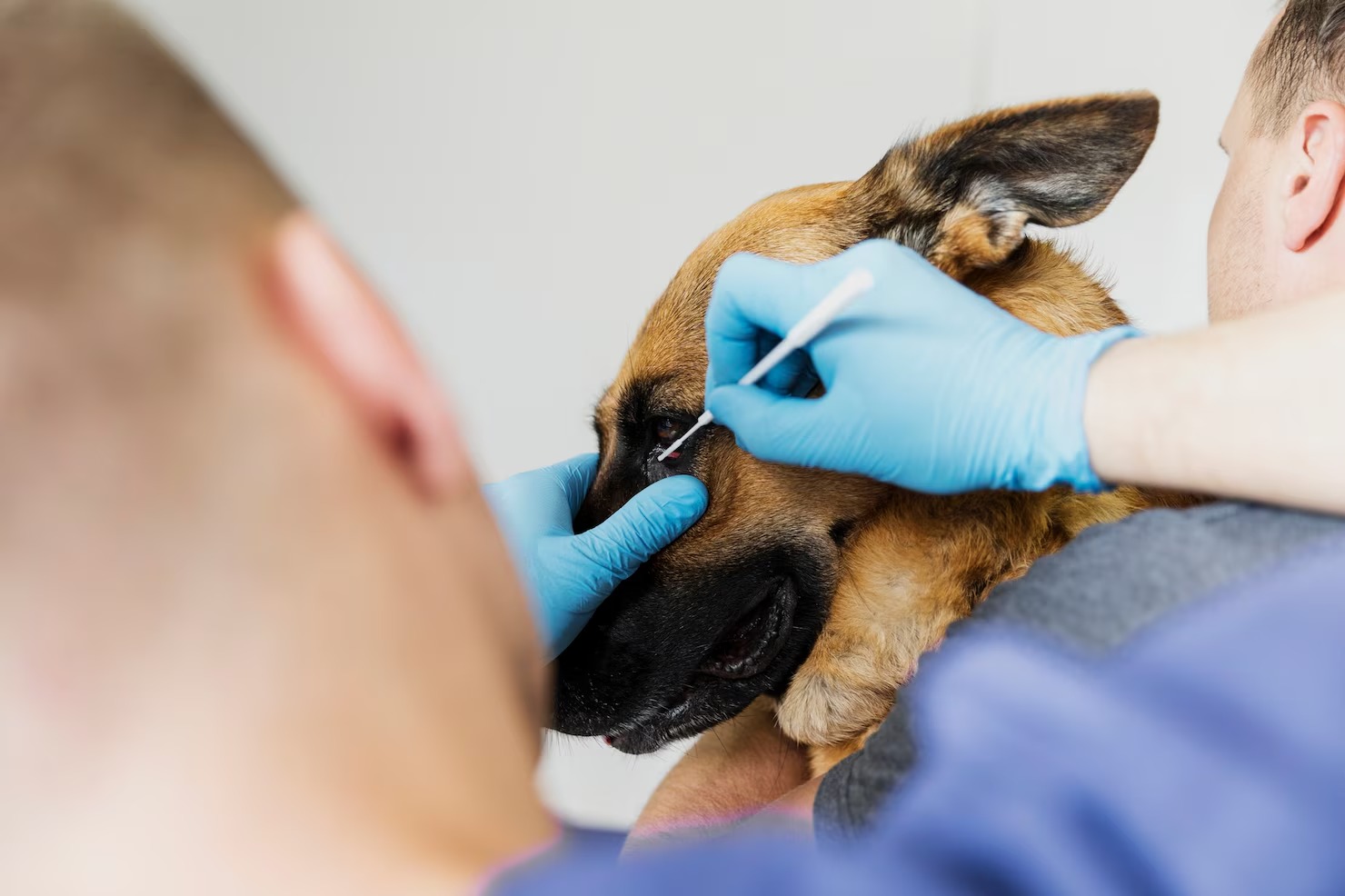
Diagnosing canine glaucoma requires specialized equipment and a meticulous ophthalmic examination. Because a red, cloudy eye can be indicative of numerous other conditions—such as severe corneal ulcers or anterior uveitis—a definitive diagnosis cannot be made by visual inspection alone. Veterinarians and board-certified veterinary ophthalmologists rely on a combination of tonometry, gonioscopy, and advanced imaging to confirm the diagnosis, determine the underlying cause, and evaluate the remaining visual potential[3].
Tonometry (Intraocular Pressure Measurement)
Tonometry is the definitive, gold-standard diagnostic test for glaucoma. It measures the exact pressure inside the eye. Modern veterinary clinics primarily utilize two types of handheld tonometers: applanation tonometers (such as the Tono-Pen) and rebound tonometers (such as the TonoVet). Applanation tonometers measure the amount of force required to flatten a small area of the cornea, which necessitates the application of prescription topical anesthetic drops. Rebound tonometers, conversely, bounce a microscopic, magnetized plastic probe off the cornea so quickly that the dog barely registers the touch, eliminating the need for anesthetics. A normal intraocular pressure for a dog is between 10 and 25 mmHg. A reading consistently above 30 mmHg, especially in conjunction with clinical signs of vision loss or pain, strongly supports a diagnosis of glaucoma. Accurate measurement is critical; the veterinarian must ensure they do not tightly grip the dog’s neck or compress the jugular veins during restraint, as this can artificially spike the pressure reading[11].
Gonioscopy
If tonometry confirms an elevated pressure, the next diagnostic step is gonioscopy, a specialized procedure used to evaluate the anatomical structure of the iridocorneal drainage angle. Because the angle is hidden behind the limbus (the junction between the clear cornea and white sclera), the veterinarian places a specialized, domed magnifying glass called a Koeppe goniolens directly onto the surface of the eye. By shining a bright slit-lamp through the lens, the veterinarian can visually inspect the pectinate ligaments. If the ligaments are thickened into solid sheets (goniodysgenesis), a diagnosis of primary angle-closure glaucoma is confirmed. Gonioscopy is usually performed on the *unaffected* fellow eye, as the glaucomatous eye is often too cloudy (due to corneal edema) to allow for internal visualization[12].
Ophthalmoscopy and Vision Testing
To determine if the optic nerve has suffered permanent damage, the veterinarian will perform a thorough fundic examination using an indirect ophthalmoscope. They will look for hallmark signs of glaucomatous damage, such as optic disc cupping, attenuation (thinning) of the retinal blood vessels, and hyperreflectivity of the tapetum (indicating retinal degeneration). Furthermore, neuro-ophthalmic reflexes must be tested. The Menace response (blinking when a hand approaches the eye) tests higher cortical vision, while the Dazzle reflex (squinting when a bright light is suddenly shined into the eye) tests subcortical retinal function. If the Dazzle reflex is completely absent, it strongly suggests that the retinal ganglion cells have died, meaning vision restoration is unlikely regardless of treatment[20].
Advanced Imaging: Ultrasonography and ERG
In cases where the cornea is entirely opaque from severe edema or hemorrhage, visual inspection of the interior eye is impossible. In these scenarios, High-Resolution Ocular Ultrasonography (or Ultrasound Biomicroscopy – UBM) is utilized. UBM utilizes high-frequency sound waves to generate detailed cross-sectional images of the anterior chamber, allowing the clinician to detect hidden intraocular tumors, luxated lenses, or massive retinal detachments that are driving the secondary glaucoma. Additionally, an Electroretinogram (ERG) may be performed prior to any expensive, vision-saving surgeries. An ERG measures the electrical activity of the photoreceptor cells in the retina in response to light flashes. If the ERG trace is flat, the retina is dead, and vision-saving surgery is contraindicated[13].
Treatment for Glaucoma in Dogs

The treatment of canine glaucoma is a complex, multifactorial endeavor that requires a strict, lifelong commitment from the pet owner. Under the guidance of veterinary ophthalmology, the immediate goals of therapy are to aggressively mitigate intraocular pressure, alleviate excruciating pain, and salvage any remaining optic nerve function. The long-term strategy is dictated by whether the eye retains visual potential or has succumbed to permanent blindness. Treatment modalities are broadly divided into emergency medical management, maintenance medical therapy, vision-saving surgeries, and salvage (comfort) surgical procedures[14].
Emergency Medical Interventions
When a dog presents with acute glaucoma, every minute counts. The veterinarian will initiate rapid-acting topical and systemic medications to “break” the pressure spike. The most effective emergency class of drugs are prescription prostaglandin analogs. When applied topically, these profound medications stimulate the alternative uveoscleral outflow pathway, bypassing the blocked trabecular meshwork. Furthermore, this class of medication induces intense miosis (constriction of the pupil), which physically pulls the base of the iris away from the drainage angle, opening up whatever space remains. If topical therapies fail, the veterinarian may administer a prescription intravenous hyperosmotic agent. This hyperosmotic treatment rapidly increases the osmotic pressure of the blood, physically drawing water out of the vitreous humor of the eye and dehydrating the globe to lower pressure rapidly[15].
Long-Term Maintenance Medication
Once the acute crisis is stabilized, the dog must be placed on a strict regimen of topical medications to maintain the IOP within normal limits. Consistency is paramount; skipping even a single dose can trigger a catastrophic pressure spike.
- Carbonic Anhydrase Inhibitors (CAIs): Prescription carbonic anhydrase inhibitors are the backbone of maintenance therapy. They inhibit the carbonic anhydrase enzyme within the ciliary body, effectively slowing down the active production of aqueous humor. By reducing the fluid entering the eye, the pressure is naturally lowered.
- Beta-Blockers: Prescription topical beta-blockers act on the sympathetic receptors in the ciliary body to further decrease aqueous production. These beta-blockers are frequently combined with carbonic anhydrase inhibitors in a single prescription drop to maximize pressure reduction while minimizing the number of drops the owner has to administer.
- Analgesics and Anti-inflammatories: Because high pressure damages tissues and causes significant secondary inflammation, prescription topical steroids or systemic veterinary nonsteroidal anti-inflammatory drugs (NSAIDs) are often prescribed. Prescription pain medication may also be utilized to combat the severe neuropathic pain associated with optic nerve compression[14].
Surgical Treatment for Visual Eyes
Medical therapy for primary glaucoma eventually fails in almost all dogs due to down-regulation of drug receptors and continued anatomical deterioration. If the eye still retains vision (evidenced by a positive Dazzle reflex and normal ERG), vision-saving surgeries may be attempted by a specialist. Endolaser Cyclophotocoagulation (ECP) is an advanced procedure where a microscopic fiber-optic camera and laser probe are inserted into the eye. The surgeon uses the diode laser to precisely burn and destroy portions of the ciliary body, permanently reducing its ability to produce aqueous humor. ECP is almost always performed alongside cataract surgery (phacoemulsification). Another option is the implantation of a gonioimplant (such as an Ahmed valve), a small silicone tube inserted into the anterior chamber that shunts fluid out of the eye to a small reservoir under the conjunctiva. However, these shunts have a high failure rate in dogs due to the aggressive fibrotic healing response that eventually scars over the drainage plate[16].
Surgical Salvage Procedures for Blind Eyes
When an eye is permanently blind and painful, keeping it medically managed is both expensive and ethically questionable, as it prolongs the animal’s suffering. In these cases, surgical salvage procedures are highly recommended to provide immediate, permanent pain relief. The most common procedure is enucleation, the complete surgical removal of the eye globe and the suturing of the eyelids closed. While emotionally difficult for the owner, dogs adapt phenomenally well to enucleation and exhibit a massive increase in energy and happiness once the source of their chronic migraine is removed.
For owners seeking a cosmetic alternative, an evisceration with an Intrascleral Silicone Prosthesis (ISP) can be performed. In this procedure, the internal contents of the eye (iris, lens, retina) are surgically scraped out, leaving only the outer white scleral shell and the cornea. A black silicone sphere is then implanted inside the shell, giving the appearance of a normal, slightly clouded eye. Alternatively, a chemical ciliary body ablation can be performed, which involves injecting prescription medications directly into the vitreous to chemically destroy the fluid-producing cells. However, this is considered a less reliable procedure with potential long-term complications, such as tumor formation. Whatever the course of action, it is vital to remember: if your dog suffers an eye injury, ensure that they receive immediate veterinary care to minimize the risk of complications. You must always consult your veterinarian before making any changes to your pet’s care[18].
How to Prevent Dogs From Having Glaucoma
Mitigating the risk of glaucoma in dogs is a multifaceted challenge. Primary glaucoma is genetically hardwired into an animal’s DNA, making absolute prevention impossible. However, proactive management, aggressive screening, and excellent general health care can delay the onset of the disease, protect the unaffected eye, and preserve the dog’s comfort and vision for as long as possible[19].
- Genetic Screening and Responsible Breeding: The most effective way to eliminate primary glaucoma from the canine population is through strict breeding practices. Breeders of high-risk dogs (such as Basset Hounds, Siberian Huskies, and Cocker Spaniels) should have their breeding stock evaluated by a board-certified veterinary ophthalmologist. Screening tools include gonioscopy to identify hidden structural abnormalities and genetic cheek-swab testing to screen for known POAG gene mutations (like the ADAMTS10 mutation in Beagles). Dogs with abnormal gonioscopy results or carrying the genetic markers must not be bred[4].
- Prophylactic Therapy for the Contralateral Eye: Primary glaucoma is a bilateral disease. When a dog presents with an acute glaucomatous crisis in one eye, the second (contralateral) eye is almost guaranteed to develop the condition eventually. Without intervention, the second eye typically fails within 8 to 10 months. However, initiating prophylactic (preventative) topical therapy on the healthy eye—usually with a prescription beta-blocker or specialized veterinary pressure-reducing drop—can significantly delay the onset of the disease, extending the dog’s visual lifespan by an average of 18 to 31 months[20].
- Routine Tonometry Screenings: For breeds predisposed to glaucoma, intraocular pressure checks should become a standard part of their annual or bi-annual wellness exams starting at 3 years of age. Catching a pressure slowly trending upward from 15 mmHg to 22 mmHg, even before it hits the critical 30 mmHg mark, allows the veterinarian to intervene medically before irreversible optic nerve cupping occurs.
- Prompt Treatment of Secondary Ocular Conditions: Secondary glaucoma is entirely preventable if the primary inciting cause is aggressively treated. If a dog develops an inflammatory condition, owners must seek immediate veterinary care to resolve the inflammation before it clogs the trabecular meshwork. Additionally, owners of terrier breeds predisposed to lens luxations should be vigilant for signs of lens instability.
- Systemic Health and Nutrition: Maintaining robust systemic health supports overall ocular function. Ensuring your dog enjoys a balanced diet, routine exercise, and a healthy weight helps prevent metabolic conditions like diabetes, which can lead to rapid-onset cataracts. Cataracts, if left untreated, can rupture and cause phacoclastic uveitis—a massive inflammatory reaction that inevitably leads to aggressive secondary glaucoma[8].
While the word “glaucoma” is frightening for any pet owner to hear, modern veterinary medicine offers more tools than ever to manage this disease. Through vigilance, early detection via routine tonometry, and immediate action at the first sign of ocular distress, owners can significantly alter the trajectory of their pet’s visual health.
Frequently Asked Questions
Is Glaucoma in dogs painful?
Yes, glaucoma in dogs is extraordinarily painful. While human glaucoma (often open-angle) can be a slow, painless progression, canine glaucoma (predominantly closed-angle) results in acute, massive spikes in intraocular pressure. This pressure stretches the sensitive tissues of the eye and compresses the optic nerve, causing a debilitating pain that is frequently compared to a severe, unrelenting migraine headache. Dogs often mask this pain, presenting instead as lethargic, depressed, or unwilling to eat. Rapid medical or surgical intervention is required to alleviate this suffering.
Can a dog live a happy life with one eye removed due to glaucoma?
Absolutely. Dogs possess an incredible ability to adapt to monocular (single-eye) vision or even complete blindness. When an eye is surgically removed (enucleated) due to end-stage glaucoma, the immediate result is the complete eradication of the chronic pain they had been enduring. Owners universally report that within days of the surgery, their dog’s energy, appetite, and playful personality return in full force. The loss of an eye does not hinder a dog’s capacity to live a long, joyful, and deeply fulfilling life.
How quickly can glaucoma cause permanent blindness in a dog?
The speed of vision loss depends on the type and severity of the glaucoma, but it can be shockingly rapid. In cases of acute angle-closure glaucoma, where the pressure spikes to 50 or 60 mmHg, the intense compression of the optic nerve and retinal blood vessels causes severe ischemia (lack of blood flow). If this pressure is not medically broken within 24 to 48 hours, the retinal ganglion cells die, and the dog will suffer irreversible, permanent blindness. This is why a red, cloudy, or squinting eye is always considered a true veterinary emergency.
Concerned About Your Pet’s Vision?
Glaucoma is a medical emergency that requires prompt intervention. If you notice any signs of eye discomfort, cloudiness, or vision loss, please schedule an appointment with a veterinarian immediately to protect your dog’s sight.
References
- Gelatt, K. N., & MacKay, E. O. Prevalence of primary breed-related glaucoma in purebred dogs in North America. Veterinary Ophthalmology, 2004.
- Pizzirani, S. Definition, classification, and pathophysiology of canine glaucoma. Veterinary Clinics: Small Animal Practice, 2015.
- Maggio, F. Glaucomas. Merck Veterinary Manual, 2015.
- Kuchtey, J., et al. Mapping of the disease locus and identification of ADAMTS10 as a candidate gene in a canine model of primary open angle glaucoma. PLOS Genetics, 2011.
- Kato, K., et al. Incidence of canine glaucoma with goniodysgenesis in Japan: a retrospective study. Journal of Veterinary Medical Science, 2006.
- Townsend, W. M., et al. Golden Retriever pigmentary uveitis: Challenges and solutions. Veterinary Medicine: Research and Reports, 2020.
- Reinstein, S. L., et al. Canine lens luxation. Journal of the American Veterinary Medical Association, 2018.
- Leiva, M., et al. Secondary glaucomas in dogs. Veterinary Ophthalmology, 2012.
- McLellan, G. J., & Miller, P. E. Canine Glaucoma. Veterinary Ophthalmology, 2021.
- Plummer, C. E., et al. Primary glaucoma in dogs. Veterinary Clinics: Small Animal Practice, 2013.
- Bras, I. D., et al. Evaluation of rebound tonometry in dogs. Journal of the American Veterinary Medical Association, 2017.
- Bentley, E., et al. Gonioscopy Guidelines. American College of Veterinary Ophthalmologists (ACVO) Publications, 2014.
- Strom, A. R., et al. Electroretinography in veterinary ophthalmology. Journal of Veterinary Medical Science, 2020.
- Alario, A. F., et al. Medical treatment of primary canine glaucoma. Veterinary Clinics of North America, 2015.
- Komáromy, A. M., et al. The future of canine glaucoma therapy. Veterinary Ophthalmology, 2019.
- Bentley, E. Surgical management of glaucoma. Veterinary Clinics of North America, 2022.
- Graham, K. L., et al. Ocular manifestations of systemic disease and neuroprotection. Veterinary Ophthalmology, 2021.
- Esson, D. W. Clinical Atlas of Canine and Feline Ophthalmic Disease. Wiley-Blackwell, 2015.
- American College of Veterinary Ophthalmologists (ACVO). Genetics of Canine Eye Diseases. ACVO Publications, 2023.
- Miller, P. E., & Bentley, E. Clinical signs and diagnosis of the canine primary glaucomas. Veterinary Clinics of North America: Small Animal Practice, 2015.


