


 March 9, 2023
 Phil Good, DVM
What are Eye Infections in Cats?
This content was prepared with AI assistance and reviewed by a licensed professional for accuracy.
Introduction
As a feline owner, witnessing your companion suffer from an eye infection, which often presents with swelling eyes, excessively irritated eyes, and deeply inflamed eyes, can be a deeply distressing experience. The feline eye is an exceptionally complex and delicate organ, heavily innervated and structurally designed to provide the extraordinary visual acuity cats rely on as obligate carnivores. When pathogenic organisms such as bacteria, viruses, or fungi breach the eye’s natural defenses, they can trigger a cascade of inflammatory responses. As a veterinary professional, I often see cats presenting with acute ocular distress that ranges from mild discomfort to severe, vision-threatening conditions. Understanding the anatomical structures involved—such as the clear cornea, the vascular conjunctiva, the sclera, and the internal uveal tract—is fundamental to grasping how these infections manifest and progress[1].
The ocular surface is an immune-privileged site, meaning it has specialized mechanisms to tolerate the introduction of antigens without inducing severe, sight-damaging inflammation. However, this delicate balance can be easily disrupted. The conjunctiva, a thin mucosal membrane that lines the inner eyelids and covers the sclera (the white of the eye), is particularly susceptible to environmental irritants and infectious agents. Furthermore, the feline eye is intimately connected to the upper respiratory tract via the nasolacrimal duct system. This anatomical relationship explains why so many feline upper respiratory infections concurrently present with severe ocular symptoms. Recognizing the onset of these infections and understanding their underlying pathophysiology is crucial for early intervention, ensuring that your cat receives the appropriate veterinary care to preserve their vision and overall quality of life[2].
It is important to differentiate between primary eye infections—where the eye itself is the initial target of the pathogen—and secondary infections, where the ocular structures become compromised due to a systemic illness or an adjacent anatomical issue, such as severe dental disease. Feline ocular health is a highly specialized field, and the array of potential pathogens is vast. While some infections are self-limiting, many require targeted antimicrobial or antiviral therapies to prevent permanent structural damage. As we explore the various types, causes, and treatments of feline eye infections, it becomes clear that proactive monitoring and swift veterinary diagnostics are the cornerstones of effective ocular health management[3].
Types of Eye Infection in Cats
The classification of an eye infection depends heavily on which specific anatomical structure within the eye is primarily affected. Accurate categorization is essential for formulating an effective treatment protocol, as different ocular tissues respond differently to pathogens and pharmacological interventions.
Conjunctivitis
Conjunctivitis is the most frequently diagnosed ocular disorder in feline medicine. It refers to the inflammation of the conjunctiva, the highly vascularized mucous membrane that coats the inner aspect of the eyelids and the anterior sclera. The conjunctiva is populated with specialized goblet cells responsible for producing the mucin layer of the tear film. When inflamed by an infectious agent, these goblet cells often overproduce mucus, leading to the thick, mucopurulent discharge commonly associated with the condition. Clinically, conjunctivitis presents with severe chemosis (swelling of the conjunctival tissue), hyperemia (redness due to engorged blood vessels), and the formation of lymphoid follicles. Unlike in dogs, where conjunctivitis is frequently allergic or immune-mediated, feline conjunctivitis is almost always infectious in origin, with Feline Herpesvirus-1 (FHV-1) and Chlamydia felis being the leading culprits[4]. Left untreated, chronic conjunctivitis can lead to permanent changes in the conjunctival tissue, scarring, and secondary tear film deficiencies.
Keratitis
Keratitis involves the inflammation of the cornea, the transparent, dome-shaped outer layer that covers the front of the eye. The cornea is comprised of multiple layers: the outer epithelium, the thick collagenous stroma, Descemet’s membrane, and the inner endothelium. In cats, keratitis can present in several unique forms. Non-ulcerative keratitis involves inflammation without the loss of the epithelial layer, often resulting in severe corneal edema, which gives the eye a cloudy or blue appearance. A specific and distinct condition in cats is feline eosinophilic keratitis, characterized by the infiltration of eosinophils (a type of white blood cell) into the cornea, resulting in raised, pink, or white vascularized plaques on the corneal surface. Another feline-specific condition is the development of a corneal sequestrum, where a portion of the corneal stroma undergoes necrosis and turns black or dark brown, often occurring secondary to chronic viral keratitis. Managing keratitis requires precise diagnostics to differentiate between viral, bacterial, and immune-mediated etiologies[5].
Uveitis
Uveitis refers to uvea inflammation, which is the heavily pigmented and highly vascular middle layer of the eye. The uveal tract consists of the iris, the ciliary body (which produces aqueous humor), and the choroid (which nourishes the retina). Uveitis represents a breakdown of the blood-aqueous barrier, allowing proteins and white blood cells to leak into the anterior chamber of the eye. This leakage creates a cloudy appearance known as “aqueous flare.” Clinical signs of uveitis include profound miosis (a constricted pupil), severe photophobia (sensitivity to light), blepharospasm (squinting), and hypotony (low intraocular pressure). In cats, uveitis is frequently an ocular manifestation of a severe systemic infectious disease, such as Feline Infectious Peritonitis (FIP), Feline Immunodeficiency Virus (FIV), Feline Leukemia Virus (FeLV), or systemic fungal infections like Toxoplasmosis. Therefore, a diagnosis of uveitis warrants an exhaustive systemic workup to identify the root cause[6].
Corneal ulcers
Corneal ulcers represent a physical breach or wound in the outer epithelial layer of the cornea, exposing the underlying sensory-rich stroma. In felines, viral infections—particularly FHV-1—are notorious for causing pathognomonic “dendritic” ulcers, which appear as branching, tree-like erosions across the corneal surface. When a superficial ulcer becomes secondarily infected by opportunistic bacteria, the bacteria can release proteolytic enzymes (collagenases) that rapidly digest the corneal stroma. This condition, known as a “melting ulcer” or keratomalacia, is an absolute veterinary emergency, as it can progress to a descemetocele (where only the innermost membrane prevents the eye from rupturing) or full ocular perforation within a matter of hours. The feline cornea is densely packed with trigeminal nerve endings, making corneal ulcers excruciatingly painful, leading to severe behavioral changes, lethargy, and anorexia in affected cats[7].
Blepharitis
Blepharitis is the localized inflammation of the eyelids. The feline eyelid is a complex structure containing specialized Meibomian glands that secrete the lipid (fatty) layer of the tear film, which prevents tear evaporation. When bacteria—typically staphylococcal species—infiltrate these glands or the surrounding hair follicles, it can lead to painful swelling, erythema (redness), and the formation of localized abscesses, commonly referred to as styes (hordeolum) or chalazions. Blepharitis can cause intense pruritus (itching), leading the cat to relentlessly rub their face against furniture or aggressively scratch at their eyes with their claws. This self-trauma frequently exacerbates the condition, introducing more bacteria and potentially causing secondary corneal abrasions. Chronic blepharitis can also lead to structural changes in the eyelid margin, resulting in entropion (inward rolling of the eyelid), which causes the eyelashes to continuously rub against the cornea, creating a vicious cycle of irritation and infection[8].
Chlamydial conjunctivitis
Chlamydial conjunctivitis in felines is caused by the obligate intracellular bacterium Chlamydia felis. Because it is an intracellular pathogen, it cannot survive for long in the environment and relies on direct, close contact between cats for transmission, making it highly prevalent in multi-cat households, catteries, and animal shelters. The hallmark presentation of a Chlamydia felis infection often begins unilaterally—affecting only one eye—before progressing to bilateral involvement within a few days. Cats infected with this pathogen typically exhibit intense conjunctival hyperemia, profound chemosis (so severe that the conjunctiva may bulge out past the eyelid margins), and a copious mucopurulent discharge. Unlike viral pathogens like FHV-1 and FCV, Chlamydia felis rarely causes significant upper respiratory signs or corneal ulceration; its pathology is strictly localized to the conjunctiva. Treatment requires specific classes of prescription antibiotics capable of penetrating host cells to reach the bacteria[9].
Feline Infectious Peritonitis (FIP)-Related Ocular Conditions
Feline Infectious Peritonitis (FIP) is a complex, often fatal systemic disease caused by a mutation of the highly common feline enteric coronavirus (FCoV). When the virus mutates into the FIPV biotype, it attacks the cat’s macrophages, leading to widespread pyogranulomatous vasculitis (inflammation of the blood vessels). The eyes are incredibly vascular organs, making them prime targets for FIP-related inflammation. Ocular manifestations of FIP typically present as severe, bilateral granulomatous anterior uveitis. A classic clinical sign during an ophthalmic exam is the presence of “mutton-fat” keratic precipitates—large, greasy-looking clumps of inflammatory cells adhering to the inner surface of the cornea. Additionally, FIP can cause hyphema (blood in the anterior chamber), fibrin deposition within the eye, retinal detachment, and optic neuritis leading to acute blindness. Ocular FIP is a grave diagnosis, though recent advancements in antiviral therapeutics have begun to alter the previously uniformly fatal prognosis[10].
Feline Immunodeficiency Virus (FIV)-Related Eye Infections
Feline Immunodeficiency Virus (FIV) is a slow-acting lentivirus, akin to human HIV, that progressively degrades the feline immune system by targeting CD4+ T-lymphocytes. While FIV itself does not typically cause direct damage to the ocular tissues, the resulting immunosuppression leaves the cat highly vulnerable to a host of opportunistic secondary eye infections. Cats with FIV are significantly more prone to severe, recurrent bouts of viral conjunctivitis, aggressive bacterial keratitis, and systemic opportunistic pathogens that cause uveitis, such as Toxoplasmosis or systemic fungal infections like Cryptococcosis. Furthermore, chronic anterior uveitis and pars planitis (inflammation of the narrowed area between the iris and the choroid) are frequently documented in FIV-positive cats. Managing eye infections in an FIV-positive feline requires a far more aggressive and prolonged therapeutic approach, as their compromised immune systems cannot effectively assist in clearing the pathogens[11].
Causes of Cat Eye Infections

The etiology of feline eye infections is vast and multifaceted. Identifying the precise causative agent is the most critical step in resolving the infection, as empirical treatments without a confirmed diagnosis can lead to treatment failure, antibiotic resistance, and prolonged suffering.
Bacterial Infections
Unlike dogs, primary bacterial eye infections in cats are relatively uncommon; they are almost always secondary to an underlying viral infection, structural abnormality, or trauma. However, when bacteria do establish a foothold, they can cause rapid and devastating tissue damage. Normal feline conjunctival flora includes low numbers of Gram-positive bacteria. When the local ocular immunity drops, pathogenic strains like Staphylococcus aureus, Streptococcus species, and particularly Pseudomonas aeruginosa can proliferate. Pseudomonas is especially feared in veterinary ophthalmology due to its rapid replication rate and ability to secrete massive amounts of collagenases, which can literally dissolve a cat’s cornea in less than 24 hours. Primary bacterial infections can occasionally be caused by Mycoplasma species, which reside on the mucosal surfaces and can trigger severe conjunctivitis characterized by heavy, purulent discharge and significant conjunctival swelling[12].
Viral Infections
Viral pathogens are unquestionably the leading cause of primary ocular disease in cats. Feline Herpesvirus-1 (FHV-1) is the most pervasive, affecting an estimated 80% to 90% of the global feline population. Once a cat is infected, FHV-1 establishes lifelong latency within the trigeminal ganglia. During periods of physiological or psychological stress—such as moving to a new home, the introduction of a new pet, or a concurrent illness—the virus recrudesces, traveling down the ophthalmic branch of the trigeminal nerve to shed on the ocular surface. This shedding triggers severe conjunctivitis and the classic dendritic corneal ulcers. Feline Calicivirus (FCV), another major viral player, primarily causes upper respiratory tract infections and oral ulcerations, but it frequently involves a mild to moderate conjunctivitis component. Differentiating between FHV-1 and FCV is clinically important, as FHV-1 has a profound affinity for corneal tissue, whereas FCV rarely causes corneal ulceration[13].
Fungal Infections
Fungal eye infections, known clinically as mycotic keratitis or mycotic uveitis, are less common in felines but represent some of the most challenging conditions to treat. They are often geographically dependent. For example, in certain regions of North America, systemic fungal organisms such as Cryptococcus neoformans, Blastomyces dermatitidis, or Histoplasma capsulatum can infect cats. Cryptococcus is particularly notable in feline medicine; it typically initiates as a severe nasal cavity infection that invades the central nervous system and the optic nerve, subsequently extending into the eye to cause granulomatous chorioretinitis and retinal detachment. Direct fungal infections of the cornea can also occur if a cat suffers an ocular scratch from a plant or tree branch contaminated with environmental fungi like Aspergillus or Candida, requiring prolonged therapy with highly specific topical antifungal medications[14].
Parasitic Infections
Parasites like mites or fleas can infect cats’ eyes, inducing irritation, inflammation, and setting the stage for aggressive secondary bacterial infections. While rare, specific parasites target the feline ocular structures. Thelazia californiensis, a nematode commonly known as the eye worm, resides directly in the conjunctival sac and tear ducts of cats, visibly wriggling across the eye and causing intense irritation and conjunctivitis. Another notable parasite is the larval stage of the Cuterebra fly, which can occasionally undergo aberrant migration, ending up in the anterior chamber of the feline eye, resulting in a devastating and blinding uveitis. Systemically, the intracellular parasite Toxoplasma gondii—for which the cat is the definitive host—can cause profound anterior and posterior uveitis when it encysts within the ocular tissues during its complex life cycle. Eradicating these parasitic infections requires targeted antiparasitic agents alongside profound anti-inflammatory therapy[15].
Allergies
Feline allergies, including environmental atopy (reactions to pollen, dust mites, or mold) and food allergies, can provoke a systemic immune response that heavily involves the ocular tissues. When a sensitized cat encounters an allergen, localized mast cells within the conjunctival tissue degranulate, releasing massive quantities of histamine and other pro-inflammatory cytokines. This rapid release results in allergic conjunctivitis, characterized by profound ocular pruritus, intense conjunctival hyperemia, and clear, watery epiphora (excessive tearing). Unlike infectious conjunctivitis, allergic ocular conditions are usually bilateral and frequently accompanied by other dermatological signs, such as miliary dermatitis or eosinophilic granuloma complex lesions. Identifying and removing the offending allergen, coupled with antihistamines and topical immunomodulators, is required to manage allergic ocular inflammation effectively[16].
Trauma or Injury
Ocular trauma is a frequent cause of sudden, severe eye infections in cats. As agile predators and territorial animals, cats frequently engage in altercations that result in sharp claw injuries directly to the face and eyes. A cat scratch to the cornea not only causes a painful laceration but physically inoculates the sterile corneal stroma with a myriad of aggressive bacteria residing on the claw, including Bartonella henselae and Staphylococcus. Additionally, outdoor cats are prone to encountering foreign bodies such as grass seeds, thorns, or plant awns, which can easily become lodged behind the nictitating membrane (the third eyelid). These retained foreign objects continuously scrape against the cornea every time the cat blinks, preventing any healing and serving as a nidus for intractable bacterial infections until they are physically removed under veterinary sedation[17].
Blocked Tear Ducts
The feline nasolacrimal apparatus is responsible for draining excess tears from the eye down into the nasal cavity. When this system becomes obstructed—a condition known as nasolacrimal duct occlusion or dacryocystitis—tears spill over onto the cat’s face, causing chronic epiphora. This constant moisture on the facial fur creates a perfect, humid microenvironment for bacteria and yeast (like Malassezia) to thrive, leading to secondary periocular skin infections and subsequent conjunctivitis. Obstructions can be congenital, such as imperforate puncta (where the drainage holes fail to develop), or acquired, often secondary to chronic inflammation from FHV-1 infections that cause scarring and stricture of the delicate drainage ducts. Resolving this often requires a veterinarian to cannulate and flush the ducts using specialized catheters and sterile saline[18].
Secondary Infections
Because the anatomical structures of the feline head are highly interconnected, an infection originating in an adjacent area can easily migrate to the eye. One of the most common culprits of secondary ocular infections is severe feline dental disease. A tooth root abscess—particularly involving the upper fourth premolar—can cause extensive inflammation and bacterial spread that breaches the thin bony plates of the maxilla, entering the retrobulbar space (the area directly behind the eye). This results in retrobulbar cellulitis or a retrobulbar abscess, which pushes the eye forward (exophthalmos), prevents the cat from closing its mouth without extreme pain, and severely comprises the ocular tissues. Similarly, severe chronic rhinosinusitis can spread infection upwards through the ethmoid bones, leading to complex, multi-systemic ocular infections[19].
Cataracts or Dry Eye
While Keratoconjunctivitis Sicca (KCS), or “dry eye,” is far more prevalent in dogs, it does occur in cats, frequently as a long-term consequence of severe viral (FHV-1) damage to the lacrimal glands. A deficiency in the aqueous layer of the tear film deprives the cornea of vital nutrients and essential immune-regulating proteins (like lysozymes and lactoferrin). This chronic desiccation causes the cornea to become thickened, vascularized, and highly susceptible to secondary bacterial infections. Additionally, the development of cataracts—whether due to genetics, aging, or diabetes—can leak modified lens proteins into the surrounding ocular chambers. The feline immune system does not recognize these internal lens proteins, triggering a profound localized immune response known as lens-induced uveitis (phacolytic uveitis), which causes immense pain and paves the way for further opportunistic infections within the eye[20].
Symptoms of Eye Infections in Cats

Recognizing the clinical signs of a feline eye infection early is paramount to achieving a successful outcome. Because cats are incredibly stoic creatures, hardwired by evolutionary biology to mask signs of pain and vulnerability, overt symptoms often indicate that the infection has already reached a significant level of severity. The clinical presentation varies dramatically based on the specific anatomical tissue involved and the pathogenic agent responsible, but several hallmark signs should alert any pet owner to an immediate problem.
The most immediate and universal sign of ocular discomfort is blepharospasm, a condition where the cat aggressively squints or holds the affected eye tightly shut. This is a direct reflex mediated by the trigeminal nerve in response to intense pain. You will frequently observe excessive tearing, medically termed epiphora, where tears spill over the eyelid margins and stain the facial fur. As the infection progresses, the character of this discharge often changes. A clear, watery discharge suggests a viral or allergic etiology, whereas a thick, yellow, or greenish mucopurulent discharge is highly indicative of a heavy bacterial burden or a Chlamydia felis infection.
Inflammation of the conjunctiva, or chemosis, will cause the pink tissues lining the eyelids to swell dramatically, sometimes protruding beyond the eyelid margins itself. The sclera (the white part of the eye) may become severely hyperemic, presenting with bright red, engorged, and tortuous blood vessels. In cases involving the cornea, you may notice a sudden cloudiness or a distinct “blue haze,” which is a sign of corneal edema. This occurs when the barrier function of the corneal epithelium or endothelium is compromised, allowing fluid to pool within the corneal stroma. Furthermore, the cat’s third eyelid (the nictitating membrane), which usually remains hidden in the medial canthus of the eye, may prolapse and cover a portion of the globe—a generalized sign of ocular pain and orbital retraction[21].
In more severe, internal infections like uveitis, owners might notice anisocoria, a condition where the pupils are of unequal sizes. The pupil of the infected eye often becomes pinpoint (miotic) due to painful spasms of the ciliary muscle. In advanced cases, you may see hyphema (pooling of blood) or hypopyon (pooling of white pus) resting at the bottom of the anterior chamber, indicating a massive breakdown of the blood-aqueous barrier. Behavioral changes are also profound; affected felines often exhibit profound photophobia, hiding in dark closets or under beds to avoid light, accompanied by lethargy, anorexia, and vocalization when the facial area is touched[22].
Diagnosis of Eye Infections in Cats

Arriving at an accurate, definitive diagnosis for a feline eye infection requires a methodical, step-by-step ophthalmic examination performed by a skilled veterinarian. Ocular conditions can deteriorate extraordinarily rapidly, so guesswork is heavily discouraged in veterinary ophthalmology. The diagnostic process always begins with a comprehensive physical examination, focusing heavily on assessing cranial nerve function through tests like the menace response, the pupillary light reflex (PLR), and the palpebral reflex, which evaluate the integrity of the neurological pathways connecting the eye to the brain.
One of the first standardized diagnostic tests performed is the Schirmer Tear Test (STT). This involves placing a tiny strip of calibrated filter paper inside the lower eyelid to measure the cat’s aqueous tear production over precisely 60 seconds. Normal feline tear production typically ranges from 11 to 23 millimeters per minute. A significantly reduced STT suggests Keratoconjunctivitis Sicca (dry eye), which severely complicates ocular healing, while a highly elevated STT is a normal physiological response to active ocular pain. Following the STT, the veterinarian will apply a specialized, non-toxic orange dye called fluorescein to the eye. When exposed to a cobalt blue light, the dye brilliantly fluoresces green. The intact, lipophilic corneal epithelium repels the dye, but if there is any ulceration or scratch, the underlying hydrophilic stroma will aggressively absorb the stain, instantly revealing the exact size, depth, and margins of a corneal ulcer[23].
Tonometry is another critical diagnostic modality. Using a highly calibrated instrument called a tonometer (either applanation or rebound style), the veterinarian measures the intraocular pressure (IOP) of the eye. Normal feline IOP is approximately 10 to 20 mmHg. An abnormally low pressure (hypotony) is a hallmark sign of uveitis, whereas a spike in pressure indicates the development of secondary glaucoma, a blinding condition that requires immediate emergency intervention. For deeper evaluations, the vet will utilize slit-lamp biomicroscopy, which provides intense, magnified, three-dimensional illumination of the anterior segment of the eye, allowing the practitioner to detect microscopic inflammatory cells floating in the aqueous humor (aqueous flare) or pinpoint minute structural defects.
When an infectious pathogen is suspected, laboratory diagnostics are employed. Cytology involves gently scraping the conjunctiva or cornea with a specialized micro-spatula to collect surface cells. These cells are stained and examined under a high-powered microscope to identify the presence of specific white blood cells, such as eosinophils (indicative of feline eosinophilic keratitis) or intracellular inclusion bodies (indicative of Chlamydia). For definitive identification, especially in suspected viral or stubborn bacterial cases, Polymerase Chain Reaction (PCR) panels are invaluable. By submitting a simple conjunctival swab to an external laboratory, PCR testing amplifies the DNA or RNA of the pathogen, accurately identifying FHV-1, FCV, Chlamydia felis, or Mycoplasma, which dictates the exact targeted pharmacological strategy[24].
Treatment for Feline Eye Infections

The pharmacological and supportive treatment strategies for feline eye infections are meticulously tailored to the specific pathogen identified and the anatomical structures compromised. Due to the rapid cellular turnover and sensitive nature of ocular tissues, adherence to the prescribed veterinary regimen is absolutely vital to halt tissue degradation and restore visual function.
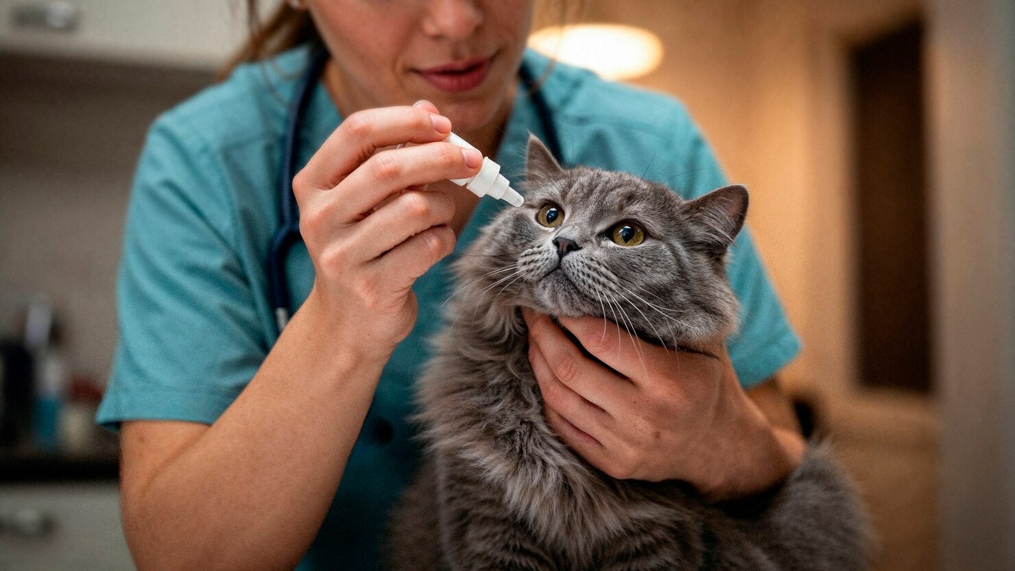
Topical Medications
The mainstay of ocular therapy involves the direct administration of topical medications onto the ocular surface. Depending on the diagnosis, veterinarians prescribe broad-spectrum or targeted prescription antibiotics to eradicate bacterial overgrowths. For active viral infections, particularly severe FHV-1 keratitis, highly specialized prescription antiviral eye medications are utilized to inhibit viral DNA replication within the host cells. It is clinically crucial that if a cat requires both a drop and an ointment, the drop must be applied first, with a mandatory five-to-ten minute waiting period before applying the ointment. Ointments have a lipid base that creates a physical barrier, preventing any subsequently applied aqueous drops from penetrating the cornea[25].
Systemic Medications
In cases where the infection is systemic or deeply embedded within tissues that topical medications cannot effectively penetrate, systemic oral therapies are necessitated. For example, a Chlamydia felis infection almost universally requires a prolonged course of oral prescription antibiotics. It is a critical clinical mandate in feline medicine that these oral medications must be immediately followed by a small syringe of water or a small meal; if a capsule becomes lodged in the esophagus, its highly acidic nature can cause severe, life-threatening esophageal strictures. Additionally, severe viral flare-ups of FHV-1 are now frequently managed with a systemic prescription antiviral medication that has shown remarkable efficacy in suppressing viral replication and alleviating severe ocular and respiratory clinical signs in felines.
Warm Compresses
Adjunctive physical therapies, such as the application of warm compresses, offer significant relief and promote faster healing. By applying a clean, warm, moist cloth gently against the closed eyelids for five to ten minutes, pet owners can induce localized vasodilation. This increased blood flow brings a higher concentration of the cat’s own white blood cells to the infected area. Furthermore, the gentle heat helps to melt and loosen hardened mucopurulent discharge and unclog obstructed Meibomian glands, facilitating the proper distribution of the lipid layer of the tear film across the cornea.
Eye Irrigation
Regular eye irrigation is often recommended to physically flush out aggressive pathogens, destructive inflammatory enzymes, and necrotic cellular debris from the conjunctival sac. Using a sterile, veterinary-approved ophthalmic saline wash restores the ocular surface to a neutral physiological pH. Removing this debris is not merely about cleanliness; thick purulent discharge can physically block topical medications from coming into contact with the actual corneal or conjunctival tissues, rendering expensive medications ineffective.
Supportive Care
Systemic supportive care ensures the feline patient remains hydrated, nourished, and pain-free, allowing their immune system to dedicate resources to fighting the infection. Severe ocular pain frequently induces anorexia in cats. Therefore, veterinarians heavily prioritize pain management, often prescribing systemic prescription pain medications administered by your veterinarian to dull the intense trigeminal nerve pain. Additionally, some practitioners advocate for the dietary supplementation of the amino acid L-lysine. While its efficacy is currently debated in modern veterinary literature, the theoretical mechanism is that L-lysine competes with arginine, an amino acid that feline herpesvirus strictly requires for replication, potentially reducing the severity of viral flare-ups.
E-collar (Elizabethan Collar)
The implementation of an Elizabethan collar (E-collar) is frequently a non-negotiable aspect of ocular therapy. Because eye infections cause intense pruritus and pain, cats will instinctively and aggressively rub their faces against rough surfaces or use their hind claws to scratch at the eye. This self-trauma can instantly convert a mild, superficial corneal ulcer into a deep, catastrophic descemetocele or cause complete globe rupture. While cats notoriously despise wearing E-collars—experiencing stress and “whisker fatigue”—the collar serves as an absolute mechanical barrier required to protect the delicate ocular structures while pharmacological treatments take effect.
Addressing Underlying Conditions
Treating the primary symptom is insufficient if the root systemic cause remains unaddressed. If an eye infection is secondary to infectious diseases such as FIV or FeLV, long-term immunomodulatory and supportive systemic therapies must be instituted to manage the cat’s overall immune status. If the infection stems from environmental or dietary allergies, the offending allergens must be identified through elimination trials and permanently removed from the cat’s environment. Because stress is the primary trigger for the recrudescence of latent feline herpesvirus, integrating environmental enrichment, synthetic feline facial pheromones, and maintaining a stable, predictable routine is just as important as the medical therapy itself. Please ensure you consult your veterinarian before making any changes to your pet’s care, as altering medical plans without professional guidance can lead to rapid ocular deterioration.
Prevention of Eye Infections in Cats
Preventative medicine is the most effective strategy for maintaining optimal feline ocular health. The foundation of prevention is adherence to a strict, veterinary-approved vaccination schedule. The core FVRCP vaccine heavily protects against Feline Viral Rhinotracheitis (caused by FHV-1) and Feline Calicivirus. While no vaccine offers 100% sterilizing immunity to prevent infection entirely, proper vaccination drastically reduces the severity of the clinical symptoms and limits the degree of viral shedding, protecting the cat from severe, vision-threatening corneal ulcerations.
Environmental management plays a pivotal role. Prevent Trauma to the Eyes: keeping your cat exclusively indoors eliminates their exposure to territorial fights with feral felines, reducing the risk of claw-induced corneal lacerations and the transmission of severe lentiviruses like FIV. Indoor living also protects them from environmental foreign bodies like plant awns and minimizes their exposure to systemic fungal spores and complex parasites. Furthermore, maintaining exceptional hygiene in the home, particularly regarding litter boxes and food bowls, limits bacterial loads.
Finally, routine veterinary wellness examinations are critical. A veterinarian can identify subtle anatomical changes—such as a mild entropion or the early stages of a tear duct obstruction—long before they escalate into an active, painful infection. Providing a high-quality, biologically appropriate diet ensures the cat maintains a robust immune system, capable of naturally fending off opportunistic pathogens before they can colonize the delicate structures of the eye.
Frequently Asked Questions
Can a cat’s eye infection go away on its own?
While some extremely mild, superficial conjunctival irritations caused by minor environmental allergens may self-resolve as the cat’s immune system clears the irritant, true infectious eye diseases rarely resolve independently without causing damage. Viral, bacterial, and fungal infections possess mechanisms to evade the feline immune system. Leaving an active infection untreated, particularly a corneal ulcer, risks the development of aggressive collagenase activity, which can literally melt the cornea and cause permanent blindness or the loss of the eye in a matter of days. Immediate veterinary assessment is always the safest course of action.
Can you treat a cat eye infection with human eye drops?
Absolutely not. You should never administer human eye medications—whether over-the-counter allergy drops, redness relievers, or leftover prescription antibiotics—to a feline. The feline ocular anatomy and physiological pH differ significantly from humans. Many human eye drops contain preservatives or active ingredients like prescription steroids that are highly toxic to cats or contraindicated. For example, applying a human steroid eye drop to a cat with an undiagnosed viral corneal ulcer will rapidly suppress local immunity, causing the virus to replicate explosively, which often results in the catastrophic rupture of the eyeball.
How long does a cat’s eye infection take to heal?
The healing timeline is entirely dependent on the specific pathogen, the precise anatomical tissue affected, and how promptly targeted therapy was initiated. Mild, uncomplicated bacterial conjunctivitis often responds to topical antibiotics within 5 to 7 days. However, severe conditions like deep stromal corneal ulcers, fungal keratitis, or Chlamydia felis infections may require aggressive, multi-modal therapy spanning several weeks to months. Viral infections like FHV-1 are lifelong; while the acute flare-up may be managed in two weeks, the virus will remain dormant in the cat’s neurological system, possessing the potential to recrudesce during periods of future stress.
Protect Your Cat’s Vision Today
Eye infections can deteriorate rapidly and cause permanent damage if left untreated. Don’t wait to seek professional care.
References
- Merck Veterinary Manual. Eye Structure and Function in Animals. Merck & Co., Inc., 2023.
- VCA Animal Hospitals. Conjunctivitis in Cats. VCA Hospitals, 2023.
- Maggs, D. J. Update on pathogenesis, diagnosis, and treatment of feline herpesvirus type 1. Clinical Techniques in Small Animal Practice, 2005.
- Merck Veterinary Manual. Conjunctivitis in Animals. Merck & Co., Inc., 2023.
- Andrew, S. E. Feline eosinophilic keratitis. Veterinary Clinics of North America: Small Animal Practice, 2005.
- VCA Animal Hospitals. Uveitis in Cats. VCA Hospitals, 2023.
- VCA Animal Hospitals. Corneal Ulcers in Cats. VCA Hospitals, 2023.
- Merck Veterinary Manual. Blepharitis in Animals. Merck & Co., Inc., 2023.
- Sykes, J. E. Feline chlamydiosis. Clinical Techniques in Small Animal Practice, 2005.
- Pedersen, N. C. A review of feline infectious peritonitis virus infection: 1963-2008. Journal of Feline Medicine and Surgery, 2009.
- English, R. V., et al. Ocular manifestations of systemic viral infections in the cat. Veterinary Clinics of North America: Small Animal Practice, 1998.
- American Veterinary Medical Association (AVMA). Disease Risks for Dogs and Cats. AVMA, 2023.
- Cornell University College of Veterinary Medicine. Feline Calicivirus. Cornell Feline Health Center, 2023.
- Ginn, J. A., et al. Systemic fungal infections in cats. Journal of the American Veterinary Medical Association, 2011.
- ASPCA. Common Cat Diseases. ASPCA, 2023.
- Merck Veterinary Manual. Atopic Dermatitis in Animals. Merck & Co., Inc., 2023.
- Veterinary Information Network (VIN). Corneal Ulceration in Cats. VIN, 2023.
- Merck Veterinary Manual. Diseases of the Nasolacrimal and Lacrimal Apparatus in Animals. Merck & Co., Inc., 2023.
- VCA Animal Hospitals. Retrobulbar Abscess in Cats. VCA Hospitals, 2023.
- Merck Veterinary Manual. Keratoconjunctivitis Sicca in Animals. Merck & Co., Inc., 2023.
- VCA Animal Hospitals. Ocular Conditions and Horner’s Syndrome. VCA Hospitals, 2023.
- Glaze, M. B. Feline ophthalmology: the interior eye. Veterinary Clinics of North America: Small Animal Practice, 2001.
- Merck Veterinary Manual. Ophthalmic Examination in Animals. Merck & Co., Inc., 2023.
- Maggs, D. J. Use of PCR in feline ophthalmology. Clinical Techniques in Small Animal Practice, 2005.
- VCA Animal Hospitals. Applying Eye Drops to Cats. VCA Hospitals, 2023.


