


 March 10, 2023
 Phil Good, DVM
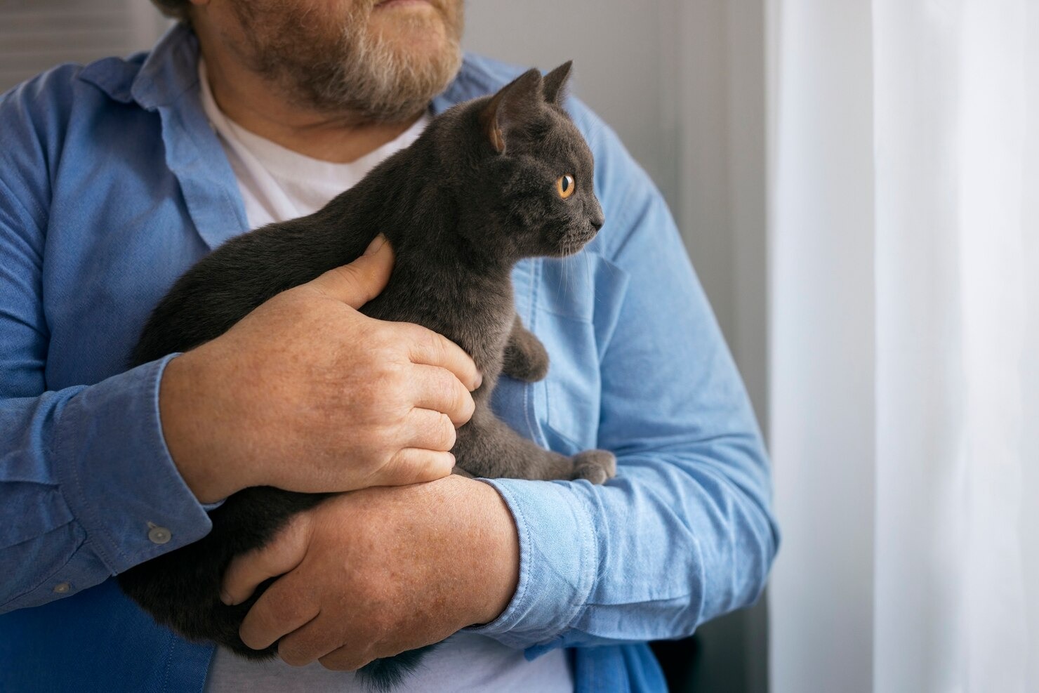
What is Melanoma in Cats?
This content was prepared with AI assistance and reviewed by a licensed professional for accuracy.
Introduction
When pet parents notice an unusual dark spot or lump on their beloved feline companion, it is completely natural to feel a sudden surge of anxiety. Navigating a potential cancer diagnosis is one of the most challenging aspects of pet ownership. Among the various types of skin cancer that can affect domestic felines, melanoma in cats represents a particularly complex and concerning diagnosis that requires immediate veterinary attention and comprehensive understanding.[1]
Melanoma is a specific type of cancer that originates from melanocytes. Melanocytes are specialized cells that reside in the skin, the mucous membranes, and the eyes. Their primary biological function is to produce melanin, the dark pigment responsible for giving color to a cat’s skin, fur, and the iris of their eyes. In a healthy animal, melanin serves an essential protective role, shielding the deeper layers of tissue from the harmful effects of ultraviolet radiation. However, when the delicate genetic programming within these melanocytes becomes damaged or corrupted, the cells can begin to divide and multiply uncontrollably. This uncontrolled cellular proliferation is the hallmark of cancer, leading to the formation of a neoplastic mass or tumor.[2]
Unlike in humans, where melanoma is strongly correlated with severe ultraviolet light exposure and is a relatively common form of skin cancer, feline melanoma is considered a rare disease. It accounts for a very small percentage of all diagnosed feline cancers. In fact, while dogs frequently develop oral melanomas, cats are far more likely to develop squamous cell carcinomas or fibrosarcomas in their oral cavities. When a cat does develop melanoma, it behaves quite differently than it does in humans or dogs. Feline melanomas can present as heavily pigmented, dark black or brown masses, but they can also be “amelanotic.” Amelanotic melanomas completely lack the characteristic dark pigment, making them appear pink, fleshy, or red. This lack of pigmentation makes visual diagnosis incredibly difficult and highlights the absolute necessity of advanced veterinary diagnostics, such as biopsies and specialized cellular staining.[3]
Understanding the intricacies of this disease is the first line of defense for a proactive pet owner. The location of the tumor plays a massive role in how the cancer will behave, the symptoms it will produce, and the treatment options that will be available. Melanoma in cats can arise on the haired skin (cutaneous melanoma), inside the mouth (oral melanoma), or within the structures of the eye (ocular melanoma). Each of these specific anatomical locations carries its own unique set of diagnostic challenges and prognostic outlooks. By arming yourself with scientifically accurate, clinically verified information, you can partner effectively with your veterinary oncologist to make the best possible medical decisions for your feline friend.[4]
Types of Melanoma in Cats
The term “melanoma” is often used as a broad umbrella term, but in veterinary oncology, distinguishing between the exact type and biological behavior of the tumor is critical. Melanocytic tumors in felines are generally classified based on their cellular characteristics (whether they are benign or malignant) and their anatomical location. Because melanocytes are distributed throughout the entire body during embryonic development, these tumors can theoretically arise almost anywhere. However, clinical data shows that they have distinct predilection sites. The behavior of the tumor—specifically its local invasiveness and its potential to metastasize (spread) to distant organs—is heavily influenced by where it first originates.[5]
Benign Melanoma (Melanocytoma)
A benign tumor of melanocytes is medically referred to as a melanocytoma. The word “benign” in oncology implies that the tumor cells, while abnormal and dividing more than they should, lack the genetic capacity to invade deeply into adjacent healthy tissues or to enter the lymphatic or circulatory systems to spread to other organs. Melanocytomas in cats are generally considered to be low-grade, slow-growing masses. They are typically well-circumscribed, meaning they have a clear, distinct border separating them from the surrounding healthy tissue, which makes surgical removal significantly easier and more successful.[6]
When examined under a microscope by a veterinary pathologist, the cells of a melanocytoma appear relatively uniform and mature. They have a low mitotic index, meaning that very few cells are actively caught in the process of dividing at any given time. There is little to no cellular atypia (abnormalities in cell shape or size), and the tumor often contains abundant amounts of melanin pigment. Despite being classified as benign, these tumors still require professional medical attention, as they can cause localized discomfort, irritation, or secondary infections if the cat scratches or traumatizes the mass. The two primary anatomical presentations of benign melanomas are:
- Cutaneous Melanocytoma: This is a benign melanocytic growth occurring on the haired skin of the cat. These are often solitary, distinct, dome-shaped nodules. They are most frequently found on the head, specifically around the eyelids, the planum nasale (the hairless part of the nose), and the pinnae (the ear flaps). They are usually heavily pigmented, appearing dark brown or black, and are generally firm to the touch. Because they do not metastasize, a complete surgical excision is usually curative, and the cat will go on to live a normal lifespan without recurrence of that specific tumor.[7]
- Ocular Melanocytoma (Limbal Melanoma): Within the feline eye, benign melanomas can arise, most notably at the limbus. The limbus is the anatomical junction where the clear cornea meets the white sclera of the eye. A limbal melanocytoma typically presents as a smooth, dark mass protruding from this junction. While benign and not known to spread to distant organs, an ocular melanocytoma can grow large enough to disrupt the delicate architecture of the eye. If left entirely unmonitored, it could theoretically cause localized inflammation or interfere with normal ocular function, though they tend to grow exceedingly slowly. Frequent monitoring by a veterinary ophthalmologist is the standard of care for these specific tumors.[8]
Malignant Melanoma
In stark contrast to their benign counterparts, malignant melanomas are highly aggressive, invasive, and potentially life-threatening neoplasms. The term “malignant” signifies that the cancer cells have accumulated enough genetic mutations to grant them aggressive biological capabilities. These cells can break through the basement membrane of the tissue, invade deep into surrounding muscles, nerves, and blood vessels, and ultimately break away from the primary tumor. Once free, these cancer cells can travel through the lymphatic system to regional lymph nodes, or through the bloodstream to distant organs—a process known as metastasis. In felines, malignant melanoma has a high propensity to metastasize to the lungs, the liver, and various regional lymph node chains.[9]
Malignant melanomas are notoriously unpredictable. When viewed under a microscope, malignant melanocytes exhibit significant cellular pleomorphism (extreme variation in size and shape), a high mitotic rate (many cells actively dividing), and prominent nucleoli. Furthermore, as mentioned earlier, malignant melanomas can be amelanotic, completely lacking pigment, which can sometimes delay diagnosis as they may be mistaken for localized infections or other types of tumors. Malignant melanomas in cats are primarily categorized by their specific anatomical origin:
- Cutaneous Malignant Melanoma: When arising on the skin, skin melanoma can manifest as a quickly expanding black mass. These lesions are rarely static; they tend to grow rapidly, and their rapid expansion often outpaces their own blood supply. This lack of blood supply causes the center of the tumor to undergo necrosis (cell death), leading to severe ulceration, bleeding, and secondary bacterial infections. These masses are often found on the lower abdomen, the head, or the scrotum in male cats. Cutaneous malignant melanomas require wide surgical margins for complete removal and carry a guarded prognosis due to their metastatic potential.[10]
- Oral Malignant Melanoma: This is perhaps the most devastating form of the disease. While less common in cats than in dogs, oral melanoma in felines is highly aggressive. These tumors typically arise on the gingiva (gums), the buccal mucosa (inside of the cheeks), or the hard and soft palate. They are highly locally invasive, aggressively eating away at the underlying maxillary or mandibular bone. This bone lysis causes severe pain, loosening of teeth, excessive drooling (ptyalism), terrible bad breath (halitosis), and a complete inability or unwillingness to eat. Because the mouth is highly vascular and rich in lymphatic drainage, oral melanomas frequently metastasize early in the disease process, making long-term survival rare.[11]
- Ocular Malignant Melanoma (Feline Diffuse Iris Melanoma – FDIM): Feline Diffuse Iris Melanoma is the most common primary intraocular tumor in cats. It begins insidiously, often starting as simple, flat, pigmented freckles on the iris (the colored part of the eye) known as iris melanosis. Over months or even years, these benign freckles can undergo malignant transformation. The pigment spreads, causing a generalized darkening of the iris, thickening of the iris tissue, and alterations in the shape of the pupil (dyscoria). As the malignant cells replicate and shed, they can physically clog the iridocorneal angle—the anatomical drainage system of the eye. This blockage prevents the normal outflow of aqueous humor, leading to a dangerous and highly painful buildup of pressure within the eye, known as secondary glaucoma. Furthermore, FDIM has a known rate of late metastasis, often spreading to the liver or lungs long after the primary tumor has been identified.[12]
- Digital Malignant Melanoma: This is a rare but highly aggressive variant that occurs on the cat’s digits (toes), specifically originating in the nail bed (subungual melanoma). These tumors often cause severe swelling of the toe, lysis (destruction) of the small phalangeal bones, and the loss of the claw. Because they mimic the symptoms of a simple torn nail or a severe bacterial/fungal nail bed infection, diagnosis is frequently delayed. Amputation of the affected digit is required, and the metastatic rate is unfortunately high.[13]
Stages of Melanoma
In veterinary oncology, establishing the stage of a cancer is an absolutely essential step before implementing any treatment protocol. Staging is a methodical, clinical process used to determine exactly how far the cancer has progressed and whether it has spread beyond its original starting point. Staging dictates the prognosis (the expected outcome of the disease) and guides the veterinarian in choosing between curative-intent treatments and palliative-intent treatments. For feline melanoma, veterinary oncologists utilize the internationally recognized TNM staging system, established by the World Health Organization (WHO) for animal tumors.[14]
The TNM system evaluates three distinct clinical parameters. “T” represents the primary Tumor, assessing its size, depth of invasion, and whether it has invaded underlying bone or tissue. “N” represents the regional lymph Nodes, determining whether cancer cells have traveled through the lymphatic vessels to colonize the local lymph glands. Finally, “M” represents distant Metastasis, indicating whether the cancer has spread through the bloodstream to distant organs like the lungs, liver, or distant skin sites. Based on the evaluation of these three factors, the melanoma is assigned a specific stage:
- Stage I: This is the earliest clinical stage of the disease. In Stage I, the primary tumor is extremely small (typically less than 2 centimeters in diameter). It is entirely localized to its tissue of origin, meaning it has not invaded deep fascial planes or underlying bone structure. Crucially, in Stage I, rigorous diagnostic testing shows absolutely no evidence that the cancer has spread to the regional lymph nodes, nor is there any evidence of distant organ metastasis. Cats diagnosed at this stage have the most favorable prognosis, and aggressive surgical intervention often yields excellent long-term control or even a cure.[15]
- Stage II: As the cancer progresses, it reaches Stage II. The primary tumor is now significantly larger, generally measuring between 2 to 4 centimeters in diameter. It may begin to show microscopic evidence of deeper tissue invasion, perhaps reaching into the underlying subcutaneous fat or muscle layers. However, despite this local growth, comprehensive testing (including fine needle aspiration of the lymph nodes) still confirms that the cancer has not yet metastasized to the local lymphatic system or to distant organs. The prognosis is more guarded than Stage I, requiring more aggressive surgical margins.[16]
- Stage III: This stage marks a critical and dangerous turning point in the biological behavior of the melanoma. In Stage III, regardless of the size of the primary tumor on the skin or in the mouth, cancer cells have successfully broken away, traveled through the lymphatic channels, and established secondary colonies within the regional lymph nodes. For example, an oral melanoma might spread to the mandibular or retropharyngeal lymph nodes in the neck. The presence of lymph node metastasis drastically alters the treatment plan, often requiring the surgical removal of the affected lymph nodes alongside the primary tumor, and strongly suggesting the need for systemic therapies like chemotherapy.[17]
- Stage IV: This is the most advanced, aggressive, and devastating stage of feline melanoma. In Stage IV, the malignant melanocytes have entered the systemic bloodstream and have seeded tumors in distant visceral organs. The most common sites for distant metastasis in feline melanoma are the lungs, resulting in thousands of tiny pulmonary nodules, and the liver. Stage IV melanoma is considered an incurable, terminal disease. Medical intervention at this stage shifts entirely away from attempting to cure the cancer and focuses strictly on palliative care—managing pain, ensuring comfort, and maintaining the highest possible quality of life for the cat’s remaining time.[18]
The staging process is not merely an academic exercise; it is the cornerstone of responsible veterinary oncology. A cat with a Stage I cutaneous melanoma may simply require a single surgery, while a cat with Stage III oral melanoma requires a multidisciplinary approach involving advanced surgery and systemic treatments. Early detection is paramount; the lower the stage at the time of initial diagnosis, the better the cat’s chances of survival.
Causes of Melanoma in Cats

When a veterinarian delivers a diagnosis of malignant melanoma, the immediate and most natural question a pet owner asks is, “Why did this happen?” Unfortunately, the precise, singular cause of melanoma in domestic felines remains highly elusive. In modern veterinary medicine, cancer is understood not as a single disease with a single trigger, but rather as a highly complex, multifactorial condition. Neoplasia (cancer) occurs when there is a catastrophic failure of the body’s natural cellular control mechanisms. Normal cells are programmed to grow, divide, perform their specific function, and then undergo programmed cell death (apoptosis) when they become old or damaged. Cancer cells ignore these signals, continuing to divide and mutate. In feline melanoma, this breakdown is believed to be the result of a complex interplay between the cat’s inherent genetic makeup and various external environmental influences.[19]
Genetic Influences
At its core, cancer is a disease of the DNA. Genetic factors play an undeniable, foundational role in predisposing certain cats to the development of melanoma, functioning similarly to how hereditary genetic traits influence human cancer risks. Every cell in a cat’s body contains specific genes known as proto-oncogenes (which promote normal cell growth) and tumor suppressor genes (which halt abnormal cell growth and repair DNA damage). If a cat is born with inherited mutations in these genes, or if these genes spontaneously mutate over the course of the cat’s life due to errors during cell division, the melanocytes can lose their regulatory brakes. For instance, in human and canine melanomas, mutations in pathways like the MAPK pathway or genes such as c-KIT and PTEN are frequently implicated. While the specific genomic mapping of feline melanoma is still an area of active, ongoing veterinary research, it is widely accepted that intrinsic genetic instability is the primary driver that allows normal feline melanocytes to transform into a malignant tumor.[20]
Sun Exposure
In human medicine, the link between chronic, unprotected exposure to ultraviolet (UV) radiation from sunlight and the development of cutaneous melanoma is an undisputed, well-documented scientific fact. UV radiation damages the DNA within human melanocytes, directly causing the mutations that lead to cancer. In veterinary medicine, the role of sunlight in feline melanoma is significantly more nuanced. Chronic sun exposure is a massive, widely recognized risk factor for a different type of feline cancer in cats known as squamous cell carcinoma (SCC), which frequently devastates the ear tips and noses of white or light-colored cats. However, the correlation between UV exposure and feline *melanoma* is considered much weaker. While cats with thin, white coats who spend excessive hours sunbathing outdoors may theoretically experience UV-induced oxidative stress and localized DNA damage that could contribute to melanocytic mutations, it is vital to remember that the most aggressive and common forms of feline melanoma—such as oral melanoma and intraocular melanoma—occur in areas completely protected from sunlight. Therefore, while sun exposure may be a minor contributing factor for cutaneous melanoma, it is definitively not the primary cause of the disease in cats overall.[21]
Persistent Inflammation or Irritation
One of the most compelling theories regarding the development of oral and ocular cancers in felines involves the concept of chronic inflammation. Persistent, long-term inflammation creates a highly toxic localized environment. When tissues are chronically inflamed, the immune system constantly floods the area with reactive oxygen species, cytokines, and growth factors in a ceaseless attempt to heal the perceived damage. Over years, this constant state of cellular turnover and immune hyper-reactivity can lead to DNA damage and errors in cellular replication. In the context of the feline mouth, severe, unmanaged dental disease—such as advanced periodontal disease or chronic feline gingivostomatitis—causes intense, lifelong inflammation of the gingival tissues. While not a direct cause, many veterinary oncologists believe that this persistent inflammatory state creates a microenvironment that is highly conducive to neoplastic transformation, potentially encouraging the development of aggressive oral melanomas.[22]
Age
Age is arguably the single most consistent and significant risk factor for the development of almost all types of cancer, including melanoma, in companion animals. Melanoma is overwhelmingly a disease of geriatric felines, typically diagnosed in cats who are 10 to 14 years of age or older. The relationship between advanced age and cancer is rooted in cellular biology. As a cat ages, its cells have undergone thousands of cycles of division. With each division, there is a minute risk of a copying error in the DNA. Over a decade or more, these microscopic errors accumulate. Furthermore, the aging process naturally leads to a gradual decline in the efficacy of the cat’s immune system, a phenomenon known as immunosenescence. A younger, robust immune system is highly efficient at identifying and destroying abnormal, mutating cells before they can form a tumor. An older immune system may fail to recognize these rogue melanocytes, allowing them to evade detection, proliferate, and eventually establish a clinical melanoma.[23]
Viral Infections
The role of viral pathogens in feline oncology is a massive area of study. Felines are uniquely susceptible to specific retroviruses that are known to have profound oncogenic (cancer-causing) capabilities. The feline leukemia virus (FeLV) and the Feline Immunodeficiency Virus (FIV) are the primary culprits. These viruses do not directly create melanoma cells; rather, they profoundly suppress the cat’s cellular immune system. FeLV can physically insert its own viral DNA into the cat’s genome, potentially disrupting normal tumor suppressor genes. By severely compromising the cat’s natural immune surveillance mechanisms, these severe infectious diseases leave the feline patient highly vulnerable to the development of numerous malignancies, including lymphomas, sarcomas, and potentially melanomas. While a direct, causative link between FeLV/FIV and melanoma is not as strongly established as it is with feline lymphoma, the profound state of immunodeficiency caused by these viruses is considered a highly significant systemic risk factor.[24]
Symptoms of Melanoma in Cats

Identifying the clinical signs of melanoma in cats requires a high degree of vigilance from the pet owner. Because felines are evolutionary survivors, they are masters at masking pain, discomfort, and illness. By the time a cat begins to show overt, undeniable symptoms of cancer, the disease has frequently progressed to an advanced stage. The symptoms of feline melanoma are not uniform; they are entirely dependent upon the anatomical location of the primary tumor, the size of the mass, the degree of local tissue destruction, and whether the cancer has metastasized to distant organs resulting in systemic illness.[25]
If the melanoma is located on the cutaneous tissue (the skin), the symptoms are generally localized and visual. A pet owner may discover an unusual, newly formed growth while petting or grooming their cat. These masses are often distinctly raised above the normal skin level, firm to the touch, and frequently exhibit hyperpigmentation, appearing dark brown, deep blue, or stark black. However, it is crucial to remember the existence of amelanotic melanomas, which may simply look like a pink, inflamed nodule. Unlike benign skin tags or fatty lipomas, malignant cutaneous melanomas often exhibit rapid, explosive growth. As they outgrow their blood supply, they may ulcerate, leaving an open, bleeding, crusty sore that fails to heal despite basic first aid or generic topical wound care. The cat may relentlessly lick or bite at the mass due to localized pruritus (itching) or pain. When evaluating skin lumps, veterinarians must differentiate melanoma from other common cutaneous malignancies; for example, the appearance can occasionally mimic the presentation of aggressive mast cell tumors, necessitating precise cytological testing.[26]
When melanoma strikes the oral cavity, the symptoms are far more debilitating and traumatic. Because oral melanomas aggressively invade the bone of the jaw, they cause exquisite pain. A cat with an oral melanoma will frequently exhibit profound dysphagia (difficulty swallowing) and a drastically reduced appetite. They may approach their food bowl eagerly, only to turn away in pain after attempting a single bite. Owners may notice the cat dropping food from its mouth, chewing exclusively on one side, or vocalizing in pain while eating. Excessive, thick ptyalism (drooling) is common, and the saliva may be blood-tinged due to the ulcerated nature of the tumor. The necrosis of the tumor tissue and the associated secondary bacterial infections result in severe, distinctive halitosis (bad breath). Upon close inspection, a dark, fleshy mass may be visible protruding from the gums, the palate, or the inner cheek. Facial asymmetry or noticeable swelling on one side of the jaw may occur as the underlying bone is destroyed.[27]
Ocular melanomas present with an entirely different set of clinical signs. In the early stages of Feline Diffuse Iris Melanoma, the only symptom may be a subtle change in the color of the eye. An owner might notice dark brown or black flat spots (freckles) appearing on an otherwise green, yellow, or blue iris. Over time, these spots coalesce, thickening the iris tissue and causing the pupil to become irregularly shaped or unresponsive to light (dyscoria). If the tumor progresses to cause secondary glaucoma, the symptoms become acute and severe. The affected eye may appear significantly enlarged or bulging (buphthalmos). The cornea may become cloudy or totally opaque due to severe edema (fluid buildup). Bleeding within the front chamber of the eye (hyphema) can occur. The cat will experience blinding pain, often presenting with constant squinting (blepharospasm), tearing, and severe lethargy, eventually culminating in total blindness in the affected eye.[28]
In advanced, Stage IV melanoma where the cancer has metastasized systemically, the cat will present with generalized, non-specific signs of severe illness. Profound, unexplained weight loss (cancer cachexia) occurs as the tumors siphon the body’s nutrients. Extreme lethargy, depression, and a total lack of grooming are common. If the melanoma has metastasized heavily to the lungs—a common occurrence—the cat may develop a persistent cough, tachypnea (rapid breathing), or severe dyspnea (difficulty breathing), indicating that the respiratory system is failing under the burden of metastatic disease.
Diagnosing Melanoma in Cats

The journey from the initial discovery of a suspicious lump to a definitive diagnosis of melanoma is a complex, multi-tiered medical process. A visual inspection alone, no matter how experienced the veterinarian, is never sufficient to officially diagnose cancer. Many benign conditions, deep fungal infections, and other types of tumors can perfectly mimic the outward appearance of a melanoma. A rigorous, scientifically structured diagnostic workup is absolutely essential. This workup not only confirms the exact cellular nature of the disease but also establishes the clinical stage, which is the foundational blueprint for all subsequent treatment decisions.[2]
Physical Assessment
The diagnostic process always begins with a comprehensive, nose-to-tail physical examination. The veterinarian will meticulously evaluate the cat’s overall physiological condition, assessing their weight, body condition score, hydration status, and baseline vital signs. The primary tumor itself will be examined in exhaustive detail. The vet will utilize calipers to accurately measure the mass in three dimensions, document its precise anatomical location, note its color and consistency, and assess whether it feels freely movable or if it is firmly anchored (fixed) to the underlying deep fascial planes or bone. A critical component of this physical assessment is the rigorous palpation of the regional lymphatic system. The veterinarian will carefully feel the submandibular, prescapular, and popliteal lymph nodes, assessing them for abnormal enlargement, asymmetry, or hardness, which would highly raise the clinical suspicion of early lymphatic metastasis. If the tumor is located in the mouth, a thorough oral examination is required; however, due to the severe pain associated with oral melanomas, this often necessitates the administration of heavy sedation or general anesthesia to allow for a safe and complete evaluation of the oral cavity and the back of the throat.[5]
Fine Needle Aspiration or Tissue Sampling
To definitively prove that a mass is comprised of malignant melanocytes, the veterinarian must obtain cellular material from the tumor. The least invasive method is a Fine Needle Aspiration (FNA). During this procedure, the vet inserts a small-gauge hypodermic needle directly into the mass, applies negative pressure with a syringe, and extracts a microscopic sample of cells. These cells are sprayed onto a glass slide, stained, and examined under a microscope—a process known as cytology. Cytology is rapid and relatively inexpensive. If the cells contain abundant, dark melanin granules and exhibit criteria of malignancy (like large nucleoli and varying cell sizes), a preliminary diagnosis of melanoma can be made. However, FNA has limitations. It does not evaluate the tissue architecture, and if the tumor is an amelanotic melanoma (lacking pigment), cytology alone is often completely inconclusive.[3]
For an ironclad, definitive diagnosis, a surgical tissue biopsy is mandatory. A biopsy involves the surgical removal of a solid piece of the tumor tissue. This can be an incisional biopsy (removing a small wedge of the mass) or an excisional biopsy (attempting to remove the entire mass during the diagnostic step). The harvested tissue is placed in formalin and sent to a specialized veterinary pathology laboratory. A board-certified pathologist performs a histopathological examination, slicing the tissue incredibly thin and evaluating the structural architecture of the tumor. Histopathology provides crucial data: the exact tumor type, the mitotic index (how aggressively it is growing), and whether the surgical margins are clean (free of cancer cells). If the pathologist suspects an amelanotic melanoma, they will utilize advanced Immunohistochemistry (IHC) testing. IHC involves applying specialized chemical stains, such as Melan-A, S-100, or PNL2, to the tissue. These specific antibodies bind exclusively to unique proteins found only on melanocytes, illuminating them under the microscope and conclusively proving the melanoma diagnosis even in the absolute absence of visible dark pigment.[7]
Imaging Procedures
Once the diagnosis of melanoma is confirmed at the cellular level, the veterinary team must utilize advanced diagnostic imaging to determine the extent of the disease and hunt for metastasis. Because melanoma frequently spreads to the lungs, high-quality, three-view thoracic radiographs (chest X-rays) are considered a mandatory minimum standard of care. The veterinarian evaluates the inflated lung fields searching for radiopaque, nodular densities that would indicate metastatic lung cancer. For oral melanomas, standard X-rays are often insufficient due to the complex superimposition of the skull bones. In these cases, a Computed Tomography (CT) scan of the head and neck is incredibly valuable. A CT scan provides highly detailed, cross-sectional, three-dimensional images, allowing the surgeon to see exactly how deeply the oral tumor has invaded into the delicate structures of the maxilla or mandible, and whether the deeply seated retropharyngeal lymph nodes are enlarged. Furthermore, an abdominal ultrasound is routinely performed to thoroughly examine the architecture of the liver, spleen, kidneys, and internal abdominal lymph nodes, ensuring that no microscopic cancer colonies have seeded within the abdominal cavity.[14]
Blood Analysis
Comprehensive blood testing is a fundamental requirement prior to embarking on any cancer treatment plan, particularly if that plan involves general anesthesia, extensive surgery, or the administration of potent chemotherapeutic agents. A Complete Blood Count (CBC) is performed to evaluate the cat’s red blood cells, white blood cells, and platelets. The CBC can reveal if the cat is anemic due to a chronically bleeding tumor, or if they have a systemic infection. A comprehensive Serum Biochemistry Panel is utilized to assess the functional capacity of the cat’s major internal organs, specifically focusing on liver enzymes and kidney values (BUN and Creatinine). Because cats must clear anesthetic drugs and chemotherapy through their liver and kidneys, these organs must be functioning adequately. A urinalysis is also standard, and in geriatric cats, a Total T4 test to check for hyperthyroidism is often included. These blood tests provide a critical window into the cat’s systemic health, ensuring they are stable enough to tolerate the rigors of oncological therapy.[23]
Disease Staging
The final, culminating step of the diagnostic process is the synthesis of all collected data—the physical exam findings, the pathology reports, the CT scans, the chest X-rays, and the bloodwork—into a cohesive clinical picture. This synthesis is the staging process. By formally assigning a TNM stage (Stage I through Stage IV), the veterinary oncologist can provide the pet owner with a realistic, evidence-based prognosis. Staging is the critical pivot point where the medical team transitions from diagnostic investigation to therapeutic action. If the staging reveals early-stage, localized disease, the focus will shift entirely toward aggressive, curative-intent surgical options. If staging reveals advanced, widespread metastasis, the veterinarian will have an honest, compassionate discussion with the family about shifting the focus to palliative care, prioritizing the cat’s comfort and quality of life over aggressive medical interventions.[17]
Treatment Options for Cats with Melanoma

The therapeutic approach to feline melanoma is highly individualized and deeply complex. There is no single “magic bullet” protocol. The optimal treatment plan is dictated by a multitude of factors: the specific anatomical location of the tumor (mouth, eye, or skin), the TNM stage of the cancer (whether it is localized or widely metastasized), the tumor’s biological grade (benign vs. malignant), and crucially, the cat’s overall age and concurrent medical conditions. Veterinary oncology frequently utilizes a multimodal approach, combining surgery, radiation, and systemic therapies to attack the cancer from multiple angles.[4]
Benign Melanoma
When histopathology definitively confirms that the tumor is a benign melanocytoma, the treatment path is relatively straightforward and carries an excellent prognosis. The treatment of choice is complete surgical excision. Because these tumors are well-circumscribed and do not invade deeply or metastasize, the veterinary surgeon only needs to take narrow “marginal” surgical borders—meaning they remove the tumor along with a very small rim of surrounding healthy tissue. Once the mass is removed, it is sent to pathology one final time to confirm that the surgical margins are “clean” (completely free of neoplastic cells). If the margins are clean, the surgery is considered completely curative, and no further adjuvant therapy is required. For benign ocular melanocytomas, particularly small limbal masses, a veterinary ophthalmologist may utilize highly localized therapies such as laser photoablation or cryotherapy (freezing the tissue) to destroy the tumor while preserving the overall structure and vision of the eye, avoiding the need for invasive surgery.[6]
Malignant Melanoma
The treatment of malignant melanoma requires a highly aggressive, deeply considered, and often multimodal strategy. Because malignant melanomas are locally invasive and possess a high risk of spreading, the medical interventions are significantly more intense, requiring specialized veterinary surgical and oncological expertise.[9]
- Surgery
Aggressive surgical resection remains the absolute primary cornerstone of treatment for any localized malignant melanoma. The goal of surgery in oncology is not just to remove the visible mass, but to completely excise the microscopic tendrils of cancer cells that extend deep into the surrounding normal-looking tissue. This requires the surgeon to take “wide margins,” often removing 2 to 3 centimeters of healthy tissue in all directions around the tumor, including a deep fascial plane beneath the mass. For cutaneous melanomas on the trunk or limbs, this wide excision is often achievable. However, when dealing with oral tumors, achieving wide margins is incredibly complex. Because oral melanomas relentlessly invade the jawbone, simple removal of the gum tissue is completely ineffective. Curative-intent surgery often requires a mandibulectomy (the surgical removal of a portion of the lower jawbone) or a maxillectomy (removal of a portion of the upper jawbone). While this sounds extreme to pet owners, felines are remarkably adaptable; many cats recover exceptionally well from partial jaw resections, relearning to eat and groom within weeks. If the melanoma is located on a toe, a complete digital amputation is required. For Feline Diffuse Iris Melanoma (ocular melanoma), once the tumor begins to alter the shape of the pupil or if secondary glaucoma develops, the definitive surgical treatment is enucleation—the complete surgical removal of the affected eye. This definitively eliminates the primary tumor source and prevents the agonizing pain of end-stage glaucoma.[11]
- Radiation Therapy
Radiation therapy utilizes high-energy megavoltage x-rays or electron beams to severely damage the DNA of cancer cells, preventing them from dividing and ultimately causing cell death. Radiation is a localized therapy and is primarily used when aggressive surgery is anatomically impossible, or when post-surgical pathology reports indicate that the tumor margins were “dirty” (microscopic cancer cells were left behind). Melanomas are generally considered to be somewhat radioresistant, meaning they do not respond well to the standard small, daily doses of radiation used for other tumors. Instead, veterinary oncologists utilize “hypofractionated” radiation protocols. This involves delivering a smaller number of exceptionally massive, highly targeted doses of radiation (often given once a week for 3 to 4 weeks). This high-dose approach is more effective at overcoming the melanoma’s inherent resistance. Radiation can also be used with palliative intent; rather than trying to cure the cancer, short courses of radiation can be incredibly effective at shrinking a painful, inoperable oral tumor, significantly reducing bleeding, and dramatically improving the cat’s daily comfort.[16]
- Chemotherapy
Chemotherapy involves the systemic administration of potent cytotoxic drugs designed to hunt down and destroy rapidly dividing cancer cells anywhere in the body. It is typically deployed when the melanoma has already metastasized (Stage III or IV), or as an adjuvant therapy following surgery for highly aggressive tumors with a known high rate of spread. Unfortunately, malignant melanoma in both cats and dogs is notoriously chemoresistant. Traditional maximum-tolerated dose chemotherapy protocols, utilizing systemic chemotherapy agents prescribed by your veterinary oncologist, generally yield disappointingly low response rates in feline melanoma patients. Because the cancer cells are exceptionally adept at repairing the specific type of DNA damage caused by these drugs, systemic chemotherapy is often considered less effective for melanoma than it is for other cancers like lymphoma. However, veterinary oncologists may still offer chemotherapy in advanced cases, carefully balancing the potential for modest tumor suppression against the risk of side effects such as bone marrow suppression or gastrointestinal distress.[18]
- Immunotherapy
Immunotherapy represents one of the most exciting, cutting-edge frontiers in veterinary oncology. The goal of immunotherapy is not to poison the cancer cells directly, but rather to biologically retrain the cat’s own immune system to recognize the melanoma cells as foreign invaders and actively destroy them. The most prominent example is a targeted veterinary melanoma vaccine. This is a highly advanced xenogeneic DNA vaccine. It contains human DNA that codes for the production of tyrosinase, an enzyme essential for melanin production in melanoma cells. Because the DNA is human, the cat’s immune system recognizes it as intensely foreign and mounts a massive immune response against it. Crucially, this robust immune response cross-reacts, causing the cat’s immune system to simultaneously attack its own tyrosinase-producing melanoma cells. While this vaccine is officially USDA-approved specifically for the treatment of canine oral melanoma, veterinary oncologists frequently use it “off-label” in feline patients. While extensive clinical trials in felines are still ongoing, many oncologists report promising results utilizing the vaccine to delay metastasis and significantly extend survival times in cats with malignant melanoma, particularly when combined with aggressive initial surgery.[20]
- Palliative Care
When diagnostic staging reveals terminal, widespread Stage IV metastasis, or if the pet owner chooses not to pursue aggressive treatments due to the cat’s advanced age or concurrent illnesses, the medical focus shifts entirely to palliative care. Palliative care is not a failure of medicine; it is a dedicated, highly compassionate branch of veterinary medicine focused strictly on alleviating suffering and maximizing the cat’s quality of life for whatever time remains. This involves rigorous, multi-modal pain management protocols, often utilizing a combination of prescription pain medication, nerve-pain medication prescribed by your veterinarian, and non-steroidal anti-inflammatory drugs. If the cat has an oral tumor, palliative care may involve the surgical placement of an esophageal feeding tube (E-tube). An E-tube allows the owner to painlessly deliver complete liquid nutrition, vital hydration, and liquid medications directly into the stomach, completely bypassing the painful oral cavity and preventing the slow, agonizing process of starvation. Veterinary-prescribed antibiotics are used to manage secondary infections in ulcerated tumors, and anti-nausea medications are provided to maintain the cat’s appetite. Always remember to consult your veterinarian before making any changes to your pet’s care, particularly when adjusting pain medications or managing end-of-life nutritional support.[25]
How to Prevent Feline Melanoma?
Because the fundamental genesis of melanoma is deeply rooted in complex, largely unavoidable genetic mutations and the inevitable cellular degradation of the aging process, there is no absolute, guaranteed method to completely prevent a cat from developing this cancer. We cannot alter a feline’s genetic blueprint. However, proactive pet ownership involves identifying known environmental risk factors and systematically mitigating them to reduce the overall cumulative risk. A multifaceted approach focused on environmental management, vigilant surveillance, and optimizing systemic health provides the best defense against cancer.[1]
Sun Exposure Management
While the direct correlative link between ultraviolet (UV) radiation and melanoma is somewhat weaker in cats compared to human beings, chronic UV exposure undeniably damages feline skin cells and contributes to a generalized risk of cutaneous neoplasia. Protecting your cat from the sun is a fundamental tenet of preventative care. This is an absolute necessity for cats with white fur, thin coats, or hairless breeds like the Sphynx, who lack the natural protective barrier of thick fur and dark pigmentation. The most effective prevention strategy is maintaining a strictly indoor lifestyle, completely removing the risk of midday sunbathing. For indoor cats who love to lounge in windowsills, owners should consider installing high-quality UV-blocking window films, which allow visible light to enter while filtering out the harmful UVA and UVB rays that damage cellular DNA. If an owner chooses to allow their cat outdoors in a secure enclosure (a “catio”), access should be strictly limited to the early morning or late evening hours, avoiding the peak solar radiation window between 10:00 AM and 4:00 PM.[21]
Routine Vet Consultations
In the realm of oncology, early detection is synonymous with survival. Cancer that is caught in Stage I is frequently curable; cancer caught in Stage IV is almost uniformly fatal. Routine veterinary consultations are the most powerful tool in the early detection arsenal. For adult cats, an annual comprehensive physical examination is essential. During this exam, the veterinarian performs a professional palpation, identifying subtle lumps, changes in lymph nodes, or minor oral abnormalities that an owner might easily overlook. Once a cat enters their geriatric years (typically over the age of 10), veterinary oncologists strongly recommend increasing these check-ups to twice a year (biannually). These senior exams should routinely include comprehensive blood panels and urinalysis to establish internal health baselines, ensuring that any subtle deviations indicating a hidden systemic disease process are identified and investigated immediately.[23]
Oral Hygiene
Given the highly aggressive and devastating nature of feline oral melanoma, maintaining pristine oral hygiene is arguably one of the most impactful preventative measures an owner can take. Chronic periodontal disease leads to a state of permanent, severe inflammation within the gingival tissues. This continuous inflammatory response floods the oral cavity with cytokines and reactive oxygen species, creating a toxic microenvironment that can theoretically foster the development of neoplastic cells over years of constant irritation. Preventative oral care must be proactive. This includes attempting daily at-home tooth brushing using feline-specific enzymatic toothpaste, providing Veterinary Oral Health Council (VOHC) approved dental diets or treats designed to mechanically reduce plaque, and most importantly, scheduling regular professional Comprehensive Oral Health Assessment and Treatment (COHAT) procedures under general anesthesia. During a COHAT, the veterinarian can thoroughly clean beneath the gum line, extract diseased teeth, and crucially, perform a detailed inspection of the entire oral cavity, identifying and biopsying any suspicious, early-stage mucosal lesions before they have the opportunity to invade the jawbone.[22]
Tobacco Smoke Avoidance
The inhalation and ingestion of environmental toxins is a massive risk factor for feline cancer. Cats living in households with active tobacco smokers are subjected to a dual threat. Firstly, they constantly inhale secondhand smoke, exposing their respiratory system to hundreds of known chemical carcinogens. Secondly, and perhaps more dangerously, they are exposed to “thirdhand smoke.” As smoke settles, the heavy particulate carcinogens coat the furniture, the carpets, and the cat’s fur. Because felines are meticulous groomers, they ingest these toxic chemicals directly into their oral cavity and gastrointestinal tract on a daily basis. Extensive epidemiological studies have strongly linked environmental tobacco exposure to a significantly increased risk of feline oral squamous cell carcinoma, and this massive influx of carcinogens likely elevates the risk for all oral malignancies, including melanoma. Maintaining a completely smoke-free environment—or strictly smoking outdoors while wearing a dedicated “smoking jacket” that is removed before interacting with the cat—is a critical lifestyle change that protects the pet’s long-term health.[19]
Healthy Lifestyle
While there is no specific “anti-melanoma” diet, maintaining a robust, optimized foundation of systemic health is crucial. The immune system is the body’s natural defense force, tasked with identifying and destroying mutating cancer cells before they can establish a clinical tumor. A healthy lifestyle ensures this immune system functions at peak capacity. This involves providing a highly digestible, biologically appropriate, commercially balanced diet that meets all of the cat’s nutritional requirements. Some veterinary nutritionists advocate for the inclusion of high-quality Omega-3 fatty acid supplements (derived from marine fish oil), as these compounds have scientifically proven anti-inflammatory properties that may help modulate systemic inflammation. Furthermore, rigorous weight management is vital; obesity is a well-documented chronic inflammatory state that negatively impacts overall immune function and significantly increases the anesthetic risk should the cat ever require oncological surgery. Keeping the cat lean, active, and mentally stimulated supports optimal immune surveillance.[15]
Changes Surveillance
Ultimately, the pet owner is the absolute first line of defense. You know your cat’s baseline normalcy better than any veterinarian. Establishing a routine of dedicated, purposeful surveillance at home is critical for early detection. Once a week, implement a “fear-free” physical exam at home while your cat is relaxed. Gently run your hands over their entire body, purposefully feeling for any new subcutaneous lumps, bumps, or crusty skin lesions. Lift their lips to visually inspect their gums, looking for any dark discoloration, bleeding, or unusual swelling. Look closely into their eyes under bright light, checking for any new brown freckles on the iris or changes in the shape of the pupil. Monitor their daily habits rigorously: any subtle change in chewing behavior, a sudden reluctance to eat dry kibble, an unexplained increase in drooling, persistent bad breath, or generalized lethargy should be treated as a major red flag. If you observe any alterations in your cat’s physical landscape or daily behavior, do not adopt a “wait and see” approach. Immediate consultation with a veterinarian is imperative. When dealing with aggressive tumors like melanoma, a delay of even a few weeks can be the difference between a curative surgery and a terminal prognosis.[27]
Frequently Asked Questions
What does a benign fatty tumor look like compared to a melanoma?
A benign fatty tumor, medically known as a lipoma, presents very differently from a cutaneous melanoma. Lipomas are incredibly common, slow-growing masses composed entirely of mature fat cells. When you palpate a lipoma, it typically feels very soft, pliable, and almost “squishy.” It is usually located just beneath the skin in the subcutaneous layer and is freely movable, meaning you can easily shift it back and forth over the underlying muscle. It does not cause the overlying skin to change color or lose hair. In stark contrast, a cutaneous melanoma is typically a firm, solid, dome-shaped nodule that is often heavily pigmented (appearing dark brown or black). Melanomas may be firmly attached to the deeper tissues, and they frequently exhibit rapid growth, leading to ulceration or bleeding. However, because some lipomas can be firm and some melanomas can lack pigment (amelanotic), a veterinarian must always perform a fine needle aspiration (FNA) to examine the cells under a microscope for a definitive diagnosis. You cannot diagnose a lump solely by how it feels.[26]
Is Feline Diffuse Iris Melanoma (FDIM) painful for the cat?
In its early, initial stages, Feline Diffuse Iris Melanoma is completely painless. It typically begins simply as flat, dark, hyperpigmented spots or “freckles” on the surface of the iris (the colored part of the eye), a condition known as iris melanosis. At this stage, the cat’s vision is unaffected, and there is no discomfort. However, as the disease progresses and undergoes malignant transformation, it becomes exceptionally painful. As the melanoma cells proliferate, they physically thicken the iris and inevitably shed into the aqueous humor (the fluid inside the eye). These shed cancer cells, along with inflammatory debris, clog the iridocorneal angle—the eye’s natural drainage system. When the fluid cannot drain, the intraocular pressure spikes dangerously high, resulting in secondary glaucoma. Glaucoma is intensely, blindingly painful. A cat with glaucoma will squint continuously, the eye may bulge or appear cloudy, and the cat will often become lethargic and lose its appetite due to the severe, radiating headache caused by the pressure.[12]
What is the prognosis and survival time for a cat with oral melanoma?
The prognosis for feline oral melanoma is, unfortunately, highly guarded to poor. Oral melanoma is an exceptionally aggressive malignancy in cats. It is characterized by rapid local invasiveness, aggressively destroying the bone of the maxilla or mandible, and a very high rate of early metastasis to the regional lymph nodes and the lungs. The survival time is heavily dependent on the clinical stage of the disease at the time of diagnosis and the aggressiveness of the chosen treatment. With palliative care alone (pain management without attempting to remove the tumor), survival times are often measured in weeks to a few short months due to severe pain and inability to eat. If the tumor is caught early and aggressive surgical intervention (such as a mandibulectomy) is combined with radiation therapy or a targeted veterinary melanoma vaccine, some cats can achieve survival times of 6 to 12 months, and rarely longer. Because of the aggressive nature of this specific cancer, working closely with a board-certified veterinary oncologist is essential to navigate the complex treatment options and carefully balance aggressive therapy with the cat’s ongoing quality of life.[11]
Protect Your Cat’s Health
If you notice any unusual lumps, changes in eye color, or signs of oral discomfort in your feline friend, don’t wait. Early detection is your best defense against complex conditions like melanoma. Schedule an appointment with your veterinarian today to ensure your pet receives a timely, accurate diagnosis and compassionate care.
References
- American Veterinary Medical Association (AVMA). “Cancer in Pets.” AVMA, 2023.
- Merck Veterinary Manual. “Tumors of the Skin in Cats.” Merck & Co., Inc., 2023.
- Goldschmidt MH, et al. “Classification and grading of canine and feline melanocytic tumors.” Vet Pathol, 2014.
- VCA Animal Hospitals. “Melanoma in Cats.” VCA Hospitals, 2023.
- Cornell Feline Health Center. “Cancer in Cats.” Cornell University College of Veterinary Medicine, 2023.
- Day MJ. “Review of thymic pathology in 30 cats and 36 dogs.” J Small Anim Pract, 2012.
- Ramos-Vara JA, et al. “Immunohistochemical evaluation of Melan-A and PNL2 in canine and feline melanocytic neoplasms.” Vet Pathol, 2011.
- Cullen, CL. “Feline intraocular tumors.” Vet Clin North Am Small Anim Pract, 2007.
- Veterinary Information Network (VIN). “Feline Oncology: Melanoma.” VIN, 2022.
- Choy, K. “Cutaneous melanoma in cats.” J Feline Med Surg, 2006.
- Krajmacz, S. “Oral malignant melanoma in cats.” J Vet Diagn Invest, 2008.
- Wiggans, KT. “Survival and metastasis in cats with feline diffuse iris melanoma.” Vet Ophthalmol, 2010.
- Wobeser, BK. “Subungual melanoma in cats.” Vet Dermatol, 2009.
- World Health Organization. “TNM Classification of Tumours in Domestic Animals.” WHO, 2021.
- American College of Veterinary Internal Medicine. “Oncology Consensus Statements.” ACVIM, 2023.
- Proulx, DR. “Radiation therapy for feline and canine melanomas.” Vet Radiol Ultrasound, 2002.
- Herring, ES. “Lymph node mapping in feline head and neck tumors.” Vet Surg, 2015.
- Brockley, LK. “Chemotherapy for metastatic melanoma in cats.” J Vet Intern Med, 2004.
- Snyder, LA. “Environmental tobacco smoke and risk of malignant lymphoma and squamous cell carcinoma in cats.” Am J Epidemiol, 2002.
- Bergman, PJ. “Development of a xenogeneic DNA vaccine program for canine malignant melanoma.” Vaccine, 2011.
- ASPCA. “Sun Safety for Pets.” ASPCA, 2023.
- Wiggs, RB. “Feline dentistry and the risk of oral neoplasia.” Vet Clin North Am Small Anim Pract, 2016.
- Bellows, J. “Aging and cancer incidence in felines.” J Am Anim Hosp Assoc, 2013.
- Hartmann, K. “Clinical aspects of feline immunodeficiency and feline leukemia virus infection.” Vet Immunol Immunopathol, 2012.
- American College of Veterinary Surgeons. “Oral Tumors in Cats and Dogs.” ACVS, 2023.
- Miller, MA. “Cutaneous neoplasms in cats: 56 cases.” J Am Vet Med Assoc, 2001.
- Stebbins, KE. “Feline oral neoplasia: A 10-year survey.” Vet Pathol, 1989.
- Kalishman, JB. “Feline primary intraocular tumors: a retrospective study.” Vet Ophthalmol, 1999.


