


 April 24, 2023
 Phil Good, DVM
Canine Parvovirus: Causes, Symptoms, Diagnosis, Treatment
This content was prepared with AI assistance and reviewed by a licensed professional for accuracy.
When an unvaccinated puppy suddenly becomes lethargic and loses its appetite, veterinary professionals immediately consider one of the most notorious and resilient pathogens in canine medicine: Parvovirus. Canine parvovirus (CPV) is a highly contagious, aggressive, and potentially fatal viral illness that primarily strikes unvaccinated dogs and puppies under six months of age. Emerging globally in the late 1970s, this virus rapidly adapted to become one of the most significant infectious diseases affecting the domestic dog. Because the pathogen attacks rapidly dividing cells within the body—most notably in the intestinal tract and the bone marrow—the physiological devastation it causes is both swift and severe. Without prompt and aggressive medical intervention, the mortality rate of dogs infected with parvovirus can exceed 90 percent. However, with modern intensive care protocols, survival rates can be dramatically improved to over 85 to 90 percent[1]. Understanding the nature of this virus, its transmission pathways, and the critical window for intervention is paramount for pet owners. In this comprehensive clinical guide, we will explore the pathophysiology of the disease, diagnostic protocols, advanced treatment methodologies, and the rigorous prevention strategies required to protect your canine companion.
Canine parvovirus is classified as a non-enveloped, single-stranded DNA virus belonging to the family Parvoviridae. The clinical disease we see today is caused by canine parvovirus type 2 (CPV-2), which has mutated over the decades into several distinct antigenic variants, notably CPV-2a, CPV-2b, and CPV-2c[2]. All three of these variants are highly virulent and capable of causing severe systemic illness. Unlike enveloped viruses, which are relatively fragile and easily destroyed by heat or basic detergents, non-enveloped viruses like CPV are extraordinarily resilient. This virus is capable of surviving in the environment—such as in soil, on flooring, and on unwashed bedding—for many months, and in some optimal conditions, for over a year. The sheer hardiness of the virus combined with the massive quantities of viral particles shed by an infected animal creates a high-risk scenario for any unprotected dog that steps foot into a contaminated environment. To manage this threat, we must dive deep into what causes the infection to take hold in the first place.
Causes of Canine Parvovirus

The primary cause of canine parvovirus infection is exposure to the CPV-2 virus, which is transmitted through the fecal-oral route. This means that a susceptible dog must ingest the viral particles, usually by sniffing, licking, or eating contaminated feces, soil, or surfaces. Because infected dogs shed the virus in enormous quantities—often millions of viral particles per gram of feces—it takes only a microscopic amount of contaminated organic material to infect an unvaccinated host[3]. Shedding of the virus can actually begin before the dog even exhibits clinical signs of illness, typically 4 to 5 days after exposure, and can continue for up to three to four weeks after the dog has clinically recovered.
The persistence of the virus in the environment cannot be overstated. Because CPV lacks a lipid envelope, it is highly resistant to many standard household disinfectants, temperature fluctuations, and ultraviolet light. The virus easily survives winter freezes and summer heatwaves. Consequently, environmental contamination plays a massive role in the spread of the disease. A dog does not need to have direct, nose-to-nose contact with an infected dog to contract parvovirus. Indirect transmission via fomites is incredibly common. Fomites are inanimate objects that can carry infectious agents. In the case of parvovirus, common fomites include human shoes, clothing, car tires, veterinary clinic floors, dog park benches, and even the hands of a person who has unknowingly touched a contaminated surface[4].
Once the virus is ingested by a dog, it immediately targets the lymphatic tissue in the throat, specifically the tonsils and regional lymph nodes, where it begins to rapidly replicate. Following this initial replication phase, which typically takes a few days, the virus enters the bloodstream, creating a state of viremia. The virus uses the bloodstream as a highway to seek out its primary targets: the rapidly dividing cells of the body. The two most critically affected areas are the crypt epithelium of the intestinal tract and the lymphopoietic tissues, including the bone marrow[5]. In young puppies, whose hearts are still rapidly developing, the virus can occasionally target the cardiac muscle, leading to a specific and highly fatal manifestation known as parvoviral myocarditis, though this is rare today due to the presence of maternal antibodies in most neonatal puppies.
Certain factors dramatically increase a dog’s susceptibility to contracting parvovirus. Age is the most significant factor; puppies between the ages of six weeks and six months are at the highest risk. This period correlates with a phenomenon known as the “immunity gap” or “window of susceptibility.” When puppies nurse from a vaccinated or naturally immune mother, they ingest colostrum, which provides them with passive maternal antibodies against parvovirus. While these antibodies temporarily protect the puppy, they gradually decline over the first few months of life. However, even as these maternal antibodies drop below the threshold needed to protect the puppy from a wild parvovirus infection, they may still remain high enough to neutralize a modified-live vaccine. This creates a dangerous window where a puppy can be vaccinated but still remain entirely vulnerable to the disease[6].
Additionally, veterinary epidemiological studies have identified breed predispositions. While any unvaccinated dog can contract the virus, certain breeds appear to be at an inherently higher risk for severe infection and experience higher morbidity and mortality rates. These breeds include Rottweilers, Doberman Pinschers, American Pit Bull Terriers, English Springer Spaniels, and German Shepherds. The exact genetic reason for this heightened susceptibility remains unknown, but it underscores the absolute necessity of rigorous vaccination protocols for these specific breeds. Concurrent factors, such as heavy intestinal parasite burdens, poor nutrition, and stressful environments (like overcrowded shelters), can further suppress the immune system and exacerbate the severity of the parvovirus infection.
Symptoms of Parvo in Dogs and Puppies
The incubation period for canine parvovirus—the time between exposure to the virus and the onset of clinical symptoms—typically ranges from three to seven days, though it can extend up to fourteen days in some instances. The clinical manifestation of the disease is a direct result of the virus systematically destroying the body’s rapidly dividing cellular networks. As the pathogen attacks the dog’s gastrointestinal system, leading to various symptoms that can vary in severity, the physiological cascade often quickly spirals into a critical emergency[7].
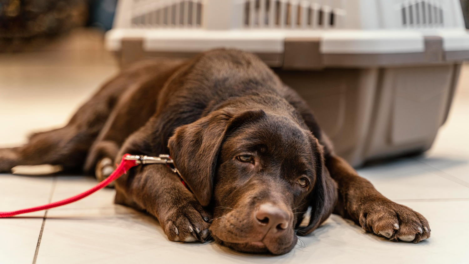
The initial symptoms of parvovirus are often nonspecific and can be easily mistaken for minor stomach upset or fatigue. The earliest sign is usually profound lethargy. A normally boisterous, playful puppy will suddenly become withdrawn, sleep excessively, and show no interest in toys or human interaction. This is typically accompanied by a complete loss of appetite (anorexia) and a high fever (pyrexia), which can sometimes exceed 104°F (40°C). As the virus rapidly replicates and the systemic inflammatory response kicks in, the dog’s condition will deteriorate, often within a matter of 12 to 24 hours.
Following the initial lethargy and fever, the gastrointestinal symptoms emerge violently. The virus specifically targets the crypts of Lieberkühn, which are the fundamental structural units of the intestinal lining responsible for cell renewal. By invading and destroying these crypt cells, the virus prevents the regeneration of the intestinal villi. The villi become blunted and ultimately slough off, leaving the intestinal tract completely denuded. This microscopic destruction translates to macroscopic disaster: the dog loses its ability to absorb nutrients and fluids, and the barrier separating the highly bacterial environment of the gut from the sterile bloodstream is completely compromised.
Consequently, the dog will begin to experience severe, intractable vomiting. This vomiting is often frothy, bilious (yellow or green), and continuous, preventing the animal from holding down any water or oral medications. Shortly after the vomiting begins, the dog will develop profuse diarrhea. Because the intestinal lining is actively sloughing and bleeding, the diarrhea is typically hemorrhagic (bloody) and is characterized by a uniquely foul, metallic, and necrotic odor that veterinary professionals instantly recognize as the “parvo smell”[8].
The combination of relentless vomiting and copious, watery, bloody diarrhea leads to catastrophic fluid loss. Dehydration sets in rapidly, clinically manifesting as sunken eyes, sticky or dry mucous membranes (gums), and a prolonged skin tent (when the skin is gently pulled up between the shoulder blades, it does not snap back into place). If the fluid loss is not aggressively replaced, the dog’s blood volume will drop precipitously, leading to hypovolemic shock. In this state, the heart struggles to pump enough blood to perfuse vital organs, leading to peripheral vasoconstriction, pale and tacky gums, a rapid and weak pulse, and ultimately, multi-organ failure.
Simultaneously, the virus is attacking the bone marrow, destroying the precursor cells responsible for creating white blood cells. This results in a condition known as severe leukopenia (a dangerously low white blood cell count). Because the intestinal barrier has been destroyed by the virus, the naturally occurring bacteria in the dog’s gut—such as Escherichia coli and Salmonella species—can freely cross into the bloodstream, a process known as bacterial translocation[9]. With no white blood cells available to fight off this bacterial invasion, the dog quickly develops sepsis (systemic bacterial infection). It is important to note that parvovirus itself rarely causes direct death; rather, it is the secondary complications of hypovolemic shock from dehydration and septic shock from bacterial translocation that prove fatal.
Diagnosing Parvo in Dogs

When a dog presents to the veterinary clinic with a history of vomiting, bloody diarrhea, and lethargy, canine parvovirus is immediately placed at the top of the differential diagnosis list, especially if the dog is young and has an incomplete vaccination history. Because the disease progresses so rapidly, early detection and treatment are crucial for improving the chances of survival for infected dogs. Diagnostics must be performed swiftly and accurately to confirm the presence of the virus and to assess the extent of the systemic damage the dog has sustained[10].
The gold standard for rapid, in-clinic diagnosis of canine parvovirus is the fecal antigen enzyme-linked immunosorbent assay (ELISA), commonly referred to as a “SNAP test.” This diagnostic tool utilizes a small swab of the dog’s feces to detect the presence of the specific parvovirus surface proteins (antigens). The test is highly valued in veterinary medicine because it provides results in less than ten minutes and has a high degree of specificity. A positive ELISA test in a dog exhibiting compatible clinical signs is generally considered definitive proof of a parvovirus infection. However, interpretation of the test requires clinical nuance.
False-negative results can and do occur with the ELISA test. If a dog is tested very early in the course of the disease (before massive viral shedding has begun) or very late in the disease (after the host’s antibodies have bound all available viral antigens in the feces), the test may not detect the virus. Therefore, if clinical suspicion remains high despite a negative SNAP test, the veterinarian will treat the dog as a parvovirus patient and may repeat the test 24 to 48 hours later. Conversely, weak false-positive results can sometimes occur if a dog has been vaccinated with a modified-live parvovirus vaccine within the preceding five to twelve days, as the vaccine virus replicates in the gut and can be temporarily shed[11].
For complicated cases or to confirm ambiguous ELISA results, molecular diagnostics such as the Polymerase Chain Reaction (PCR) test can be utilized. Fecal PCR testing is incredibly sensitive and detects the actual genetic material (DNA) of the virus. While PCR is highly accurate and can differentiate between wild-type virus strains and vaccine strains, it usually requires sending the sample to an external reference laboratory, meaning results take several days to return. Therefore, PCR is more commonly used for epidemiological tracking or confirming complex cases rather than for immediate, point-of-care triage.
Diagnosing the virus itself is only the first step; the veterinarian must also perform blood work to diagnose the secondary, life-threatening complications. A Complete Blood Count (CBC) is critically important. As the virus attacks the bone marrow, the CBC will often reveal profound leukopenia, specifically neutropenia (a severe drop in neutrophils, the primary white blood cells responsible for fighting bacterial infections). The severity of the neutropenia is an important prognostic indicator; a dog with an almost nonexistent white blood cell count is at an extreme risk for fatal sepsis.
Additionally, a comprehensive serum chemistry panel will be evaluated. This blood test checks the dog’s organ function and helps quantify the severity of the metabolic derangements caused by the illness. Dogs with severe parvovirus frequently suffer from hypoglycemia (dangerously low blood sugar) due to anorexia, vomiting, and consumption of glucose by circulating bacteria (sepsis). They also frequently present with hypokalemia (low potassium) from massive gastrointestinal losses, and hypoalbuminemia (low blood protein) because the damaged intestinal lining allows vital proteins to leak out of the blood and into the gut (a protein-losing enteropathy)[12]. Understanding these specific biochemical imbalances allows the veterinary team to formulate a highly customized and precise treatment plan.
Treatment Options for CPV

It is crucial to understand that there is no antiviral drug available that directly kills the canine parvovirus. Treatment for CPV is entirely supportive and symptom-driven. The goal of medical intervention is to support the dog’s vital organ systems, correct profound dehydration, and fight off secondary bacterial infections long enough for the dog’s own immune system to rally, produce antibodies, and clear the virus. Because the physiological collapse associated with vomiting and diarrhea is a significant concern in dogs, the standard of care requires intensive, multi-day hospitalization in a specialized isolation ward to prevent the spread of the virus to other vulnerable patients[13].
The cornerstone of parvovirus treatment is aggressive intravenous (IV) fluid therapy. An IV catheter is placed to provide direct access to the venous system, and the dog is administered isotonic crystalloid fluids (such as Lactated Ringer’s Solution or Plasmalyte). The veterinary team carefully calculates the dog’s fluid requirements based on their estimated percentage of dehydration, their daily maintenance needs, and their ongoing losses from continuous vomiting and diarrhea. In severe cases where the dog has developed hypoalbuminemia, the blood loses its oncotic pressure, meaning fluids leak out of the blood vessels and pool in the tissues (edema). In these critical scenarios, the veterinarian may utilize synthetic colloids or administer fresh frozen plasma transfusions to replenish vital blood proteins, provide clotting factors, and offer immediate passive immunity via donor antibodies.
Because the damaged intestinal tract allows gut bacteria to translocate into the bloodstream, establishing broad-spectrum antibiotic coverage is mandatory to prevent or treat septic shock. IV antibiotics are required since the gut cannot absorb oral medications. Veterinarians typically employ a multi-drug approach, pairing a beta-lactam antibiotic (such as ampicillin or cefazolin) with a fluoroquinolone (such as enrofloxacin) or an aminoglycoside to ensure comprehensive coverage against both gram-positive and gram-negative bacteria, particularly anaerobes and enterobacteriaceae[14].
Controlling nausea and preventing vomiting are critical components of care, not only to improve the dog’s comfort but also to prevent further fluid loss and the risk of aspiration pneumonia. Powerful, centrally-acting antiemetics are administered intravenously. Maropitant citrate (a neurokinin-1 receptor antagonist) and ondansetron (a serotonin 5-HT3 receptor antagonist) are highly effective at suppressing the vomiting reflex. Gastric protectants, such as proton pump inhibitors (pantoprazole), may also be utilized to reduce stomach acid and soothe the inflamed upper gastrointestinal tract.
Historically, veterinary protocols mandated withholding all food and water (keeping the patient “NPO”) until the vomiting ceased, to allow the gut to “rest.” However, modern veterinary literature has proven that early enteral nutrition is vital for survival. The cells lining the intestines (enterocytes) receive their nutrition directly from the food passing through the gut lumen, not from the bloodstream. Therefore, feeding the dog micro-meals, even if they are still experiencing some nausea, helps regenerate the intestinal lining faster, improves immune function, and decreases hospital stay duration. If the dog refuses to eat, veterinarians will often place a nasogastric (NG) tube—a small, flexible feeding tube passed through the nose, down the esophagus, and into the stomach—to deliver liquid, highly digestible critical care nutrition continuously[15].
While intensive inpatient hospitalization offers the highest survival rate (typically 85-90%), it is undeniably expensive due to the required 24-hour monitoring, medications, and isolation protocols. For pet owners facing severe financial constraints where euthanasia is the only alternative, veterinary medicine has developed evidence-based outpatient protocols. Pioneered by institutions like Colorado State University, outpatient parvovirus treatment involves administering large volumes of subcutaneous (under the skin) fluids daily, providing long-acting injectable antibiotics (like cefovecin), and administering potent anti-nausea injections. The owner must rigorously monitor the dog at home, manage body temperature with heating pads, and syringe-feed the dog. While outpatient therapy is highly labor-intensive and carries a lower survival rate (around 70-80%) compared to hospitalization, it serves as a vital, life-saving alternative when inpatient care is financially inaccessible.
Prevention for Parvovirus in Dogs
Because the treatment of canine parvovirus is financially demanding, emotionally exhausting, and carries a high risk of patient mortality, rigorous prevention is the absolute best medicine. Protecting your dog requires a multi-pronged approach focused on appropriate immunization protocols, understanding the window of susceptibility, and employing strict environmental management. The core tenet of prevention is ensuring your pet will receive appropriate vaccinations and booster shots as recommended by your veterinarian.
The parvovirus vaccine is considered a “core” vaccine by the American Animal Hospital Association (AAHA) and the World Small Animal Veterinary Association (WSAVA). It is generally administered in a combination shot commonly abbreviated as DAPP or DHPP (Distemper, Adenovirus, Parainfluenza, and Parvovirus). To overcome the dangerous “immunity gap” caused by maternal antibodies, puppies require a carefully timed series of vaccinations rather than a single shot. The AVMA and AAHA strongly recommend initiating the vaccine series when the puppy is between 6 and 8 weeks of age. Subsequent booster vaccines must be administered every 3 to 4 weeks until the puppy reaches a minimum of 16 weeks of age. In high-risk environments, or for highly susceptible breeds like Rottweilers and Dobermans, veterinarians often recommend extending the vaccination series, giving a final booster at 18 to 20 weeks of age to ensure maternal antibodies have completely waned and the puppy’s own immune system has responded effectively to the vaccine[16].
Following the successful completion of the initial puppy series, a booster vaccination must be administered exactly one year later. This adult booster provides long-lasting, robust immunity. After the one-year booster, current veterinary guidelines suggest that the DHPP/DAPP vaccine should be administered every three years, though some owners may opt for titer testing (a blood test that measures antibody levels) to confirm adequate immunity. Until a puppy has received its complete series of vaccinations (meaning two weeks have passed since their final 16-week or 20-week shot), owners must practice strict environmental avoidance. Unvaccinated puppies should never be allowed to walk in dog parks, visit pet stores, or interact with dogs of unknown vaccination status. Socialization is incredibly important, but it must be done safely in controlled environments with fully vaccinated, healthy adult dogs.
If a dog is diagnosed with parvovirus in a home environment, aggressive decontamination is required to protect future canines. Because the virus is non-enveloped, standard household cleaners like ammonia or quaternary ammonium compounds will not kill it. The most effective, widely accessible disinfectant for parvovirus is a diluted bleach solution. The Environmental Protection Agency (EPA) recommends a dilution of 1 part household bleach (5.25% sodium hypochlorite) to 30 parts water (approximately half a cup of bleach per gallon of water). All contaminated surfaces—including hard floors, crates, and food bowls—must be thoroughly cleaned of organic debris first, then soaked in the bleach solution for a minimum contact time of 10 to 15 minutes before being rinsed. Alternatively, accelerated hydrogen peroxide products are highly effective and safer for some surfaces. Bedding should be washed in hot water with bleach, though heavily soiled items are often best discarded. Yards and grass cannot be effectively disinfected with bleach without killing the vegetation; therefore, natural sunlight and the passage of time (often several months to a year) are required to reduce the viral load in outdoor environments[17].
By strictly adhering to vaccination schedules and practicing diligent hygiene, pet owners can effectively shield their pets from this devastating virus. However, medical decisions regarding vaccine protocols and disease management must always be customized to your specific pet. Please consult your veterinarian before making any changes to your pet’s care, as they can provide regional epidemiological insights and tailor a preventative health plan perfectly suited for your dog.
Frequently Asked Questions
Can a dog survive parvo at home?
A dog can survive parvovirus at home, but attempting to manage the disease without veterinary intervention carries a significantly higher mortality rate. Parvovirus causes profound dehydration, electrolyte imbalances, and severe secondary bacterial infections (sepsis) that are incredibly difficult to manage without intravenous fluids and injectable medications. If hospitalization is financially impossible, veterinarians can provide a specialized “outpatient protocol.” This involves teaching the owner how to administer subcutaneous fluids, providing long-acting injectable antibiotics, and dispensing potent anti-nausea medications. However, this is still a rigorous medical treatment plan guided by a professional. Home remedies, such as feeding raw eggs or over-the-counter pedialyte alone, are vastly insufficient and generally result in the animal suffering a painful death.
How long does parvo last in the environment?
Canine parvovirus is notoriously resilient and can survive in the environment for exceptionally long periods. Indoors, at room temperature, the virus can remain infectious on hard surfaces, unwashed bedding, and clothing for at least two months. Outdoors, protected from direct sunlight (such as in shaded soil or feces), the virus can easily survive for five to seven months, and under ideal conditions (such as freezing winter temperatures, which preserve the virus), it can survive well over a year. Because it lacks a lipid envelope, CPV is resistant to heat, cold, drying, and many common household cleaners. Effective eradication from an environment requires the removal of all organic matter followed by disinfection with a 1:30 bleach-to-water solution with a ten-minute contact time, or the use of commercial accelerated hydrogen peroxide disinfectants.
Can humans or cats catch canine parvovirus?
No, humans cannot catch canine parvovirus. The virus is highly species-specific. Humans are affected by a completely different strain of parvovirus known as Parvovirus B19, which causes a mild rash illness known as “Fifth Disease” in children. Similarly, canine parvovirus cannot infect humans. However, the situation with felines is slightly more complex. While the original CPV-2 strain that emerged in the 1970s did not infect cats, the newer mutated strains (CPV-2a, 2b, and 2c) have been shown to infect felines and can cause clinical signs similar to feline panleukopenia virus (FPV). Fortunately, the standard feline core vaccine (FVRCP) provides strong cross-protection against these canine parvovirus variants, keeping vaccinated cats safe from infection.
References
- American Veterinary Medical Association (AVMA). Canine Parvovirus. AVMA, 2023.
- Miranda, C., & Thompson, G. Canine parvovirus: the worldwide occurrence of antigenic variants. Journal of General Virology, 2020.
- Merck Veterinary Manual. Canine Parvovirus. Merck & Co., Inc., 2022.
- Cornell University College of Veterinary Medicine. Canine Parvovirus. Cornell University, 2021.
- Decaro, N., & Buonavoglia, C. Canine parvovirus—A review of epidemiological and diagnostic aspects, with emphasis on type 2c. Veterinary Microbiology, 2012.
- American Animal Hospital Association (AAHA). AAHA Canine Vaccination Guidelines. AAHA, 2022.
- VCA Animal Hospitals. Parvovirus in Dogs. VCA Hospitals, 2023.
- Goddard, A., & Leisewitz, A. L. Canine parvovirus. Veterinary Clinics of North America: Small Animal Practice, 2010.
- Prittie, J. Canine parvoviral enteritis: a review of diagnosis, management, and prevention. Journal of Veterinary Emergency and Critical Care, 2004.
- Mylonakis, M. E., et al. Canine parvoviral enteritis: an update on the clinical diagnosis, management, and prevention. Veterinary Medicine: Research and Reports, 2016.
- Proksch, A. L., et al. Diagnostic value of a point-of-care test for canine parvovirus-2 detection. Veterinary Journal, 2018.
- Otto, C. M., et al. Evaluation of a comprehensive protocol for the treatment of canine parvovirus. Journal of the American Veterinary Medical Association, 1997.
- Venn, E. C., et al. Evaluation of an outpatient protocol in the treatment of canine parvoviral enteritis. Journal of Veterinary Emergency and Critical Care, 2017.
- Macintire, D. K. Treatment of canine parvoviral enteritis. Veterinary Clinics of North America: Small Animal Practice, 2008.
- Mohr, A. J., et al. Effect of early enteral nutrition on clinical outcome in dogs with severe parvoviral enteritis. Journal of the American Veterinary Medical Association, 2003.
- ASPCA. Common Dog Diseases: Parvovirus. ASPCA, 2023.
- Patterson, E. V., et al. Efficacy of disinfectants against canine parvovirus in environmental settings. Journal of Small Animal Practice, 2014.


