


 March 3, 2023
 Phil Good, DVM
What is ACL Tear in Dogs?
This content was prepared with AI assistance and reviewed by a licensed professional for accuracy.
Introduction
An acl tear in dogs is widely recognized by veterinary professionals as one of the most common, painful, and complex orthopedic conditions encountered in clinical practice. To understand this condition fully, one must first recognize the anatomical terminology. While humans possess an Anterior Cruciate Ligament, in veterinary medicine, these are injuries to the ligaments of the stifle joint, which is the canine equivalent of the human knee. These specific knee injuries are medically referred to as Cranial Cruciate Ligament (CCL) ruptures or tears. Regardless of the terminology used—whether a pet owner calls it an ACL tear or a veterinarian diagnoses it as a CCL rupture—the resulting joint instability, severe inflammation, and chronic pain are identical, fundamentally altering a dog’s biomechanics and quality of life.[1]
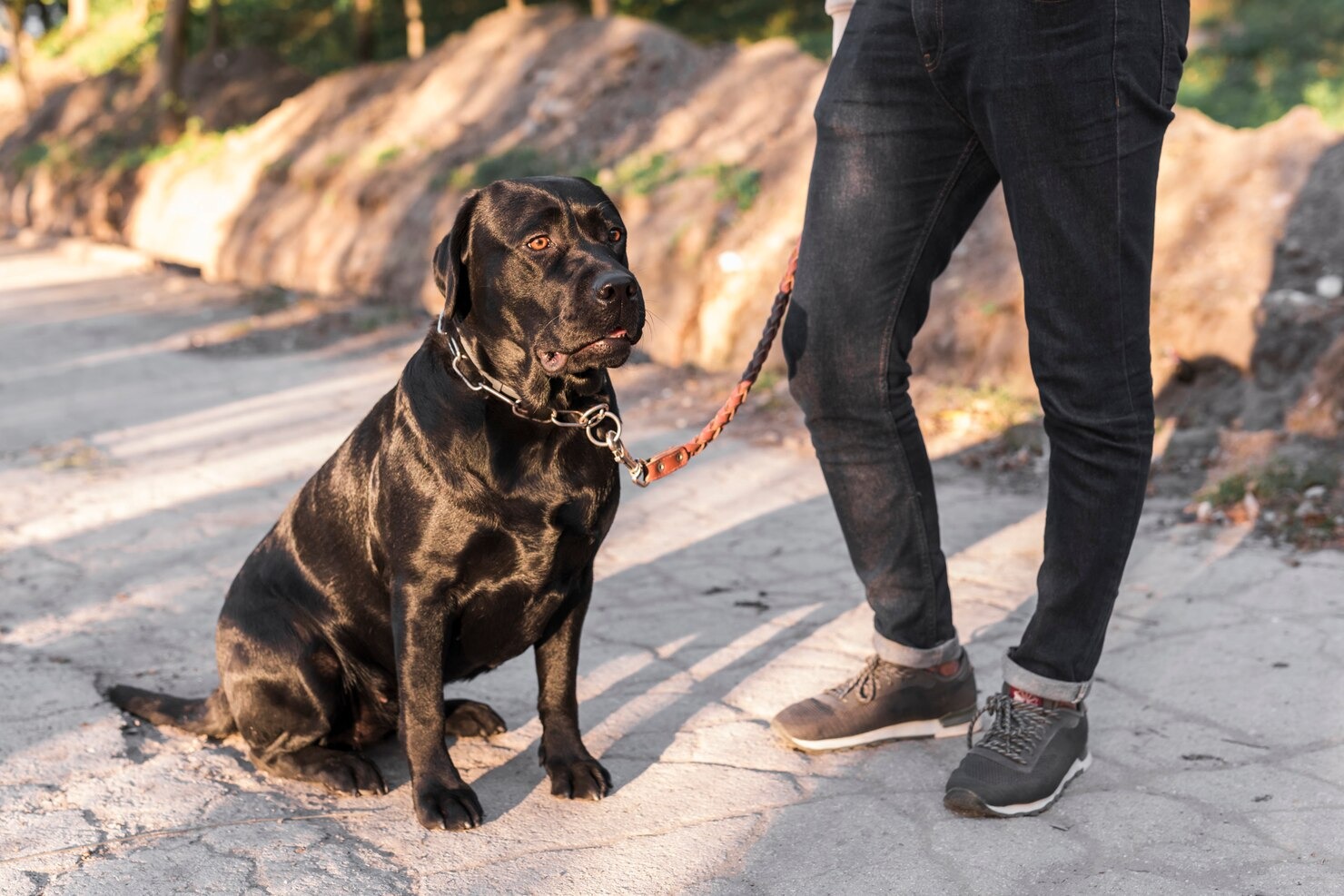
Consider the classic case of a patient named Titan. For years, Alex and his loyal Rottweiler, Titan, had enjoyed many vigorous outdoor adventures, exploring new, rugged trails and vast dog parks every weekend. Titan was a highly athletic, robust canine weighing over one hundred pounds. One afternoon, while playing an intense and energetic game of frisbee on an uneven grassy field, Titan launched himself into the air to catch the disc. As he landed and planted his left hind leg to pivot, he suddenly yelped in sharp, unmistakable pain. He immediately pulled the leg up against his abdomen, holding it in a flexed position, and began limping severely, entirely unable to put weight on his hind leg. Worried about his beloved, usually stoic companion, Alex quickly realized that Titan was suffering from an acute orthopedic emergency and required immediate professional veterinary care. Upon arriving at the animal hospital, the clinical presentation was a textbook example of a ruptured cruciate ligament.[2]
After conducting a careful, detailed physical and orthopedic examination—including a thorough gait analysis, palpation of the joint for effusion, and specific manipulative tests designed to evaluate ligamentous integrity—the veterinarian definitively diagnosed Titan with a cranial cruciate ligament tear. This is an incredibly common but exquisitely painful injury in dogs, representing a significant source of hindlimb lameness across various canine populations. In this comprehensive, medically focused article, we will rigorously discuss what an ACL tear is, explore the underlying pathophysiological causes, detail the diverse clinical symptoms, and meticulously outline the modern diagnostic criteria and advanced treatment options. By deeply understanding the biomechanical and biological realities of this condition, you will be significantly better prepared to support your canine friend’s long-term orthopedic recovery and help them return to a healthy, active, and pain-free lifestyle.[3]
A cranial cruciate ligament injury represents a severe biomechanical impairment and structural damage to the dog’s internal knee joint architecture. The stifle joint is a complex, delicate hinge mechanism consisting of the distal femur (thigh bone), the proximal tibia (shin bone), the patella (kneecap), and the fabellae (small sesamoid bones). The cranial cruciate ligament is a robust band of fibrous connective tissue linking the femur to the front (cranial aspect) of the tibia. Its primary physiological functions are threefold: it prevents the tibia from thrusting forward relative to the femur during weight-bearing (a phenomenon known as cranial tibial thrust), it prevents excessive internal rotation of the tibia, and it prevents the extreme hyperextension of the stifle joint. Consequently, the dog’s ACL is absolutely integral to comfortable, stable movement, dynamic agility, and all routine weight-bearing activities.[4]
Such a structural injury introduces a myriad of cascading clinical issues, leading to severe localized discomfort, mechanical joint instability, and profound functional disability in our canine friends. While it is highly prevalent in large breed dogs—such as Labrador Retrievers, Mastiffs, and Rottweilers—it is also increasingly diagnosed in smaller breeds like the Bichon Frise, the Miniature Poodle, and the Yorkshire Terrier. The symptoms of a torn cruciate ligament range widely depending on the chronicity of the injury, spanning from a subtle, intermittent lameness and mild joint inflammation to noticeable, vocalized discomfort and absolute refusal to bear weight on the distressed leg. The joint capsule becomes distended with inflammatory synovial fluid, causing palpable swelling and radiating pain.[5]
The pathogenesis of this injury is uniquely different in canines compared to humans. In human sports medicine, an ACL tear usually happens abruptly due to a sudden, catastrophic traumatic event—like a football tackle or a skiing accident. However, in dogs, while an acute traumatic rupture is possible, the condition far more frequently evolves progressively due to long-term wear and tear, immune-mediated joint degradation, and chronic micro-trauma. The ligament literally undergoes a slow process of structural degeneration, losing its tensile strength over months or even years before finally failing under a normal, everyday load. Dogs afflicted with this type of progressive injury may struggle with daily mobility, demonstrate subtle signs of pain or stiffness in the knee after resting, and seem hesitant to apply full, confident weight to the impacted limb during turns or stairs.[6]
Two Primary Types of ACL Tear in Dogs
In veterinary orthopedic medicine, there are two primary clinical classifications of ACL tears in dogs: partial tears and complete ruptures. These tears can result from a sudden, acute biomechanical overload or, far more commonly, from the gradual, insidious wear and tear on the ligament’s collagen matrix. Understanding the distinction between these two classifications is critical, as the severity, presentation, and type of the tear will profoundly influence the diagnostic process and determine the most appropriate, effective treatment plan for your specific dog.[7]
Partial ACL Tear
A partial tear occurs when the cranial cruciate ligament is structurally damaged and partially torn, but the fibers are not completely severed. To understand how a partial tear functions, one must examine the micro-anatomy of the ligament itself. The canine CCL is composed of two distinct functional bands: the craniomedial band and the caudolateral band. During the normal range of motion, the craniomedial band remains continuously taut during both the flexion (bending) and extension (straightening) of the knee joint. Conversely, the caudolateral band is only taut in extension and actually relaxes when the knee is flexed. Because the craniomedial band is constantly under biomechanical tension regardless of the joint’s position, it is significantly more vulnerable to micro-trauma and is almost always the first section of the ligament to tear.[8]
This type of localized partial tear can cause an agonizing cycle of pain, synovial swelling (effusion), and mild to moderate instability in the knee joint. However, because the caudolateral band remains somewhat intact, the joint may still retain a degree of basic function, making the diagnosis challenging. Dogs with a partial tear often present with a “warm-up” lameness; they may appear stiff and limp noticeably when rising from a prolonged rest, but their gait may seemingly improve as they walk and the joint fluid warms up. Unfortunately, a partial tear creates a highly inflammatory environment within the joint. Inflammatory enzymes, such as matrix metalloproteinases, are released into the synovial fluid, and these enzymes actively degrade the remaining healthy collagen fibers. Consequently, a partial tear almost inevitably progresses to a complete, catastrophic tear if it is not appropriately, proactively managed. Treatment for early-stage partial tears may occasionally involve rigorous conservative management, including strict crate rest, multi-modal pain management, targeted weight control, and professional physical therapy, though the prognosis for returning to high athletic function without surgery remains heavily guarded.[9]
It is vital for pet owners to recognize that the ligamentous tissue inside the stifle has a notoriously poor blood supply, meaning it lacks the physiological ability to heal itself in the way that skin or muscle tissue does. Some dogs with partial tears may eventually require major orthopedic surgery if conservative treatments fail to provide sustained relief, or when the inevitable mechanical failure occurs, and the tear worsens over time into a complete rupture. Relying solely on rest often results in profound muscle atrophy in the affected hind limb, further destabilizing the joint complex.[10]
Complete ACL Tear
A complete tear occurs when the anterior cruciate ligament is entirely severed, ruptured, or mechanically obliterated, resulting in catastrophic, immediate loss of stability and severe biomechanical dysfunction in the knee joint. When the ligament fails completely, the femur and tibia are no longer tethered together appropriately. Every time the dog attempts to bear weight on the affected limb, the natural slope of the tibial plateau causes the femur to slide backward, effectively thrusting the tibia forward. This abnormal, shearing motion is called “cranial tibial thrust,” and it is incredibly damaging to the internal structures of the joint. Dogs with a complete ACL tear often exhibit severe, sudden-onset lameness, adopting a three-legged gait, alongside intense pain and palpable, warm swelling in the affected stifle.[11]
One of the most severe secondary complications of a complete cranial cruciate ligament tear is the concurrent damage to the medial meniscus. The meniscus is a C-shaped wedge of fibrocartilage that sits between the femur and the tibia, acting as a vital shock absorber and load distributor. Because the medial meniscus is firmly anchored to the tibial plateau by strong coronary ligaments, it moves forward with the tibia during cranial tibial thrust. As the femur slides backward, the crushing sheer force acts like a rolling pin, catching the caudal horn of the medial meniscus and tearing or crushing it. This meniscal damage amplifies the pain exponentially and accelerates the development of debilitating osteoarthritis. In most cases of a complete tear, prompt surgical intervention is considered medically necessary to repair or biomechanically replace the torn ligament, inspect and treat any meniscal tearing, and definitively restore stability to the joint.[12]
In modern veterinary surgery, several advanced surgical techniques are widely available, each with specific biomechanical philosophies. These include the traditional extracapsular repair (also known as the lateral fabellar suture technique), the highly regarded tibial plateau leveling osteotomy (TPLO), and the tibial tuberosity advancement (TTA). The choice of surgical procedure is highly nuanced and depends heavily on critical factors such as the dog’s exact body weight, tibial plateau angle geometry, age, anticipated athletic activity level, and the board-certified veterinary surgeon’s specific clinical preference. The primary goal of these procedures is not actually to “repair” the ligament—which is impossible—but rather to alter the biomechanics of the joint so that the cruciate ligament is no longer necessary for stability during weight-bearing.[13]
It is profoundly essential to immediately consult with your veterinarian if you even remotely suspect your dog has suffered an ACL tear. Delayed intervention allows chronic inflammation to ravage the articular cartilage, leading to irreversible, crippling osteoarthritis. Early diagnosis, accurate staging of the joint disease, and the implementation of appropriate, evidence-based treatment protocols can help prevent catastrophic further joint damage, relieve chronic pain, preserve muscle mass, and drastically improve your dog’s long-term quality of life.[14]
What Causes Canine Cranial Cruciate Ligament Tears?

The etiology of cranial cruciate ligament disease in canines is notoriously multifactorial and highly complex. Unlike human ACL injuries, which are predominantly the result of a single, acute traumatic sporting event, canine CCL tears are usually the culmination of a chronic, degenerative biological process combined with biomechanical stress. Various distinct factors can independently or synergistically lead to canine ACL or CCL injuries, spanning traumatic incidents, chronic progressive ligament degeneration, and inherent predispositions tied to the dog’s breed, sheer physical size, biological age, and specific skeletal structure. Let us meticulously explore these well-documented, common causes of ACL tears in dogs in much greater clinical detail:[15]
- Acute biomechanical trauma: While less common than the degenerative pathway, a sudden, forceful twisting motion or extreme hyperextension bending of the dog’s knee joint during high-energy, explosive activities—like sprinting after a squirrel, jumping to catch a frisbee, or engaging in playful, erratic antics with other dogs—may precipitate an acute ACL tear. Dogs that participate in intense, energetic athletic endeavors, for example, competitive agility training, flyball, dock diving, or relentless fetching on uneven terrain, are often more susceptible to this specific type of traumatic injury due to the immense shear forces placed upon the stifle joint during rapid acceleration and deceleration.[16]
- Progressive wear and tear (Degenerative Cruciate Disease): This is the most prevalent cause of CCL ruptures in the canine population. Persistent, repetitive biomechanical stress on the ACL, combined with an insidious, sterile immune-mediated inflammatory process within the joint, can progressively weaken the ligament’s collagen fibers, leading to its eventual tear over time. This process is often compared to a thick rope slowly fraying strand by strand until it inevitably snaps under a normal load. This degenerative scenario is highly likely in dogs involved in repetitive high-impact activities that exert constant micro-pressure on the knee joint, such as relentless running or jumping on hard, unforgiving terrains. Furthermore, a chronically inflammatory synovial environment accelerates this structural decay, compromising the tensile strength of the ligament long before the dog ever shows the first sign of a limp.[17]
- Breed predisposition and Genetics: Extensive veterinary orthopedic research has definitively proven that certain dog breeds possess a powerful genetic inclination towards ACL tears due to both the unique skeletal anatomy of their hind limbs and hereditary predispositions to ligamentous degeneration. For instance, breeds such as Labrador Retrievers, powerful Rottweilers, heavy-set Golden Retrievers, majestic Newfoundlands, and intensely active German Shepherds often demonstrate statistically higher incidences of ACL tears. Recent genome-wide association studies have even begun to isolate specific collagen-coding genetic markers that may dictate a dog’s inherent risk of developing cruciate disease, suggesting that the structural integrity of the ligament is largely coded in their DNA from birth.[18]
- Size, weight, and the Adipokine effect: Larger, more robust, giant-breed dogs naturally face an elevated biomechanical risk of ACL injuries, given that their massive size places augmented, constant stress on their weight-bearing joints. However, the impact of weight extends far beyond simple gravity. overweight dogs are more prone to injuries as the extra weight overburdens the delicate ligament connecting the femur to the shin bone. Furthermore, adipose (fat) tissue is not merely inert storage; it is a highly active endocrine organ that secretes potent inflammatory hormones called adipokines. These circulating inflammatory mediators travel systemically and actively degrade the collagen structures within the stifle joint. Thus, an obese dog is suffering from both mechanical overloading and chemical degradation simultaneously.[19]
- Age and Senescence: The ligaments and joints of older dogs may weaken due to age-related degeneration, thereby significantly increasing their susceptibility to ACL tears. As a dog ages, the blood supply to the core of the ligament diminishes, reducing the tissue’s ability to repair everyday micro-traumas. The collagen fibers become less elastic, more brittle, and highly prone to mechanical failure during routine activities such as navigating stairs or rising from a slippery floor.[20]
- Physical structure and Conformation: Dogs possessing specific, naturally occurring anatomical characteristics—such as a pronounced curvature in their legs (bow-leggedness), straight stifles (upright hind limb conformation often seen in Chows and Akitas), or exceptionally steep tibial plateau angles—are at a phenomenally greater risk of ACL injuries. A steep tibial plateau means the “hill” the femur rests on is very sharp, requiring the cruciate ligament to work exponentially harder just to keep the femur from sliding down the slope during normal standing. This severely modified biomechanics and uneven stress distribution guarantee premature ligament failure. Additionally, the timing of spaying and neutering plays a critical role; pediatric altering delays the closure of the tibial growth plates, resulting in longer bones and a drastically altered, steeper joint angle that predisposes the dog to orthopedic trauma later in life.[21]
To successfully avert or delay the onset of ACL tears, it is absolutely crucial to maintain a lean, healthy body weight for your dog, ensure regular, consistent, low-intensity exercise to build supportive muscle mass, and carefully avoid uncontrolled activities that could instigate sudden, violent stress or twisting of the knee joint. Furthermore, if your dog is naturally, genetically prone to ACL tears due to their specific breed heritage or unique physical structure, proactively consulting with your veterinarian about possible physical therapy conditioning strategies, joint-protective dietary supplements, and identifying the early clinical warning signs would be highly beneficial to their longevity.[22]
Symptoms of Torn ACL in Dogs

The clinical indications and behavioral manifestations of a canine ACL tear—also deeply known in the medical literature as a cranial cruciate ligament rupture—might differ drastically based on the exact chronicity and intensity of the injury. An acute, catastrophic rupture will look vastly different from a slow, fraying partial tear. However, pet owners must be hyper-vigilant in observing their dog’s gait and resting posture. Here are the profoundly typical, scientifically documented signs to diligently watch out for:[23]
- Limping or profound signs of lameness: This is the most universal symptom. The lameness may be sudden and non-weight-bearing (holding the leg up entirely), or it may be a subtle, intermittent limp that worsens significantly after periods of heavy exercise and temporarily improves with rest, often confusing owners into thinking it is a minor sprain.
- Presence of palpable swelling (Joint Effusion): As the ligament tears, the joint capsule fills with inflammatory synovial fluid and blood. This swelling is particularly noticeable on the inside (medial aspect) of the knee, feeling firm and warm to the touch. Chronic tears will develop a thick, fibrous scar tissue lump called a “medial buttress.”
- Visible, vocalized pain: While dogs are notoriously stoic to mask vulnerability, a torn ACL is highly painful. A dog may yelp when the knee is accidentally touched, groan when lying down, or display behavioral changes such as sudden aggression, lethargy, or a decrease in appetite due to the systemic stress of chronic pain.
- Severe stiffness in movement: The inflammatory cascade within the joint causes the surrounding soft tissues and joint capsule to thicken and fibrose, leading to a stiff, stilted, unyielding gait, especially noticeable after a long nap or first thing in the morning.
- Diminished range of motion: The dog will actively resist flexing or extending the stifle fully because stretching the inflamed joint capsule triggers pain receptors. The stride length on the affected side will be noticeably shorter.
- Reluctance to participate in play or exercise: A previously energetic dog may suddenly refuse to jump onto the bed, struggle significantly to navigate stairs, or sit down repeatedly during a normally easy walk. This exercise intolerance is a direct protective mechanism to avoid joint loading.
- Sitting with the leg extended outward (The “Sit Test”): This is a pathognomonic behavioral sign. When a healthy dog sits, they tuck their hind legs neatly under their body in full flexion. A dog with a torn ACL will sit with the affected leg kicked out loosely to the side, avoiding the painful flexion of the knee joint entirely.
- Observable mechanical instability in movement: If the tear is complete, an astute observer might actually see the tibia shifting forward abnormally when the dog places weight on the leg. This visual “wobble” represents the physical failure of the joint’s tethering system.
- Rapid, severe signs of muscle atrophy: Because the dog is instinctively protecting the painful leg by offloading their weight onto the healthy limbs, the muscles of the affected thigh—specifically the quadriceps and hamstring groups—will begin to waste away and shrink dramatically within a matter of mere weeks.
Suppose any combination of these distressing symptoms becomes apparent in your dog’s daily behavior. In that case, it is absolutely crucial to seek a licensed veterinarian’s medical assistance promptly for a comprehensive, accurate diagnostic workup and the formulation of a highly customized, suitable treatment plan, which, in the vast majority of medium to large-breed dogs, will unequivocally include complex orthopedic surgery to restore biomechanical function and halt the rapid progression of osteoarthritis.[24]
Diagnosing ACL Tears in Dogs

Diagnosing an anterior (cranial) cruciate ligament tear in dogs is an exacting, multi-step clinical process that requires deep anatomical knowledge. It involves a strategic combination of a detailed patient history, a dynamic physical gait examination, meticulous hands-on orthopedic manipulation, and sophisticated advanced imaging techniques. Veterinary professionals conduct these methodical steps precisely to definitively confirm an ACL injury and rule out other confounding pathologies, such as patellar luxation, Achilles tendon ruptures, or bone neoplasia (cancer). Here is a highly detailed, clinical overview of the rigorous diagnostic procedures involved.[25]
Detailed History
The critical first step in diagnosing an ACL tear in dogs is taking a thorough, exhaustive, and detailed medical history. The veterinarian will methodically ask the pet owner specific questions about the exact onset timeline of the dog’s lameness. They will inquire if there was a known, witnessed traumatic injury (like slipping on winter ice or falling into a ditch), if the lameness is worse in the morning, and if the condition has improved temporarily with over-the-counter pain medications or worsened over weeks. Understanding the chronicity of the limp helps the clinician differentiate between a sudden catastrophic rupture and a slow, degenerative partial tear.[26]
Physical Examination
The standard physical examination fully and holistically assesses the dog’s overall systemic health, with particular, focused attention paid to the limbs. During this dynamic exam, the veterinarian will carefully observe the dog at a walk and a trot in the hallway, looking for subtle weight-shifting patterns, head-bobbing (which indicates weight being thrown to the front limbs to spare the painful hindquarters), and the specific phase of the stride where the dog hesitates. They will look for outward signs of lameness, postural compensation, and visual swelling in the knee, as well as performing a complete neurological screening to ensure the weakness is not originating from a spinal cord compression issue, such as intervertebral disc disease.[27]
Orthopedic Examination
The specialized orthopedic examination is significantly more detailed and strictly focuses on the biomechanics of the dog’s skeletal and ligamentous system. The veterinarian will actively look for definitive signs of an ACL tear by performing highly specific manipulative maneuvers on the dog’s knee to check for pathological, abnormal movement. The hallmark diagnostic test is the “Cranial Drawer Test.” The veterinarian places one hand on the femur (using the patella and lateral fabella as anatomical landmarks) and the other hand on the tibia (grasping the tibial tuberosity and fibular head). They then attempt to push the tibia forward relative to the femur. If the ligament is torn, the tibia will slide forward like a drawer opening—a movement that is impossible in a healthy joint. Another crucial maneuver is the “Tibial Compression Test” (or cranial tibial thrust test), which simulates the weight-bearing forces of standing to see if the joint subluxates dynamically.[28]
Radiographs (X-rays)
Radiographs, or digital X-rays, are an indispensable tool to visualize the bony architecture and the geometric space within the stifle joint. While an X-ray cannot directly show soft tissues like a torn ACL—ligaments are radiolucent and do not appear clearly on standard radiographs—it can vividly show the devastating secondary signs of the injury. A key indicator is the “infrapatellar fat pad sign,” where the normal dark triangle of fat at the front of the knee is compressed and obscured by a massive influx of inflammatory joint fluid (effusion). Furthermore, radiographs allow the clinician to measure the specific tibial plateau angle, evaluate the joint for early signs of osteoarthritis (such as osteophyte formation along the femoral condyles or tibial plateau), and conclusively rule out concurrent issues like tibial fractures or osteosarcoma.[29]
Advanced Imaging
In highly complex or ambiguous cases, particularly when trying to definitively diagnose an early, subtle partial tear or evaluate the exact condition of the menisci before surgery, advanced imaging techniques such as Magnetic Resonance Imaging (MRI) and Computed Tomography (CT) scans may be utilized. These powerful modalities provide incredibly detailed, high-resolution, three-dimensional images of the soft tissues around and inside the knee, including the ACL, the joint capsule, and the delicate cartilage. As a result, these advanced techniques can give an unmistakably clearer picture of the precise extent of the injury, allowing for immaculate pre-surgical planning.[30]
Arthroscopy
Arthroscopy is an advanced, highly specialized, minimally invasive surgical diagnostic procedure that uses a microscopic, illuminated high-definition camera inserted directly into the fluid-filled joint space through a tiny portal incision. This cutting-edge technology allows the veterinary surgeon to visually magnify and inspect the internal structures within the joint in real-time. It provides the absolute gold-standard, definitive diagnosis of an ACL tear. Furthermore, it allows the surgeon to use tiny, specialized probes to test the integrity of the medial meniscus and instantly treat any cartilage tears using minimally invasive shavers without having to cut the joint wide open, drastically reducing post-operative pain and recovery time.[31]
Surgical Exploration
Traditional surgical exploration, known as an open arthrotomy, is typically reserved for advanced cases where a definitive diagnosis cannot be made using less invasive methods, or when arthroscopic equipment is unavailable. This highly invasive procedure involves making a large surgical incision through the skin, fascia, and thick joint capsule to open up the knee joint entirely, allowing the surgeon to visualize the frayed or ruptured ACL directly, debride the necrotic tissue, manually inspect the medial and lateral menisci, and physically prepare the joint environment for whichever stabilizing surgical procedure will immediately follow.[32]
Remember that these complex diagnostic steps are generally performed in a logical, step-wise combination, and the ultimate need for advanced imaging like MRI or direct surgical exploration will depend entirely on the unique nuances of the individual case, the dog’s compliance during physical palpation, and the attending veterinarian’s extensive clinical assessment and experience.
Treatment Options for Dog ACL Tears

The intelligently chosen treatment path for a dog’s ACL tear fundamentally hinges on a vast array of clinical variables: the absolute severity of the ligamentous injury (partial versus complete), the dog’s specific physical size and body weight, their natural energy and activity levels, their chronological age, and the presence of any additional concurrent orthopedic injuries or systemic conditions (such as advanced heart disease that might complicate anesthesia). Highly disciplined conservative management and robust surgical intervention—which encompasses several distinct biomechanical ACL surgery techniques—represent the two primary, divergent treatment methodologies available in modern veterinary medicine for effectively dealing with debilitating canine ACL tears.[33]
Conservative Management
This strictly non-surgical treatment route is generally, though cautiously, suggested only for very small, lightweight dogs (typically under 20 pounds), strictly geriatric patients who are exceedingly poor anesthetic risks, or in rare cases where the tear is highly partial, incredibly subtle, and not causing significant daily discomfort or dangerous mechanical instability. The foundational principles of conservative, medical management are intense and demand rigorous owner compliance. They include:
- Strict rest and rigorously restricted activity: Curtailing your dog’s movement is the absolute bedrock of non-surgical care. This means strict crate rest or confinement to a small, carpeted room for 6 to 8 weeks, with zero access to stairs, furniture, or slippery floors. Dogs are only allowed outside on a short, non-retractable leash strictly for bathroom purposes. This extreme restriction helps avert additional catastrophic mechanical damage to the joint and surrounding tissues, theoretically facilitating the formation of dense scar tissue around the joint capsule to provide a pseudo-stability.
- Multi-modal pain management: Effective pain medication is mandatory to ensure humane welfare. This usually centers around veterinary-specific prescription non-steroidal anti-inflammatory drugs (NSAIDs) administered by your veterinarian, which actively inhibit the cyclooxygenase pathways to help manage severe discomfort and blunt the joint-destroying inflammation. This is often paired with nerve-pain medication prescribed by your veterinarian to address the complex, wind-up pain associated with chronic joint disease.
- Aggressive weight management: It is clinically vital to ensure your dog maintains an exceptionally lean, healthy weight to alleviate the sheer mechanical stress exerted on the mechanically compromised affected joint. The amplified joint strain makes Overweight dogs more prone to ligament injuries, and carrying excess body fat severely diminishes any chance of conservative management succeeding.
- Professional physical therapy and rehabilitation: This involves a highly structured, medically supervised rehabilitation program encompassing passive range of motion (PROM) exercises, therapeutic soft-tissue massage, targeted muscle strengthening routines, and the utilization of cutting-edge modalities such as Class IV photobiomodulation (laser) therapy and underwater treadmill hydrotherapy. The ultimate goal is to build massive compensatory muscle strength in the quadriceps and hamstrings to help stabilize the knee dynamically, alongside a remarkably slow, strictly gradual reintroduction to normal activity to enhance joint function. Regenerative medicine options, such as Platelet-Rich Plasma (PRP) injections and intra-articular stem cell therapies, are also increasingly utilized to promote healing and reduce synovial inflammation.
Surgical Treatment
Surgical intervention unequivocally becomes the most viable, medically recommended option when conservative management inevitably fails to restore function, or overwhelmingly as the first-line defense for medium, large, giant-breed, or highly energetic athletic dogs. Surgery represents the only definitive method to mechanically stabilize the joint, inspect for and treat excruciating meniscal tears, and significantly slow the aggressive progression of osteoarthritis. The specific surgical technique meticulously chosen depends heavily on the dog’s size, age, tibial geometry, and unique lifestyle needs. Standard, globally accepted procedures may include:[34]
- Lateral suture or extracapsular repair (ECR): In this traditional technique, a heavy-gauge, robust monofilament suture material—acting as a prosthetic “fishing line”—is surgically anchored around the lateral fabella (a small bone behind the femur) and threaded through a hole drilled in the tibial crest. This incredibly strong artificial band substitutes the mechanical function of the ruptured internal ligament, manually restoring stability to the joint by physically tethering the bones together while the body naturally builds thick, stabilizing fibrous scar tissue over several months.
- Tibial plateau leveling osteotomy (TPLO surgery): Widely considered the gold standard in canine orthopedic surgery, the TPLO completely alters the biomechanics of the knee rather than replacing the ligament. In this highly advanced technique, the surgeon uses a specialized curved saw blade to make a semi-circular slice through the top of the tibia. The cut portion of the bone containing the joint surface (the tibial plateau) is physically rotated to level the slope from a steep angle (e.g., 25 degrees) down to a flat, neutral angle of approximately 5 degrees. The bone is then rigidly secured in this new, flat alignment with a customized, surgical-grade stainless steel or titanium bone plate and screws. By leveling the plateau, the femur can no longer slide backward, completely neutralizing the shearing cranial tibial thrust, fostering a vastly more stable, highly functional joint, and entirely obviating the physiological need for the ruptured cruciate ligament.[35]
- Tibial tuberosity advancement (TTA): In this alternative biomechanical altering procedure, the front portion of the tibia (the tibial tuberosity, where the patellar tendon attaches) is surgically sliced longitudinally and physically shifted forward. A specialized titanium cage spacer and a tension-band plate are inserted to hold the bone in this advanced position. By moving the patellar tendon forward, the geometry of the knee biomechanics is radically modified so that the compressive forces of standing naturally pull the tibia backward, perfectly counteracting the forward thrust and stabilizing the joint entirely without relying on the torn ligament. Post-CCL surgery, regardless of the method utilized, your dog will strictly require comprehensive pain medication, exceptionally limited physical activity, and a highly specific, months-long physical rehabilitation program to support proper, uncomplicated bone and soft tissue healing and regain comprehensive, athletic joint functionality.
Depending on the specific surgical treatment chosen, the size and age of the individual dog, and the presence of meniscal damage, the arduous recovery process could safely span anywhere from 8 to 16 weeks, with a full return to unrestricted, explosive athletic activity generally taking upwards of four to six months of dedicated care.
Prevention for ACL Tears in Dogs
Successfully preventing ACL tears, a notoriously common and financially burdensome injury in the modern dog population, requires immense proactive dedication to maintaining your pet’s holistic, lifelong physical health and actively minimizing the known risk elements that directly contribute to such catastrophic ligament traumas. Although it may be scientifically impossible to entirely eradicate the genetic or degenerative risk of cruciate disease, as a deeply responsible pet owner, you can meticulously execute the following daily measures to dramatically lessen the statistical odds of your dog—be it rapidly growing younger dogs, highly competitive athletic dogs, or cherished dogs of all sizes—suffering an agonizing ACL tear:[36]
- Rigorous Weight management: Ensure your dog diligently maintains an exceptionally lean, healthy weight and an ideal body condition score (BCS of 4 or 5 out of 9) to heavily alleviate the massive mechanical stress exerted daily on their delicate joint structures. As veterinary research proves, the amplified joint strain makes overweight dogs more prone to ligament injuries, and the inflammatory hormones released by excess fat actively degrade the cruciate ligament’s collagen from the inside out. Regular weigh-ins and strict portion control are paramount.
- Consistent, smart exercise: Engage your dog in highly routine, regulated, and purposeful exercise to preserve robust muscle strength, enhance core proprioception, and maintain dynamic joint stability. Refrain strictly from sudden, high-intensity, erratic activities that could induce violent, excess joint stress in an unconditioned dog—the classic “weekend warrior” syndrome. Instead, routinely choose excellent, low-impact, muscle-building exercises like sustained swimming, controlled incline leash walks, navigating cavaletti rails, or gentle treks through woods that force the dog to mindfully engage their hindquarters.
- Nutritional balance and strategic supplementation: Deliver a highly balanced, premium diet that scientifically fortifies your dog’s systemic joint health. For customized advice on appropriate prescription food and high-quality nutraceutical supplements—such as highly bioavailable glucosamine, chondroitin sulfate, avocado/soybean unsaponifiables (ASU), and particularly high doses of Omega-3 fatty acids (EPA and DHA) from marine sources, which powerfully boost joint health and decrease the systemic inflammatory risk of injury—always consult your veterinarian directly.
- Avoid abrupt, dangerous movements: Actively deter and prevent activities involving sharp, uncontrolled twisting or rapid, violently turning motions on poor footing. Activities such as aggressively chasing tennis balls on slippery hardwood floors, leaping to catch high-flying frisbees, or scrambling down steep, icy embankments can impose massive, undue biomechanical stress on your dog’s stifles, leading directly to acute rupture.
- Gradual integration of new activities: When eagerly introducing your dog to new, demanding physical exercises, long hiking trails, or competitive dog sports (like agility or flyball), you must gradually, methodically escalate the intensity and duration of the workload over several months. This vital conditioning strategy helps your dog slowly build dense muscle strength, cardiovascular endurance, and tendon conditioning, drastically mitigating the sudden risk of catastrophic orthopedic injury.
- Routine, comprehensive veterinary check-ups: Regularly scheduled, biannual veterinary visits are absolutely essential to closely oversee your dog’s overall musculoskeletal health. A skilled veterinarian can identify subtle gait abnormalities, palpate early joint effusion, and address any predisposing anatomical concerns to ACL injuries or early-stage cruciate ruptures long before they become complete, debilitating tears.
Proactive, early medical intervention can significantly prevent the rapid progression of arthritic joint problems and minimize the overarching risk of further severe ligament damage. If your dog has tragically already suffered from a torn ACL, rigorously applying these preventative measures post-surgery can also massively contribute to a faster, smoother recovery, promoting overall excellent joint health in the injured leg and, crucially, helping to protect the opposite, healthy leg (as 40-60% of dogs will eventually tear the other ACL). It’s crucial to thoroughly understand that while not all ACL tears can be completely averted due to powerful genetic factors, these deliberate steps can significantly, statistically reduce your pet’s risk of injury, whether they are breeds known for dogs with ACL issues or even dogs and cats dealing with complex chronic conditions. Remember, unwavering, regular post-operative care and strict lifestyle management are universally crucial regardless of the specific type of surgery performed. Always consult your veterinarian before making any changes to your pet’s care, diet, or exercise regimen.
Frequently Asked Questions
Can a dog live comfortably with a torn ACL without surgery?
While a very small percentage of dogs—specifically those weighing under 20 pounds with highly inactive lifestyles—might achieve a degree of acceptable, functional mobility through incredibly strict conservative management and extensive physical rehabilitation, the vast majority of dogs cannot live comfortably without surgical intervention. A torn cranial cruciate ligament creates profound, permanent mechanical instability in the knee joint. This constant shearing motion during weight-bearing causes severe, chronic pain, frequently leads to agonizing tears of the medial meniscus, and accelerates the rapid, irreversible progression of debilitating osteoarthritis. Without surgery to mechanically stabilize the joint, medium and large-breed dogs will typically experience severe muscle atrophy, lifelong lameness, and a drastically reduced quality of life due to chronic, unrelenting joint pain.
What is the typical recovery time for ACL surgery in dogs?
The total recovery timeline following canine ACL surgery—whether a TPLO, TTA, or extracapsular repair—is a lengthy and highly structured process that demands strict owner compliance. The initial, critical soft-tissue and bone-healing phase generally takes 8 to 12 weeks. During this rigorous period, the dog’s physical activity must be heavily restricted to short, highly controlled leash walks strictly for bathroom purposes, completely avoiding any stairs, running, jumping, or playing. Follow-up radiographs are typically taken around the 8-week mark to confirm adequate bone healing (osteotomy union in the case of TPLO or TTA). Once the veterinarian confirms structural healing, a gradual, highly supervised reintroduction to normal activity and formal physical therapy begins. Full recovery, where the dog can safely return to unrestricted athletic activities, running off-leash, and playing robustly, typically requires four to six months of dedicated, progressive rehabilitation to fully rebuild the atrophied leg muscles and restore elite cardiovascular conditioning.
Will my dog eventually tear the ACL in their other leg?
Unfortunately, the statistical likelihood is exceptionally high. Because canine cranial cruciate ligament disease is primarily a chronic, degenerative, and often genetically predisposed condition rather than a solitary, acute traumatic event, the identical biological and biomechanical factors that caused the ligament to fail in the first knee are actively present in the opposite, healthy knee. Extensive veterinary studies consistently show that approximately 40% to 60% of dogs who suffer a ruptured CCL in one leg will inevitably go on to rupture the cruciate ligament in the opposite leg within a 12 to 24-month period. This risk is further amplified because, during the painful recovery from the first tear, the dog naturally shifts a massive amount of their body weight onto the “good” leg, accelerating the wear and tear on that remaining ligament. Maintaining a strictly lean body weight and utilizing joint-protective supplements are the best strategies to delay or prevent this secondary rupture.
Need Professional Help for Your Dog’s Mobility?
If you suspect your dog has suffered an ACL tear or is struggling with unexplained lameness, prompt professional care is essential. Schedule an appointment with a veterinarian to ensure your pet receives an accurate diagnosis and a safe, effective treatment plan.
References
- American College of Veterinary Surgeons (ACVS). Cranial Cruciate Ligament Disease. ACVS, 2023.
- American Veterinary Medical Association (AVMA). Canine Cruciate Ligament Injury. AVMA, 2022.
- Merck Veterinary Manual. Cranial Cruciate Ligament Rupture in Dogs. Merck & Co., Inc., 2023.
- Griffon, D. J. A review of the pathogenesis of canine cranial cruciate ligament disease as a basis for future preventive strategies. Veterinary Surgery, 2010.
- VCA Animal Hospitals. Cruciate Ligament Rupture in Dogs. VCA, 2022.
- Comerford, E. J., et al. Update on the aetiopathogenesis of canine cranial cruciate ligament disease. Veterinary and Comparative Orthopaedics and Traumatology, 2011.
- Slatter, D. Textbook of Small Animal Surgery, 3rd Edition. Saunders, 2003.
- Arnoczky, S. P., & Marshall, J. L. The cruciate ligaments of the canine stifle: an anatomical and functional analysis. American Journal of Veterinary Research, 1977.
- Wucherer, K. L., et al. Short-term and long-term outcomes for overweight dogs with cranial cruciate ligament rupture treated surgically or nonsurgically. JAVMA, 2013.
- Veterinary Information Network (VIN). Pathophysiology of the Stifle Joint. VIN, 2020.
- Jerram, R. M., & Walker, A. M. Cranial cruciate ligament injury in the dog: pathophysiology, diagnosis and treatment. New Zealand Veterinary Journal, 2003.
- Thieman-Mankin, K. M., et al. Meniscal tears in dogs with cranial cruciate ligament rupture. Compendium on Continuing Education for the Practicing Veterinarian, 2014.
- Krotscheck, U., et al. Long-term functional outcome of tibial plateau leveling osteotomy versus extracapsular repair in a heterogeneous population of dogs. Veterinary Surgery, 2016.
- Lazar, T. P., et al. Long-term radiographic comparison of tibial plateau leveling osteotomy versus tibial tuberosity advancement for cranial cruciate ligament rupture in the dog. Veterinary Surgery, 2005.
- Duval, J. M., et al. Breed, sex, and body weight as risk factors for rupture of the cranial cruciate ligament in young dogs. JAVMA, 1999.
- Zink, M. C., et al. Evaluation of the risk and age of onset of cancer and behavioral disorders in gonadectomized Vizslas. JAVMA, 2014.
- Baird, A. E., et al. Evaluation of specific genetic mutations in relation to cranial cruciate ligament rupture in the dog. Veterinary Surgery, 2014.
- Baker, L. A., et al. Genome-wide association analysis in dogs implicates 99 loci as risk variants for anterior cruciate ligament rupture. PLoS One, 2017.
- ASPCA. Dog Obesity and Joint Health. ASPCA Pet Care, 2021.
- Torres, B. T., et al. Aging and its effect on ligamentous tensile strength in the canine stifle. Veterinary Journal, 2018.
- Whitehair, J. G., et al. Epidemiology of cranial cruciate ligament rupture in dogs. JAVMA, 1993.
- Zink, C. Spay/Neuter Considerations in Canine Sports Medicine. Canine Sports Productions, 2019.
- Conzemius, M. G., et al. Effect of surgical technique on limb function after surgery for cranial cruciate ligament rupture in dogs. JAVMA, 2005.
- Millis, D. L., & Levine, D. Canine Rehabilitation and Physical Therapy, 2nd Edition. Elsevier, 2013.
- Muir, P. Advances in the Canine Cranial Cruciate Ligament. Wiley-Blackwell, 2010.
- ACVS. Osteoarthritis in Dogs. American College of Veterinary Surgeons, 2022.
- Innes, J. F., et al. Systematic review of treatments for canine cranial cruciate ligament rupture. Veterinary Surgery, 2000.
- Evans, R. B., et al. Diagnostic accuracy of physical examination techniques for canine cranial cruciate ligament tear. Veterinary Record, 2012.
- Arighi, M. Radiographic assessment of the canine stifle. Veterinary Clinics of North America: Small Animal Practice, 2008.
- Böttcher, P., et al. MRI evaluation of the canine stifle joint. Veterinary Radiology & Ultrasound, 2010.
- Beale, B. S., et al. Small Animal Arthroscopy. Saunders, 2003.
- Bojrab, M. J. Current Techniques in Small Animal Surgery, 5th Edition. Teton NewMedia, 2014.
- Vandekerckhove, P., et al. Medical management of cranial cruciate ligament rupture in dogs. Veterinary and Comparative Orthopaedics and Traumatology, 2011.
- Gordon-Evans, W. J., et al. Comparison of tibial plateau leveling osteotomy and lateral fabellar suture for treating cranial cruciate ligament rupture. JAVMA, 2013.
- Slocum, B., & Slocum, T. D. Tibial plateau leveling osteotomy for repair of cranial cruciate ligament rupture in the canine. Veterinary Clinics of North America, 1993.
- Merck Veterinary Manual. Nutritional Requirements for Joint Health. Merck & Co., 2023.


