


 March 4, 2023
 Phil Good, DVM
What is an Eye Infection in Dogs?
This content was prepared with AI assistance and reviewed by a licensed professional for accuracy.
Introduction
When considering the overall health and well-being of our canine companions, understanding the complexities of an eye infection is paramount for any responsible pet owner. The canine eye is an incredibly delicate, highly specialized organ system comprising various intricate structures, including the protective cornea, the vascular uveal tract, the sensory retina, and the supportive adnexa, which includes the eyelids and the lacrimal (tear-producing) glands. An eye infection occurs when pathogenic microorganisms—such as aggressive bacteria, resilient viruses, invasive fungi, or even parasitic organisms—successfully breach the eye’s natural defensive barriers and proliferate within or upon these vulnerable ocular tissues[1].
The eye’s primary line of defense is the precorneal tear film, a complex three-layered fluid barrier consisting of an innermost mucin layer, a middle aqueous layer, and an outermost lipid layer. This tear film not only physically washes away debris and invading microbes but also contains essential antimicrobial proteins, lysozymes, and local antibodies that neutralize potential threats before they can establish a foothold. However, when this natural defense mechanism is compromised due to injury, underlying systemic illness, anatomical abnormalities, or environmental stressors, pathogens can bind to the ocular surface and initiate an inflammatory response[2]. The resulting inflammation, characterized by the dilation of local blood vessels and the influx of white blood cells, is what ultimately causes the hallmark signs of redness, swelling, and purulent discharge commonly associated with an infected eye.
It is crucial to recognize that an eye infection in a dog is rarely an isolated, spontaneous event; it is frequently a secondary complication arising from a primary, underlying ocular or systemic abnormality. For example, a dog suffering from untreated dry eye syndrome (keratoconjunctivitis sicca) lacks the necessary aqueous tear volume to flush away environmental bacteria, inevitably leading to chronic, recurrent bacterial conjunctivitis. Similarly, a microscopic scratch on the clear surface of the cornea—perhaps sustained while running through heavy brush—can rapidly become colonized by opportunistic environmental bacteria, leading to a sight-threatening bacterial corneal ulcer[3]. Understanding the intricate pathophysiology of these infections highlights why symptomatic treatment with over-the-counter human eye drops is often ineffective and potentially dangerous. The specific pathogen must be identified, and the underlying predisposing factor must be corrected to achieve true clinical resolution.
Furthermore, the clinical progression of canine eye infections can be astonishingly rapid. Because the cornea is entirely avascular (lacking blood vessels to maintain transparency), it relies entirely on the surrounding tear film and the internal aqueous humor for its metabolic nutrition and immune defense. Once a bacterial infection establishes itself within the deep stromal layers of the cornea, the lack of a direct blood supply hinders the body’s ability to quickly deliver immune cells to the site of the infection. Certain highly virulent bacteria, such as Pseudomonas aeruginosa, can secrete aggressive enzymes known as collagenases and proteases. These enzymes rapidly digest the structural collagen of the cornea in a process clinically termed a “melting ulcer,” which can lead to a full-thickness rupture of the globe within a mere 24 to 48 hours if aggressive, targeted antimicrobial therapy is not immediately instituted by a veterinary professional[4]. Therefore, recognizing the early signs of an eye infection is not just a matter of ensuring a dog’s comfort; it is a critical component of preserving their vision and preventing catastrophic structural damage to the eye.
As we delve deeper into the specific types, causes, symptoms, and treatments of canine eye infections, it is essential to approach the topic with a comprehensive understanding of ocular anatomy and the specialized field of veterinary ophthalmology. Prompt intervention, accurate diagnostic testing, and the implementation of highly targeted, prescription-grade therapeutic regimens are the cornerstones of effective ocular disease management. By educating themselves on the nuances of canine eye infections, pet owners can partner effectively with their veterinarians to ensure their dogs maintain optimal ocular health, visual acuity, and an exceptional quality of life throughout their years.
Common Types of Eye Infections in Dogs
Canine eye infections are generally categorized based on the specific anatomical structure of the eye that is predominantly affected by the invading pathogen. Because the eye is composed of multiple distinct, interconnected tissues, an infection in one area can easily spread to adjacent structures if left untreated. Understanding these specific classifications is essential for grasping the potential severity of the condition and the required targeted therapeutic approach.
Conjunctivitis (Pink Eye): Conjunctivitis represents the most frequently diagnosed ocular condition in veterinary medicine. It is defined as the active inflammation of the conjunctiva, the thin, highly vascularized, and semi-transparent mucous membrane that lines the inner surface of the upper and lower eyelids (palpebral conjunctiva), covers the surface of the third eyelid (nictitating membrane), and reflects back to cover the anterior sclera or the “white” of the eye (bulbar conjunctiva)[5]. When a bacterial or viral pathogen colonizes this tissue, the immune system responds by dramatically dilating the extensive network of conjunctival blood vessels, leading to the characteristic bright red or “pink” appearance of the eye. Primary, spontaneous bacterial conjunctivitis is surprisingly rare in adult dogs; it is almost invariably a secondary complication arising from another issue, such as dry eye, allergic irritation, or physical trauma from an inward-rolling eyelid (entropion)[6]. Clinical signs frequently include intense pruritus (itching), significant tissue swelling (chemosis), and the production of a thick, yellow-green mucopurulent discharge as white blood cells combat the infection.
Keratitis: Keratitis refers to the serious inflammation and potential infection of the cornea, the clear, dome-shaped outer window positioned at the very front of the eye. The cornea is a highly organized, avascular tissue composed of multiple precise layers: the outer epithelium, the thick middle stroma, Descemet’s membrane, and the inner endothelium. An infection here is a true veterinary emergency. Bacterial keratitis typically occurs when the protective epithelial layer is physically breached by a scratch or foreign body, allowing commensal bacteria from the eyelids to invade the vulnerable underlying stroma[7]. If the infection is not rapidly controlled, the bacteria and the dog’s own inflammatory cells release matrix metalloproteinases that aggressively degrade the corneal collagen, leading to an ulcerative keratitis or a “melting ulcer.” This condition causes extreme pain, profound reflex squinting (blepharospasm), excessive tearing, and a noticeable, opaque cloudiness across the typically clear surface of the eye due to severe corneal edema.
Blepharitis: Blepharitis specifically denotes the inflammation and secondary infection of the eyelids themselves, encompassing both the outer skin and the specialized glandular structures embedded within the eyelid margins. The eyelids are lined with dozens of microscopic Meibomian glands, which are responsible for secreting the crucial lipid (oil) layer of the tear film that prevents rapid evaporation of the underlying aqueous tears. When opportunistic bacteria, most commonly Staphylococcus species, invade these glands or the surrounding hair follicles, it leads to a localized bacterial pyogranulomatous blepharitis[8]. This condition manifests as markedly swollen, erythematous (red), and intensely pruritic eyelids. The dog may develop distinct, painful, pimple-like abscesses along the eyelid margin, clinically referred to as hordeola (styes) or chalazia. Chronic blepharitis can lead to permanent architectural changes of the eyelid margin, misdirected eyelashes that constantly abrade the cornea, and a significant disruption of the tear film stability, perpetuating a vicious cycle of ocular surface disease.
Uveitis: Uveitis is an incredibly painful and potentially blinding condition characterized by inflammation within the uveal tract, the densely vascularized, pigmented middle layer of the eye that includes the iris, the ciliary body, and the choroid. While uveitis is often an immune-mediated or sterile inflammatory response, it can frequently be the direct result of a severe bacterial, fungal, viral, or tick-borne infection circulating within the dog’s systemic bloodstream (septicemia) that subsequently localizes in the highly vascular uveal tissues[9]. Infectious uveitis breaks down the protective blood-aqueous barrier inside the eye, allowing inflammatory proteins and white blood cells to leak into the normally clear fluid of the anterior chamber, a phenomenon known as “aqueous flare.” Clinical signs are severe and include a profoundly constricted, pinpoint pupil (miosis), deep redness of the scleral blood vessels (episcleral injection), extreme sensitivity to light (photophobia), and a cloudy, “muddy” appearance to the iris. Left untreated, uveitis frequently leads to secondary glaucoma, irreversible cataract formation, and permanent blindness.
Canine Distemper Virus: Ocular manifestations are a frequent and highly destructive component of Canine Distemper Virus Infection. This highly contagious, systemic paramyxovirus exhibits a strong affinity for epithelial, lymphatic, and neurological tissues throughout the dog’s entire body. When the virus attacks the delicate epithelial cells of the conjunctiva and the cornea, it severely disrupts the local immune defenses and the natural tear film production, leading to a profound, rapid-onset viral conjunctivitis and keratitis. Furthermore, the immunosuppressive nature of the virus makes the dog incredibly susceptible to secondary, aggressive bacterial eye infections. In advanced stages of the disease, the distemper virus can invade the central nervous system and directly attack the optic nerve and the retina, leading to a viral retinitis and optic neuritis that can cause sudden, irreversible blindness even if the dog survives the systemic respiratory and neurological complications of the infection[10].
Canine Herpesvirus Infection (CHV-1): Canine herpesvirus-1 is a widely distributed viral pathogen that is particularly devastating to neonatal puppies, but it can also cause specific, recurrent eye infections in adult dogs. Like all herpesviruses, CHV-1 has the unique ability to establish a lifelong, latent infection within the dog’s sensory nerve ganglia, particularly the trigeminal nerve that innervates the eye. During periods of physiological stress, systemic illness, or treatment with immunosuppressive medications like corticosteroids, the virus can reactivate, travel down the nerve fibers, and actively replicate on the surface of the eye[11]. This reactivation typically causes a recurrent viral conjunctivitis and a highly characteristic form of corneal disease known as dendritic keratitis, where branching, tree-like ulcers form on the corneal surface. Because it is a viral infection, traditional antibiotic eye drops are completely ineffective; managing the condition requires specialized topical antiviral medications and diligent monitoring by a veterinary ophthalmologist to prevent deep corneal scarring.
Glaucoma (Secondary to Infection): While primary glaucoma is an inherited anatomical defect of the eye’s fluid drainage pathways, secondary glaucoma is a frequent and devastating complication of severe, untreated intraocular infections. The eye maintains a constant, spherical shape by continuously producing a clear fluid called aqueous humor, which must subsequently drain out of the eye at an equal rate through a microscopic sieve-like structure known as the iridocorneal angle. When a dog suffers from severe infectious uveitis or an overwhelming bacterial infection within the eye (endophthalmitis), the sheer volume of inflammatory proteins, white blood cells, and infectious debris can physically clog this delicate drainage angle[12]. As fluid continues to be produced but cannot escape, the intraocular pressure (IOP) skyrockets to dangerous levels. This elevated pressure physically crushes the delicate retinal cells and the optic nerve at the back of the eye, causing agonizing, migraine-like pain and rapid, irreversible cell death. If the pressure is not medically or surgically reduced within a matter of hours to days, permanent blindness is the guaranteed outcome.
Not All Canine Eye Issues Stem From Infections
When a pet owner observes a red, weeping, or painful eye in their dog, the immediate, logical assumption is often that a bacterial or viral infection is to blame. However, in the expansive and highly nuanced realm of veterinary ophthalmology, it is an established clinical fact that a significant majority of acute ocular presentations are not primary infections at all. The eye possesses a limited repertoire of responses to irritation, injury, or systemic disease; whether the insult is a pathogenic bacteria, a foreign grass seed, or an autoimmune attack, the eye will invariably become red, swollen, and prone to excessive tearing or discharge. Misinterpreting these uniform clinical signs as a simple “infection” and arbitrarily applying over-the-counter antibiotic drops can drastically delay appropriate diagnosis, allowing the true underlying condition to deteriorate, often with catastrophic consequences for the dog’s vision.
One of the most common non-infectious ocular diseases in dogs is Keratoconjunctivitis Sicca (KCS), frequently referred to as “dry eye.” KCS is primarily an immune-mediated disease wherein the dog’s own white blood cells inappropriately target and destroy the lacrimal (tear-producing) glands. As tear production plummets, the cornea and conjunctiva become critically dehydrated, severely inflamed, and deprived of vital oxygen and nutrients. To compensate for the lack of aqueous tears, the eye overproduces the mucin component of the tear film, resulting in a thick, sticky, yellowish-green discharge that owners routinely mistake for a bacterial infection[13]. While secondary bacterial infections inevitably occur due to the compromised tear film, treating only the bacteria with antibiotics is a futile endeavor; the underlying autoimmune destruction of the tear glands must be halted with specialized, prescription immunomodulatory therapies, such as topical cyclosporine or tacrolimus, to save the eye from permanent scarring and blindness.
Similarly, canine allergic disease is a massive contributor to chronic, frustrating ocular issues that mimic infections. Environmental allergies (atopic dermatitis) and dietary sensitivities frequently manifest not just with generalized skin itching, but with severe, localized periocular inflammation. The skin around the eyelids becomes deeply erythematous and intensely pruritic. When dogs with allergies violently rub their faces against carpets or scratch their eyes with their paws to relieve the allergic itch, they cause severe mechanical trauma to the conjunctiva and eyelids. This self-inflicted trauma leads to a robust inflammatory response, prominent conjunctival hyperemia (redness), and significant serous (watery) discharge. While these traumatized tissues are certainly at a high risk for developing a secondary bacterial infection, the root cause is a systemic allergic hypersensitivity. Simply prescribing an antibiotic eye drop will do absolutely nothing to suppress the underlying histamine release and allergic cascade driving the dog’s intense discomfort.
Furthermore, structural and anatomical abnormalities of the eyelids and eyelashes are incredibly common culprits behind non-infectious ocular inflammation. Conditions such as entropion (an inward rolling of the eyelid margins) or distichiasis (abnormal eyelashes growing directly out of the Meibomian glands and pointing toward the eye) result in coarse, abrasive hairs constantly raking across the highly sensitive, heavily innervated surface of the cornea with every blink[14]. This chronic, unrelenting mechanical friction leads to severe reflex tearing, profound squinting, and eventual ulceration of the cornea. To an observer, the eye appears red, painful, and actively infected, but no amount of antimicrobial medication will resolve the issue. The only definitive cure for these structural pathologies is precise, corrective ophthalmic surgery to physically alter the eyelid anatomy and remove the offending hairs, permanently eliminating the source of the mechanical irritation and protecting the globe from further damage.
Causes of Dog Eye Infections

Bacterial and Viral Infections
The ocular surface of the dog, much like the skin and the gastrointestinal tract, is not a sterile environment; it naturally harbors a resident population of commensal microorganisms. However, when the eye’s complex defensive mechanisms are compromised by physical trauma, inadequate tear production, or underlying viral immunosuppression, opportunistic pathogenic bacteria can rapidly colonize and overwhelm the tissues. The most common bacterial culprits isolated from canine eye infections include Staphylococcus pseudintermedius, various Streptococcus species, Escherichia coli, and Pseudomonas aeruginosa[15]. Pseudomonas, in particular, is a highly aggressive, opportunistic, Gram-negative bacterium heavily associated with deep, complicated corneal ulcers. It possesses advanced virulence factors, including the ability to rapidly form protective biofilms and secrete devastating proteases and collagenases that can literally “melt” through the thick stromal layers of the cornea in a matter of hours, leading to a catastrophic rupture of the eye if not countered with immediate, aggressive, and highly targeted bactericidal therapy.
Viral pathogens also play a significant and highly destructive role in canine ocular infections. The Canine Adenovirus Type 1 (CAV-1), historically responsible for Infectious Canine Hepatitis, can cause a severe, immune-mediated anterior uveitis and profound corneal edema, famously referred to in veterinary literature as “blue eye,” as the virus directly attacks the delicate endothelial cells lining the back of the cornea. Additionally, the Canine Parainfluenza virus and the widespread Canine Respiratory Coronavirus, both major components of the highly contagious Canine Infectious Respiratory Disease Complex (kennel cough), frequently cause significant, acute viral conjunctivitis alongside their classic upper respiratory symptoms[16]. Because these are viral pathogens, they do not respond to antibiotic therapies; the treatment focus must be directed toward rigorous supportive care, managing painful inflammation, and vigilantly preventing opportunistic secondary bacterial infections while the dog’s innate immune system mounts a defense against the virus.
Fungal and Yeast Infections
While significantly less common than bacterial or viral etiologies in general canine practice, fungal and yeast infections of the eye are generally far more insidious, challenging to diagnose, and notoriously difficult to treat. Ocular fungal infections (oculomycosis) frequently occur as a devastating secondary manifestation of a massive, systemic fungal disease. Deep, dimorphic environmental fungi, such as Blastomyces dermatitidis, Histoplasma capsulatum, and Coccidioides immitis, are typically inhaled from contaminated soil. Once the fungal spores enter the lungs, they disseminate widely through the bloodstream, frequently lodging in the highly vascularized choroid and uveal tract of the eyes[17]. This causes a profound, pyogranulomatous panuveitis, characterized by massive inflammation within the eye, retinal detachment, and rapid, irreversible blindness. Because the primary infection is systemic, topical antifungal eye drops are entirely insufficient; treatment requires months to years of aggressive, expensive, and potentially hepatotoxic oral antifungal medications, such as itraconazole or fluconazole, and the visual prognosis is often exceptionally poor.
Conversely, yeast infections of the ocular adnexa, primarily caused by the commensal yeast Malassezia pachydermatis, are incredibly common, particularly in canine breeds with deep facial skin folds, such as Bulldogs, Pugs, and Shar-Peis. The deep, warm, heavily folded skin around the eyes creates a dark, moist, and poorly ventilated microenvironment that traps tears, skin oils, and cellular debris—an absolute paradise for aggressive yeast proliferation. This overgrowth leads to a severe, intensely pruritic periocular dermatitis and secondary blepharoconjunctivitis. The affected skin becomes violently red, thickened, hairless, and produces a highly characteristic, pungent, musty odor[18]. Successfully resolving this type of infection demands a multimodal approach: utilizing specific topical antifungal wipes or ointments, addressing any underlying allergic triggers that initially inflamed the skin, and implementing rigorous, daily anatomical hygiene to alter the microenvironment and prevent the yeast from repopulating.
Foreign Bodies
The introduction of any foreign, particulate material into the delicate environment of a dog’s eye is a highly common trigger for acute, severe ocular inflammation and secondary infection. Because dogs explore their environments predominantly with their faces leading the way—sniffing vigorously through dense brush, digging aggressively in dirt, or enthusiastically hanging their heads out of moving car windows—they are exceptionally vulnerable to capturing high-velocity debris. Common ocular foreign bodies include airborne dust, grains of sand, microscopic fiberglass shards, wood splinters, and most notoriously, sharp, barbed plant materials such as grass awns or foxtails. When a foreign object strikes the eye, it can immediately lacerate the protective corneal epithelium, creating an open, jagged wound that invites instantaneous bacterial colonization and the rapid onset of a severe infection[19]. Taking specific precautions and being aware of the environment for your dog can prevent foreign bodies from causing these traumatic, sight-threatening ocular emergencies.
What makes ocular foreign bodies particularly insidious is their tendency to become deeply trapped in the hidden, inaccessible crevices of the eye. A small, sharp grass awn can easily migrate behind the large third eyelid (the nictitating membrane) situated in the inner corner of the eye, or it can become lodged deep within the conjunctival fornices (the deep pockets where the eyelid lining meets the eyeball). Once trapped, the object acts as a constant, abrasive physical irritant, deeply scratching the cornea with every single blink, while simultaneously serving as a highly contaminated nidus for massive bacterial growth[20]. This triggers a massive, localized immune response characterized by intense reflex blepharospasm (squinting), extreme pain, and copious, thick purulent discharge. No amount of topical antibiotic therapy will resolve this type of infection until a veterinarian performs a thorough, sedated ophthalmic examination, physically locates the hidden material, and manually extracts the offending foreign body from the ocular tissues.
Trauma
Physical trauma to the globe or the surrounding ocular adnexa is a primary and highly critical mechanism that directly precipitates severe eye infections. The structural integrity of the canine eye is its foremost defense against the billions of environmental pathogens the dog encounters daily; once that physical barrier is mechanically breached, infection is an almost inevitable secondary complication. Blunt force trauma, such as a dog being struck by a fast-moving object, colliding heavily with a blunt surface, or suffering a severe bite wound to the face, can cause massive, crushing damage to the eyelids, the supporting orbital bones, and the internal structures of the eye itself. This intense, acute trauma instantly triggers a profound, destructive inflammatory cascade within the eye, breaking down the crucial blood-aqueous barrier, causing traumatic uveitis, and creating highly vulnerable, bruised, and necrotic tissue beds that opportunistic bacteria rapidly exploit and colonize[21].
Sharp, penetrating trauma is even more immediately devastating. Cat scratch injuries to a dog’s eye are notoriously dangerous and represent a true, high-priority veterinary emergency. A cat’s sharp, curved claws are heavily coated with highly pathogenic oral and environmental bacteria, including Pasteurella multocida, Bartonella henselae, and various aggressive anaerobic species. When a claw cleanly punctures the dog’s cornea, it directly inoculates these virulent bacteria deep into the sterile stromal layers, or worse, directly into the internal anterior chamber of the eye. This penetrating injury not only creates a severe, deep-seated bacterial infection (endophthalmitis) but can also physically lacerate the lens capsule, triggering an incredibly aggressive, immune-mediated inflammatory response known as phacoclastic uveitis. Without immediate, aggressive, broad-spectrum systemic and topical antimicrobial intervention, and potentially emergency ophthalmic surgery to repair the corneal laceration, a penetrating injury will frequently result in the rapid, permanent loss of the eye.
Underlying Health Conditions
It is a fundamental principle of veterinary medicine that the eye is not an isolated organ, but rather a direct reflection of the dog’s overall systemic health. Numerous generalized medical conditions, chronic metabolic diseases, and widespread immune system disorders profoundly compromise the dog’s natural defenses, leaving the ocular tissues highly susceptible to chronic, recurrent, and difficult-to-treat infections. Endocrine disorders are classic examples. Dogs suffering from poorly regulated Diabetes Mellitus frequently develop rapid-onset diabetic cataracts. As the lens proteins denature and the lens physically swells, it can leak inflammatory proteins into the eye, causing a chronic, low-grade phacolytic uveitis that weakens the eye’s localized immune defenses. Similarly, dogs afflicted with Hyperadrenocorticism (Cushing’s disease) or Hypothyroidism suffer from generalized, systemic immunosuppression and poor tissue healing, making them significantly more vulnerable to opportunistic bacterial and fungal infections, both systemically and ocularly[22].
Autoimmune conditions represent another major category of underlying health issues that directly drive ocular infections. Beyond Keratoconjunctivitis Sicca (dry eye), conditions such as Chronic Superficial Keratitis (commonly known as Pannus, predominantly seen in German Shepherds and Greyhounds) involve the dog’s own immune system launching a destructive, progressive attack on the clear cornea. This autoimmune inflammation leads to extensive corneal scarring, heavy vascularization (blood vessel ingrowth), and massive pigment deposition across the eye’s surface. This profoundly diseased, altered corneal tissue loses its natural protective barriers and tear film stability, making the eye a prime target for secondary bacterial colonization. Managing these complex cases requires a delicate, highly tailored balancing act: utilizing powerful, targeted topical immunosuppressive medications (like corticosteroids or cyclosporine) to halt the autoimmune destruction of the tissue, while simultaneously remaining highly vigilant and prepared to aggressively treat the opportunistic bacterial infections that inevitably attempt to exploit the medically suppressed local immune environment.
Inadequate Grooming
While it may seem simplistic, subpar, inconsistent, or inappropriate anatomical grooming is a massive, highly preventable factor contributing to a high incidence of eye infections, particularly in specific canine breeds. Breeds endowed with long, continuously growing facial furnishings—such as the Shih Tzu, the Lhasa Apso, the Yorkshire Terrier, and the Old English Sheepdog—are at an exceptionally high risk. If the dense hair surrounding the eyes is not meticulously and regularly trimmed or safely tied back, it acts as a constant, abrasive physical irritant, continuously brushing against the sensitive cornea and conjunctiva with every movement the dog makes. This chronic, unrelenting friction leads to profound reflex tearing, chronic mechanical conjunctivitis, and microscopic corneal abrasions, creating multiple physical entry points for environmental bacteria to easily invade the tissues[23].
Furthermore, in many brachycephalic (flat-faced) breeds like the Pug or the French Bulldog, or in breeds with inherently shallow eye sockets and prominent globes like the Poodle, there is a common anatomical issue where normal tears cannot drain properly through the tear ducts and instead continuously spill over the lower eyelids and down the face (a condition known as epiphora). It is highly advantageous if the dog’s breed is prone to tearing to implement a rigorous, daily facial hygiene routine. The tears contain a compound called porphyrin, which turns a dark, rusty red color when exposed to oxygen and sunlight. More importantly, this constant, heavy moisture accumulation in the dense hair and deep skin folds of the face creates a warm, perpetually damp, and nutrient-rich incubator—the absolute ideal microenvironment for the explosive, rapid overgrowth of aggressive local bacteria and pathogenic yeast. This lack of hygiene quickly results in a severe, malodorous periocular dermatitis that inevitably spreads directly upward to infect the conjunctiva and the eye itself.
Issues with Tear Ducts
The canine lacrimal system is a beautifully complex and highly essential plumbing network responsible for maintaining the health and clarity of the eye. The lacrimal glands, located above the eye and at the base of the third eyelid, continuously produce fresh aqueous tears that wash across the cornea, delivering vital oxygen, removing microscopic debris, and deploying crucial antimicrobial enzymes. Crucially, these used tears must then efficiently drain away from the eye through microscopic openings at the inner corner of the eyelids called the lacrimal puncta, flow down through the nasolacrimal ducts, and eventually exit out through the dog’s nose. When this intricate drainage system experiences a physical blockage, a developmental malfunction, or an active infection—a condition clinically termed dacryocystitis—the health of the entire eye is placed in immediate jeopardy[24].
Without an open, functional drainage pathway, the tears, along with all the captured environmental dust, dead cellular debris, and opportunistic bacteria they contain, have absolutely nowhere to go. They stagnate and pool heavily at the bottom of the eye and continuously spill over the eyelid margins. This stagnant pool of nutrient-rich fluid rapidly becomes a massive breeding ground for bacterial proliferation. An infection originating deep within the nasolacrimal duct itself will continuously pump highly concentrated, infectious bacterial organisms directly back up onto the surface of the eye, causing a chronic, incredibly frustrating, and seemingly untreatable recurrent conjunctivitis. Furthermore, severe dental disease, particularly massive abscesses involving the deep roots of the upper premolar teeth, can physically impinge upon, inflame, or directly rupture the adjacent nasolacrimal duct, creating a direct, highly contaminated fistula that allows virulent oral bacteria to flood the eye’s drainage system. Resolving these complex infections frequently requires specialized veterinary procedures, such as heavily sedating the dog to pass tiny cannulas into the puncta and forcefully flushing the nasolacrimal ducts with sterile antimicrobial solutions to dislodge the physical blockage and eradicate the deep-seated infection.
Symptoms of Dog Eye Infections

Recognizing the diverse, often subtle, and rapidly evolving symptoms of an eye infection is a critical responsibility for any pet owner, as early detection is the absolute key to preventing profound pain and irreversible structural damage to the eye. The clinical presentation can vary drastically depending on the specific anatomical structure involved, the virulence of the invading pathogen, and the dog’s individual physiological pain tolerance. However, the first and most universal indicator that an ocular problem is occurring is almost always a distinct, noticeable change in the dog’s daily behavior. A dog experiencing the acute, sharp pain of a corneal ulcer or the deep, throbbing ache of internal uveitis will frequently display profound signs of localized discomfort. They may exhibit intense blepharospasm (tightly holding the eye shut), vigorously and repeatedly paw at their face, violently rub their eyes against carpets, furniture, or the owner’s legs, or display an unusual, excessive licking of their lips and nose, which is a common canine manifestation of severe pain and nausea.
Beyond these generalized behavioral changes, there are several highly specific, observable clinical signs that strongly point to an active, potentially severe eye infection. It is critical for owners to closely monitor for the following symptoms:
- Profound Hyperemia and Swelling: A deep, angry redness affecting the tissues surrounding the eye. This can involve conjunctival hyperemia (the superficial blood vessels in the white of the eye becoming highly engorged and prominent) or episcleral injection (a deeper, straighter, dark red vascular pattern that often indicates severe internal disease). Accompanying chemosis, or dramatic, balloon-like swelling of the conjunctival tissue, is also highly indicative of an active, severe inflammatory response.
- Abnormal Ocular Discharge: While a small amount of clear, watery tearing (epiphora) or a tiny, dark crust in the corner of the eye upon waking can be normal, an infection typically produces a copious, highly abnormal discharge. This exudate may be thick, stringy, and opaque, ranging in color from a bright, vivid yellow to a dark, purulent green, directly indicating the massive presence of dead white blood cells and actively reproducing bacteria.
- Blepharospasm and Photophobia: Squinting, tightly shutting the eyelids, or a complete inability to open the eye voluntarily is a definitive, undeniable sign of extreme, acute ocular pain. This is frequently accompanied by photophobia, an intense, painful sensitivity to bright environments or direct sunlight, causing the dog to actively seek out dark, unlit rooms or hide under furniture.
- Corneal Edema and Opacity: The healthy cornea is perfectly clear and transparent. An infection, particularly one that damages the protective outer epithelium or the internal endothelial pumps, allows aqueous fluid to massively flood the corneal stroma. This fluid accumulation (edema) causes the entire eye to take on a highly abnormal, cloudy, opaque, milky-blue, or heavily frosted appearance, severely obstructing the dog’s vision.
- Overt Anatomical Changes: Severe infections can cause rapid, highly visible changes to the structures within the eye. This includes the sudden appearance of a cloudy, white precipitate pooling at the bottom of the anterior chamber (hypopyon, indicating massive internal pus formation), an irregular, constricted, or completely unresponsive pupil, or severe, bulging swelling of the entire globe itself.
It is absolutely imperative to understand that these clinical symptoms are highly dynamic and can progress from a state of mild, seemingly benign irritation to a catastrophic, vision-destroying emergency in a shockingly brief period—sometimes within a matter of hours. The symptoms of a minor, easily treatable conjunctivitis can be virtually indistinguishable from the early stages of a devastating, deeply infected, melting corneal ulcer or a rapid-onset, pressure-spiking secondary glaucoma to the untrained eye. As a dedicated pet owner, the safest and most responsible course of action is to immediately contact your veterinary clinic if your dog is exhibiting any degree of eye sensitivity, abnormal redness, or unusual discharge. Do not attempt to “wait and see,” and never administer over-the-counter or old prescription eye drops without explicit veterinary direction, as applying the wrong medication (such as a steroid drop to an ulcerated eye) can rapidly accelerate the infection and ensure the permanent loss of the eye.
Diagnosis of eye infections in dogs

Achieving an accurate, definitive diagnosis of a canine eye infection—and crucially, identifying any underlying, predisposing factors—requires a highly systematic, multi-step, and technically precise ophthalmic examination by a licensed veterinary professional. Because the eye is a complex, interconnected system, a superficial glance is entirely insufficient. Veterinarians and board-certified veterinary ophthalmologists employ an amalgamation of specialized diagnostic tools, specific chemical stains, and advanced, magnification-based instrumentation to meticulously evaluate every individual structure of the eye, from the outer epidermal layers of the eyelids back to the deep neurological tissues of the optic nerve and retina. This rigorous diagnostic process is the only way to formulate a safe, effective, and highly targeted therapeutic treatment plan.
Visual Inspection
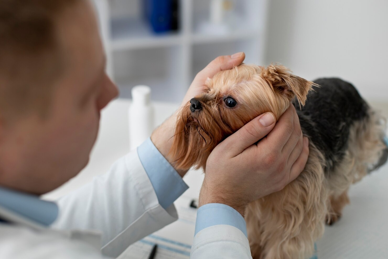
The diagnostic process invariably begins with a comprehensive, hands-off visual assessment of the dog in the exam room, observing their general facial symmetry, blink rate, and visual tracking ability. The veterinarian then proceeds to a detailed, close-up examination in a darkened room, utilizing a specialized, high-intensity focal light source, such as a Finoff transilluminator. This focused beam allows the clinician to brightly illuminate the eye and critically assess the localized structures. They will meticulously evaluate the architecture of the eyelids for abnormal hairs or anatomical rolling, assess the color and swelling of the conjunctival tissues, and closely examine the character of the ocular discharge. Furthermore, they will perform specific neurological assessments, including the pupillary light reflex (PLR) to check the functional integrity of the iris and the optic nerve, the menace response to evaluate basic visual perception, and the dazzle reflex to assess deep retinal function even through a cloudy, opaque cornea[25]. Utilizing a direct or indirect ophthalmoscope, complete with specialized magnifying lenses, the veterinarian can perform a dilated fundoscopy, casting light deep through the pupil to meticulously examine the health of the retina, the highly vascular choroid, and the optic disc at the very back of the eye.
Schirmer Tear Test
Because an adequate, healthy tear film is the eye’s primary, absolute defense against environmental pathogens, quantifying the dog’s natural tear production is a mandatory, foundational step in diagnosing almost any ocular surface infection. The Schirmer Tear Test (STT-1) is the standard diagnostic tool used to measure the exact volume of the aqueous fluid produced by the lacrimal glands. The veterinarian carefully places a specialized, standardized strip of highly absorbent filter paper directly into the ventral conjunctival fornix (the pocket just beneath the lower eyelid). The strip remains in place for exactly one minute, during which the dog’s natural basal and reflex tears wick up the paper, activating a distinct blue dye line. A normal, healthy canine tear production reading is typically between 15 to 25 millimeters per minute. A significantly low reading (less than 10-15 mm/min) definitively diagnoses Keratoconjunctivitis Sicca (dry eye), immediately indicating that the current infection is a direct, secondary result of profound tear deficiency, which dictates a completely different, highly specific, long-term treatment protocol[26].
Slit Lamp Examination
To evaluate the deeper, microscopic structures of the eye, veterinary ophthalmologists and well-equipped general practitioners utilize a highly specialized, advanced piece of equipment known as a slit lamp biomicroscope. This sophisticated instrument provides extreme, adjustable magnification and a brilliant, highly focused, slit-shaped beam of light that can optically “slice” through the transparent tissues of the cornea and the internal chambers of the eye. By precisely adjusting the angle and the width of the light beam, the clinician can evaluate the exact, microscopic depth of a corneal ulcer, perfectly assessing whether it has dangerously penetrated down to Descemet’s membrane (a true emergency known as a descemetocele). Crucially, the slit lamp allows the veterinarian to peer deep into the anterior chamber (the fluid-filled space between the cornea and the iris) to detect the subtle, microscopic presence of floating inflammatory proteins and individual white blood cells. This phenomenon, known as the Tyndall effect or “aqueous flare,” is the absolute definitive diagnostic hallmark of active, internal uveitis, a severe condition that requires immediate, aggressive systemic and topical intervention[27].
Cytology and Culture
When a dog presents with a severe, atypical, rapidly progressing, or highly chronic eye infection that has completely failed to respond to initial, empirical broad-spectrum antibiotic therapies, obtaining a precise, specific microbiological diagnosis becomes absolutely critical. The veterinarian will typically apply a drop of rapid-acting topical anesthetic (such as proparacaine) to deeply numb the eye. They will then use a sterile, microscopic cytobrush or a blunt surgical spatula to gently but firmly scrape the active edge of the infected conjunctiva or the margins of a corneal ulcer. This highly cellular sample is then rolled onto a glass microscope slide, specially stained, and immediately examined under a high-powered microscope. This crucial cytological evaluation allows the clinician to instantly identify the specific type of inflammatory response (e.g., neutrophils indicating bacteria, eosinophils indicating severe allergies) and visually detect the actual presence of specific bacterial morphotypes (rods or cocci), fungal hyphae, or budding yeast organisms. Simultaneously, a sterile swab of the deep infected tissue is carefully collected and submitted to an external diagnostic laboratory for formal aerobic and anaerobic bacterial culture and comprehensive antimicrobial susceptibility testing. This advanced testing determines the precise, specific species of the invading pathogen and exactly which specific, targeted antibiotics will effectively kill it, completely eliminating the dangerous guesswork from the treatment plan.
Additional Diagnostic Tests
A complete, thorough ophthalmic workup frequently demands additional, highly specialized diagnostic modalities. Fluorescein staining is an absolutely mandatory test performed on almost every single red or painful eye. The veterinarian applies a few drops of a brilliant, neon-green fluorescent dye to the surface of the eye, followed by a sterile saline flush. The healthy, lipid-rich, intact corneal epithelium heavily repels the water-soluble dye; however, if there is a scratch, abrasion, or deep infectious ulcer present, the exposed, hydrophilic collagen of the underlying corneal stroma acts like a sponge, instantly absorbing the dye and glowing a bright, unmistakable apple-green under a specialized cobalt blue ultraviolet light. This explicitly highlights the exact size, shape, and severe depth of the corneal injury.
Furthermore, accurately measuring the intraocular pressure (IOP) is a critical, life-saving component of the exam, typically performed using a highly precise electronic rebound tonometer (such as a TonoVet) or an applanation tonometer (such as a Tono-Pen). Normal canine IOP strictly ranges from 10 to 25 mmHg. A significantly elevated pressure reading definitively diagnoses secondary glaucoma, while a significantly decreased reading (less than 10 mmHg) strongly indicates active, severe internal uveitis. For highly complex or persistent eye conditions, or when an underlying, systemic metabolic issue is strongly suspected, the veterinarian may highly recommend comprehensive, full-panel blood tests to evaluate organ function, or advanced imaging studies, such as specialized high-resolution ocular ultrasound, to visualize the deep, hidden structures behind a cloudy, opaque, or severely swollen eye.
Treatment Options for Eye Infections in Dogs

The therapeutic management of canine eye infections is a highly precise, heavily nuanced, and strictly targeted medical endeavor. Because the eye is structurally isolated and protected by the blood-ocular barrier, achieving effective drug concentrations within the actual tissues is remarkably challenging. Treatment protocols diverge considerably based on the specific location of the infection (surface versus deep internal structures), the exact classification of the identified pathogen, and the concurrent presence of any predisposing, underlying diseases. A generic, one-size-fits-all approach is never appropriate in veterinary ophthalmology; every single therapeutic plan must be carefully customized by the attending veterinarian.
Antimicrobial Therapy
The absolute cornerstone of treating the vast majority of bacterial ocular infections involves the rigorous, scheduled application of powerful, prescription-grade topical antimicrobial medications, typically formulated as highly concentrated liquid eye drops or thick, viscous ointments that adhere to the ocular surface. The specific choice of antibiotic is dictated by the severity of the infection and the results of cytological testing. For routine, uncomplicated, superficial bacterial conjunctivitis, veterinarians frequently prescribe broad-spectrum, triple-antibiotic combinations (such as neomycin, polymyxin B, and bacitracin) or basic aminoglycosides (like tobramycin). However, for severe, deep-seated, rapidly melting corneal ulcers often caused by aggressive Gram-negative organisms like Pseudomonas, the veterinarian must deploy incredibly potent, deep-penetrating fluoroquinolones, such as ofloxacin or ciprofloxacin. In these critical, vision-threatening cases, the application frequency is incredibly intense; owners may be required to administer the drops as frequently as every 1 to 2 hours, around the clock, to rapidly halt the enzymatic destruction of the cornea[28].
Beyond traditional antibiotics, specific pathogens require highly specific pharmaceutical agents. If a deep fungal keratitis is diagnosed—often following a severe agricultural injury—topical antifungal medications like natamycin, miconazole, or compounded voriconazole are utilized, often necessitating weeks or even months of strict, dedicated therapy. Viral infections, particularly those caused by the Canine Herpesvirus (CHV-1), are entirely unresponsive to antibacterial medications. These challenging cases require the frequent application of specialized topical antiviral drops, such as idoxuridine or cidofovir, or the administration of systemic oral antiviral medications like famciclovir, to effectively suppress the viral replication occurring within the corneal tissues.
Crucially, controlling the massive, destructive inflammation associated with the infection is just as vital as killing the pathogen itself. Veterinarians frequently utilize topical Non-Steroidal Anti-Inflammatory Drugs (NSAIDs), such as flurbiprofen or diclofenac, to safely reduce severe swelling and mitigate deep pain. In very specific, carefully selected cases—such as severe, internal uveitis or chronic, autoimmune Pannus—topical corticosteroids (like dexamethasone or prednisolone acetate) are deployed for their profound, unmatched immunosuppressive capabilities. However, corticosteroids are strictly, absolutely contraindicated if there is any damage, scratch, or ulceration present on the cornea. Applying a steroid to an ulcerated eye will completely shut down the local immune response, massively accelerate the destructive collagenase enzymes, and virtually guarantee the rapid, catastrophic rupture of the eyeball.
Tear Replacement Therapy
When an eye infection is diagnosed as a direct, secondary consequence of underlying Keratoconjunctivitis Sicca (KCS) or general tear film instability, simply eradicating the surface bacteria is entirely insufficient; the primary, foundational lack of moisture must be aggressively and permanently addressed. Tear replacement therapy is a critical, ongoing component of the treatment plan. Veterinarians frequently recommend the frequent, daily application of high-quality, long-lasting artificial tears containing advanced viscous agents, such as hyaluronic acid, carbomer gels, or cross-linked hydroxypropyl methylcellulose. These sophisticated lubricants physically coat the eye, significantly supplement the deficient aqueous and mucin layers, heavily soothe the severely irritated, inflamed tissues, and physically wash away accumulated debris and opportunistic bacteria.
However, basic artificial tears merely treat the symptoms of dry eye; they do absolutely nothing to stop the underlying autoimmune destruction of the tear glands. To achieve true, long-term disease control and prevent chronic, recurrent infections, the veterinarian will prescribe powerful, topical immunomodulatory medications, most notably cyclosporine (often formulated as a 0.2% ophthalmic ointment) or tacrolimus. These advanced, highly specific drugs work deeply within the tissues to directly suppress the inappropriate, destructive local immune response (T-cell activation) targeting the lacrimal glands. Over time, these medications not only drastically reduce the severe tissue inflammation but also physically stimulate the damaged glands to resume their natural, healthy tear production. This highly specialized therapy is almost always a lifelong commitment for the dog, requiring strict, daily administration to maintain the health of the ocular surface and prevent the devastating return of the dry eye state and its associated, severe secondary infections.
Surgery
While the vast majority of simple ocular infections are successfully managed with aggressive, targeted topical medical therapy, severe, complicated, or anatomically driven cases frequently necessitate highly advanced, specialized surgical intervention. Ophthalmic surgery is typically performed by an experienced veterinarian or a board-certified veterinary ophthalmologist, utilizing operating microscopes and specialized microsurgical instruments while the dog is under deep, carefully monitored general anesthesia. Surgery becomes the primary, absolute treatment of choice when a severe infection has caused massive, irreversible structural damage to the eye, when a deeply embedded, dangerous foreign body must be physically extracted, or when an underlying anatomical defect of the eyelids is the root cause of the chronic, recurrent infections.
The potential surgical interventions are highly diverse and precisely tailored to the specific pathology. For dogs suffering from chronic, incredibly painful infections secondary to entropion (inward-rolling eyelids) or severe macrostomia (excessively large eyelid openings), surgeons perform a highly delicate procedure known as a modified Hotz-Celsus. This precise surgery involves meticulously removing a calculated, elliptical section of the excess skin adjacent to the eyelid margin, permanently everting the eyelid to its normal, healthy anatomical position, and completely eliminating the chronic, abrasive mechanical trauma to the cornea.
In cases of critical, rapidly melting corneal ulcers that are threatening to completely rupture the globe despite aggressive medical therapy, surgeons may perform a life-saving conjunctival pedicle graft. This highly advanced microsurgery involves carefully dissecting a thin flap of healthy, highly vascularized conjunctival tissue from the white of the eye and delicately suturing it directly over the deep, infected ulcer defect using microscopic sutures. This graft acts as an immediate, physical structural patch to prevent the eye from bursting, and it crucially delivers a massive, direct, and continuous supply of fresh blood, healing serum, white blood cells, and systemic antibiotics directly to the avascular, infected site, dramatically accelerating the healing process. In extreme, devastating circumstances where a severe, uncontrollable infection has completely destroyed the internal structures of the eye, resulting in irreversible blindness and intractable, agonizing pain (such as end-stage secondary glaucoma or massive, deep intraocular infection), a complete enucleation—the surgical removal of the entire diseased eye and the closure of the eyelids—is the most humane, definitive, and necessary procedure to permanently eliminate the dog’s suffering and prevent the severe infection from spreading systemically.
Complementary Treatments
In conjunction with the rigorous, mandatory application of prescription medications and the potential necessity for surgical intervention, there are several highly important, supportive complementary therapies that owners must strictly implement at home to maximize the dog’s comfort and ensure successful, uncomplicated healing. The single most critical, absolutely non-negotiable complementary treatment for any dog suffering from an eye infection or a corneal ulcer is the immediate, continuous, and strict use of a hard plastic Elizabethan collar (the “cone of shame”). Because eye infections are inherently, intensely painful and profoundly pruritic, the dog’s natural, overwhelming instinct is to violently rub and scratch at the eye with their paws or against the furniture. Just one aggressive, poorly timed scratch can effortlessly turn a minor, superficial healing ulcer into a catastrophic, full-thickness, surgical emergency rupture of the globe. The E-collar must remain on the dog 24 hours a day, 7 days a week, until the veterinarian explicitly confirms through diagnostic staining that the eye is completely, fully healed.
Additionally, maintaining meticulous, gentle hygiene around the infected eye is essential. Using clean, warm, exceptionally soft compresses applied gently to the closed eyelids can significantly help soothe the inflamed, aching tissues, gently loosen hard, dried crusts, and facilitate the safe, painless removal of the thick, heavy, infectious discharge that frequently glues the eyelids shut. However, owners must ensure they use completely separate, clean materials for each individual eye to absolutely prevent the dangerous cross-contamination and accidental spreading of the infection from a diseased eye to a healthy one. These supportive care measures, while seemingly simple, play a massive, critical role in the overall success of the comprehensive treatment plan. Always consult your veterinarian before making any changes to your pet’s care or introducing any new, unprescribed treatments or over-the-counter remedies.
Prevention of Eye Infections in Dogs
While it is practically impossible to completely, one-hundred-percent shield an active, curious dog from every conceivable environmental pathogen or minor accidental trauma, there are numerous, highly effective, proactive, and practical strategies that responsible pet owners can diligently implement to significantly minimize the risk of their dog developing a painful, vision-threatening eye infection. Prevention is deeply rooted in maintaining excellent general hygiene, recognizing early, subtle warning signs, protecting the eyes during high-risk environmental activities, and ensuring the dog receives comprehensive, regular, and thorough veterinary care.
Regular Eye Checks
The foundation of proactive ocular health is the establishment of frequent, routine, at-home visual inspections. Owners should make it a specific, weekly habit to closely examine their dog’s eyes in a brightly lit environment. You are looking for the baseline “normal”: the eyes should be bright, completely clear, wide open, and free of any significant redness, swelling, or heavy discharge. The pupils should be perfectly equal in size and highly responsive to changes in lighting. By establishing what your dog’s eyes look like when they are perfectly healthy, you become instantly, acutely aware of even the most subtle, early changes—such as a slight increase in tearing, a mild, new squint, or a faint, concerning cloudiness appearing on the cornea. The earlier a potential issue is visually detected and immediately brought to a veterinarian’s attention, the faster it can be accurately diagnosed and effectively treated, often preventing a minor, superficial irritation from evolving into a massive, deep-seated, and complicated infection.
Maintaining Proper Hygiene
Maintaining meticulous, proper hygiene around the delicate periocular area is an absolutely vital component of preventing serious ocular surface infections, particularly for specific breeds that are anatomically predisposed to heavy tear staining, deep facial folds, or excessive facial hair. If your dog’s breed is prone to tearing, establishing a rigorous, daily cleaning routine is non-negotiable. Owners must use a soft, clean, highly absorbent cloth or specifically formulated, veterinary-approved ocular hygiene wipes to gently, carefully clear away the accumulated daily tears, the oxidized porphyrin stains, and any trapped environmental debris from the corners of the eyes and the surrounding deep skin folds. This critical action prevents the dark, moist, and nutrient-rich tears from stagnating on the skin, thereby eliminating the ideal breeding ground for the explosive overgrowth of opportunistic bacteria and aggressive yeast. When bathing the dog, extreme, hyper-vigilant care must be taken to prevent highly irritating, chemical-laden shampoos, harsh soaps, or potent topical flea-control products from accidentally entering the eyes, as these harsh chemicals can instantly strip the protective tear film, cause severe, acute chemical conjunctivitis, and leave the deeply irritated tissues highly vulnerable to secondary bacterial invasion.
Taking Protective Measures
Physical protection of the ocular structures is absolutely essential, particularly for highly active, energetic, working dogs or those that enthusiastically accompany their owners on vigorous, outdoor adventures. When a dog is allowed to hang their head out the window of a fast-moving vehicle, their eyes are directly, violently exposed to a barrage of high-velocity environmental hazards, including sharp dust particles, tiny pebbles, large insects, and heavily desiccating, high-speed winds that rapidly strip the tear film. This activity is a massive, highly preventable cause of severe corneal trauma and subsequent infection. Keeping the car windows safely rolled up is the simplest, most effective preventative measure. Furthermore, if your dog frequently engages in activities like deep-brush hunting, aggressive hiking through dense, thorny undergrowth, or running through tall, dry fields filled with hazardous grass awns, investing in high-quality, specialized protective canine eyewear (such as Doggles) is an incredibly wise, highly protective decision. These durable, shatterproof goggles act as a physical, impenetrable shield, ensuring your dog can prevent foreign bodies from ever reaching the eye, drastically reducing the risk of severe, sight-threatening trauma and the massive infections that inevitably follow.
Healthy Diet
While there is no single, magical food ingredient that can guarantee a dog will never experience an eye infection, the provision of a highly balanced, nutritionally complete, and antioxidant-rich diet plays a massive, foundational role in supporting the overall systemic health of the animal, the robust function of their immune system, and the specific, highly demanding metabolic needs of the ocular tissues. The tissues of the eye, particularly the highly active retina and the rapidly regenerating corneal epithelium, require a constant, rich supply of specific essential nutrients to function optimally and maintain their structural defenses.
Diets rich in high-quality, bioavailable Omega-3 fatty acids (specifically EPA and DHA, commonly sourced from cold-water marine fish oils) are incredibly beneficial for ocular health. Omega-3s possess potent, natural systemic anti-inflammatory properties that can significantly help mitigate the severity of ocular surface inflammation, and they are absolutely crucial for maintaining the healthy, functional composition of the lipid layer of the tear film, drastically reducing tear evaporation and preventing the onset of dry eye. Furthermore, ensuring adequate, balanced intake of specific antioxidants—such as Vitamin A (critical for the health and maintenance of the corneal epithelium and retinal function), Vitamin C, Vitamin E, and crucial carotenoids like Lutein and Zeaxanthin—provides the cellular tissues of the eye with the necessary tools to combat the massive, daily oxidative stress caused by constant exposure to ultraviolet sunlight and normal metabolic processes. Always consult with your veterinarian to select a high-quality, scientifically formulated diet that appropriately meets your dog’s specific, individual nutritional requirements based on their age, breed, and activity level.
Regular Vet Visits
The importance of routine, comprehensive veterinary care cannot be overstated in the realm of preventative ocular health. Annual or bi-annual wellness examinations provide the veterinarian with the crucial opportunity to perform a thorough, professional ophthalmic assessment, utilizing specialized equipment to detect the incredibly subtle, early signs of developing ocular diseases—such as early-onset cataracts, slowly decreasing tear production, or mild, early-stage glaucoma—long before these conditions produce obvious, visible symptoms or trigger massive, secondary infections. Furthermore, these regular veterinary visits allow for the early, proactive identification and strict management of underlying, systemic health conditions, such as Diabetes or Cushing’s disease, which drastically increase the dog’s overall susceptibility to severe, opportunistic infections. Maintaining a strong, consistent relationship with a trusted veterinarian is your dog’s absolute best defense against the development of serious, preventable eye conditions.
Vaccination
Finally, adhering to a strict, highly comprehensive, veterinary-recommended vaccination protocol is a deeply fundamental, crucial component of preventing severe, systemic viral diseases that frequently feature catastrophic, sight-threatening ocular complications. Maintaining current, active immunity against the highly contagious Canine Distemper Virus (CDV) and Canine Adenovirus Type 1 (CAV-1) is absolutely essential. These are profoundly devastating, often fatal systemic viral pathogens that directly attack and destroy the delicate ocular tissues, causing massive, unmanageable viral conjunctivitis, profound, painful corneal edema, and irreversible neurological blindness. By ensuring your dog is fully, properly immunized, these vaccines can help prevent diseases from ever establishing an infection, completely protecting the dog from both the systemic illness and the severe, associated ocular manifestations. Furthermore, it is important to understand the broader implications of ocular health; while relatively rare, certain highly specific pathogens associated with dog eye infections can be contagious and potentially spread to humans, particularly zoonotic bacteria like Leptospira or certain resistant strains of Staphylococcus. Practicing excellent hygiene, such as washing your hands thoroughly after treating your dog’s eyes, and maintaining a robust, proactive preventative healthcare plan protects not only your dog’s vision but the health of your entire family.
Frequently Asked Questions
Can a dog eye infection spread to humans?
While the vast majority of routine canine eye infections are highly species-specific and pose no threat to humans, there are certain specific, zoonotic bacterial and parasitic pathogens that can potentially be transmitted from an infected dog to a human. For instance, highly aggressive bacterial strains like Staphylococcus pseudintermedius or specific environmental pathogens introduced through trauma can, under the right circumstances, infect a human, particularly if the person is immunocompromised or has open wounds on their hands. Therefore, it is absolutely essential to practice strict, rigorous hygiene. Always wash your hands thoroughly with antibacterial soap and warm water immediately after handling your dog, cleaning their ocular discharge, or administering any prescribed ophthalmic medications, and avoid touching your own eyes or face during the treatment process.
How long does it take for antibiotic eye drops to work in dogs?
The speed at which prescription antibiotic eye drops produce a noticeable, clinical improvement in a dog is highly dependent on the severity of the specific infection, the depth of the tissue involvement, and the exact virulence of the targeted bacteria. For a mild, uncomplicated, superficial bacterial conjunctivitis, owners frequently observe a significant, rapid reduction in redness, swelling, and discharge within 24 to 48 hours of initiating the strict, prescribed treatment schedule. However, for a deep, complicated, melting corneal ulcer, it may take several days of intense, around-the-clock therapy to simply halt the rapid progression of the tissue destruction before any actual healing begins. Crucially, even if the eye appears completely normal after a few days, you must meticulously administer the medication for the entire, full duration prescribed by your veterinarian to ensure the complete eradication of the bacteria and prevent a severe, highly resistant relapse.
Will an eye infection go away on its own?
In veterinary ophthalmology, it is highly recommended to never assume that an eye infection will simply resolve on its own without professional medical intervention. While a very minor, highly transient, superficial irritation from a tiny speck of dust might clear up as the dog’s natural tear film flushes the object away, a true, established bacterial, viral, or fungal infection will rapidly and aggressively worsen if left untreated. Delaying professional, targeted treatment allows the pathogenic organisms to multiply exponentially, potentially leading to deep, destructive corneal ulcerations, severe internal inflammation, intense agony for the dog, and a highly significant risk of permanent scarring or irreversible blindness. If you suspect an eye infection, seeking immediate, comprehensive veterinary care is the only safe and responsible course of action.
References
- Merck Veterinary Manual. Ophthalmology Introduction. Merck & Co., Inc., 2023.
- American Veterinary Medical Association (AVMA). Eye Care for Pets. AVMA, 2022.
- VCA Animal Hospitals. Corneal Ulcers in Dogs. VCA, 2023.
- Ledbetter, E. C., et al. “Bacterial Keratitis in Dogs.” Veterinary Ophthalmology, 2017.
- ASPCA. Common Canine Eye Issues. ASPCA, 2021.
- VCA Animal Hospitals. Conjunctivitis in Dogs. VCA, 2023.
- Merck Veterinary Manual. Corneal Diseases in Small Animals. Merck & Co., Inc., 2023.
- Maggs, D. J. “Diseases of the Eyelids.” Slatter’s Fundamentals of Veterinary Ophthalmology, 2018.
- Veterinary Information Network (VIN). Canine Uveitis. VIN, 2019.
- American Veterinary Medical Association (AVMA). Canine Distemper. AVMA, 2022.
- Ledbetter, E. C. “Canine Herpesvirus-1 Ocular Diseases.” Veterinary Ophthalmology, 2012.
- Merck Veterinary Manual. Glaucoma in Small Animals. Merck & Co., Inc., 2023.
- VCA Animal Hospitals. Keratoconjunctivitis Sicca (Dry Eye) in Dogs. VCA, 2023.
- Grahn, B. H. “Eyelid Abnormalities in Dogs.” Veterinary Clinics of North America: Small Animal Practice, 2015.
- Wang, L., et al. “Microbial Profile of Canine Ocular Infections.” Journal of Veterinary Internal Medicine, 2019.
- American Veterinary Medical Association (AVMA). Canine Infectious Respiratory Disease Complex. AVMA, 2022.
- Centers for Disease Control and Prevention (CDC). Blastomycosis in Pets. CDC, 2021.
- Peano, A., et al. “Malassezia pachydermatis in Veterinary Medicine.” Veterinary Dermatology, 2014.
- Merck Veterinary Manual. Ophthalmic Emergencies. Merck & Co., Inc., 2023.
- VCA Animal Hospitals. Eye Injuries in Dogs. VCA, 2023.
- Paulsen, M. E. “Traumatic Ocular Injuries.” Veterinary Clinics of North America: Small Animal Practice, 2016.
- Merck Veterinary Manual. Hyperadrenocorticism. Merck & Co., Inc., 2023.
- American Kennel Club (AKC). Dog Eye Infections: Causes, Symptoms, and Treatment. AKC, 2021.
- Giuliano, E. A. “Diseases of the Lacrimal System.” Veterinary Clinics of North America: Small Animal Practice, 2011.
- Veterinary Information Network (VIN). The Neuro-Ophthalmic Examination. VIN, 2020.
- Williams, D. L. “The Schirmer Tear Test.” Veterinary Ophthalmology, 2005.
- VCA Animal Hospitals. Testing for Eye Problems in Dogs. VCA, 2023.
- Merck Veterinary Manual. Ophthalmic Antibiotics and Antifungals. Merck & Co., Inc., 2023.


